. 2014 Jun 12;5(6):e1288.
doi: 10.1038/cddis.2014.248.

Genetic deficiency of neuronal RAGE protects against AGE-induced synaptic injury

Affiliations

Genetic deficiency of neuronal RAGE protects against AGE-induced synaptic injury

Hongju Zhang et al. Cell Death Dis. .

Abstract

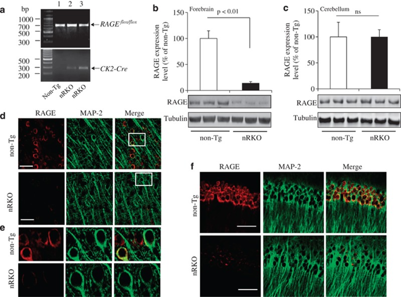
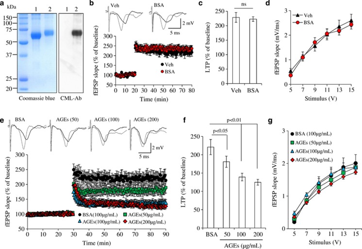
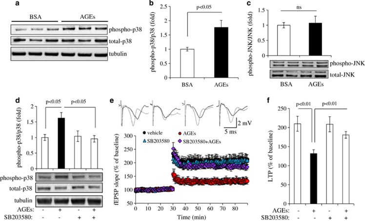
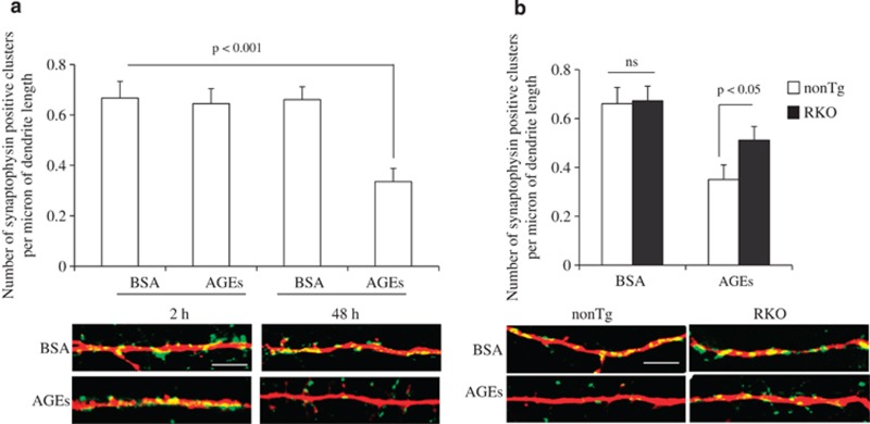
Synaptic dysfunction and degeneration is an early pathological feature of aging and age-related diseases, including Alzheimer's disease (AD). Aging is associated with increased generation and deposition of advanced glycation endproducts (AGEs), resulting from nonenzymatic glycation (or oxidation) proteins and lipids. AGE formation is accelerated in diabetes and AD-affected brain, contributing to cellular perturbation. The extent of AGEs' involvement, if at all, in alterations in synaptic structure and function is currently unknown. Here we analyze the contribution of neuronal receptor of AGEs (RAGE) signaling to AGE-mediated synaptic injury using novel transgenic neuronal RAGE knockout mice specifically targeted to the forebrain and transgenic mice expressing neuronal dominant-negative RAGE (DN-RAGE). Addition of AGEs to brain slices impaired hippocampal long-term potentiation (LTP). Similarly, treatment of hippocampal neurons with AGEs significantly decreases synaptic density. Such detrimental effects are largely reversed by genetic RAGE depletion. Notably, brain slices from mice with neuronal RAGE deficiency or DN-RAGE are resistant to AGE-induced LTP deficit. Further, RAGE deficiency or DN-RAGE blocks AGE-induced activation of p38 signaling. Taken together, these data show that neuronal RAGE functions as a signal transducer for AGE-induced synaptic dysfunction, thereby providing new insights into a mechanism by which the AGEs-RAGE-dependent signaling cascade contributes to synaptic injury via the p38 MAP kinase signal transduction pathway. Thus, RAGE blockade may be a target for development of interventions aimed at preventing the progression of cognitive decline in aging and age-related neurodegenerative diseases.


Figures

Figure 1
Figure 1
Targeted construct cassette. Schematic representation of the strategy used for conditional knockout of the RAGE gene by excision of exons 2–4 of the mouse RAGE gene. (a) Annotated (MacVector 9.5.3) display of the locus identifying the region targeted for homologous recombination. The targeting vector was constructed from three fragments, the 5′ homology arm, two loxP sites and the 3′ homology arm. PGK-neo selection cassette is inserted downstream of exon 1. The PGK-neo cassette is flanked by FRT sites and exons 2–4 are flanked by two loxP sites. (b) RAGE flox/flox mice were crossed with neuronal target CK2 mice to generate nRKO mice (RAGEflox/flox/CK2-Cre). Two loxP sites flanking RAGE exons 2 to 4 to allow for Cre-mediated deletion using Cre-recombinase. Excision of exons 2–4 by Cre recombinase leads to a frame shift and early stop codon from the mouse RAGE sequence to block RAGE expression
Figure 2
Figure 2
Characterization of transgenic nRKO mice with RAGE deletion in cortical neurons. (a) Transgenic nRKO mice were identified from tail DNA based on PCR amplication using primers for flox (700 bp) in upper panel, and CK2-Cre (300 bp) in the lower panel. Lane 1 indicates RAGEflox/flox (non-Tg ) mice with negative Cre transgene and lanes 2-3 denotes nRKO mice carried with both flox and Cre (RAGEflox/flox/CK2-Cre ). (b and c) Immunoblotting of cortical homogenates (b) and cerebellum (c) from the indicated Tg mice for RAGE. Tubulin was used as a protein-loading control. (df) Representative images of double immunostaining for RAGE and MAP2 in cortex (d and e) and hippocampus (f). Confocal microscopy images showing RAGE deletion in cortical neurons labeled with positive MAP2 as a neuronal marker. Enlarged large images of neurons derived from the corresponding cells are shown in the frame of panel (e). Scale bar=50 μm
Figure 3
Figure 3
AGE-impaired long-term potentiation (LTP). (a) Determination of the degree of CML in AGE preparation. Protein (2 μg) from BSA was incubated with glucose 6-phosphate for 8 weeks at 37 °C and then subjected to SDS-PAGE. Protein bands were stained by Coomassie Blue (left). Glycation was detected by immunoblotting with antibody to CML (right). Lane 1, BSA; lane 2, AGE-modified BSA. (b) BSA alone did not affect LTP. Inserts show representative traces of fEPSP in slices treated with vehicle or 100 μg/ml BSA before θ-burst stimulation (black line) and at the end of 1 h recording (gray line). (c) Residual potentiation from the fEPSP slopes occurring over the last 5 min of LTP recordings. (d) BSA did not affect basal synaptic transmission (BST). (e) Effect of AGE on LTP. Hippocampal slices were perfused with AGE (50, 100, and 200 μg/ml) or BSA (100 μg/ml) for 1 h and then recorded LTP. N=8–11 slices from 3 to 5 mice. (f) Residual potentiation from the fEPSP slopes occurring over the last 5 min of LTP recordings. (g) The BST of hippocampal synapses was recorded from the indicated groups of mice
Figure 4
Figure 4
Effect of RAGE depletion on AGE-induced LTP impairment. (ac) LTP was recorded in hippocampal slices from non-Tg, RKO (a), nRKO (b), and DN-RAGE (c) mice after 1 h perfusion with BSA or AGEs (100 μg/ml). RAGE deletion alone did not alter LTP in the presence of BSA. In contrast, LTP was increased in slices from RKO, nRKO, and DN-RAGE mice compared with non-Tg slices in the presence of AGE. (d) Representative traces of fEPSP in slices treated with vehicle or 100 μg/ml BSA before θ-burst stimulation (black line) and after 1 h (gray line). (e) Residual potentiation from the fEPSP slopes occurring over the last 5 min of LTP recordings. (f) There were no significant differences in BST among indicated groups. N=8–11 slices from 3 to 5 mice
Figure 5
Figure 5
Effect of AGEs on p38 activation. Brain slices were perfused with BSA or AGEs (100 μg/ml) for 1 h and then subjected to immunoblotting for phosphorylation and total p38 (a and b) or JNK (c). (a) Immunoblotting of brain homogenates from AGE- or BSA-treated slices for phosphorylation and total p38. Tubulin was used as the neuronal protein-loading control. (b) Densitometry of the combined phospho-p38 immunoreactive bands relative to total p38. (c) Densitometry of phospho-JNK immunoreactive bands relative to total JNK in slices treated with BSA or AGE. Data are expressed as fold increase relative to the BSA-treated control group. The lower panel shows representative immunoblots for the indicated proteins. N=3–5 per group of treatment. (d) Effect of p38 inhibitor (SB203580) on AGE-induced p38 phosphorylation. Densitometry of phospho-p38 immunoreactive bands relative to total p38 in non-Tg slices perfused with AGEs with or without SB203580. Representative immunoblots for the indicated protein are shown in the lower panel. (e) Effects of p38 inhibitor (SB203580) on AGE-induced LTP deficits in hippocampal slices. Slices were perfused with vehicle, AGEs (100 μg/ml), SB203580 (1 μM), or AGEs plus SB203580 for 1 h before LTP recording. n=8–11 slices per group of treatment. Upper panel shows representative traces of fEPSP in slices with the indicated treatments before θ-burst stimulation (black line) and at the end of 1 h (gray line). (f) Residual potentiation from fEPSP slopes occurring over the last 5 min of LTP recording
Figure 6
Figure 6
Effect of RAGE deficiency on AGE-induced p38 activation. Hippocampal slices from the indicated Tg mice and non-Tg mice were perfused with AGEs for 1 h and then subjected to immunoblotting for phospho- and total-p38. (a) Immunoblots for phospho- and total-p38 with tubulin used as protein-loading control. (b) Densitometry of phospho-p38 immunoreactive bands normalized by total p38 using the NIH Image J software. N=3–5 mice per group
Figure 7
Figure 7
Effect of RAGE deficiency on AGE-induced synaptic loss. (a) Non-Tg neurons were treated with 100 μg/ml AGEs or BSA for 2 and 48 h and then subjected to immunostaining with synaptophysin and MAP2. Quantification of positive synaptophysin clusters per micron of dendrites was significantly decreased in AGE-treated cells compared with BSA-treated cells. (b) RAGE-deficient neurons largely protected AGE-induced loss of synaptophysin-positive clusters compared with non-Tg neurons in the presence of AGEs. Lower panel shows representative images of synaptic staining. Synapses were visualized by synaptophysin staining (green) and dendrites by MAP2 staining (red). Scale bar=50 μm

References

    1. 1Li J, Liu D, Sun L, Lu Y, Zhang Z. Advanced glycation end products and neurodegenerative diseases: mechanisms and perspective. J Neurol Sci 2012; 317: 1–5. - PubMed
    1. 2Munch G, Thome J, Foley P, Schinzel R, Riederer P. Advanced glycation endproducts in ageing and Alzheimer's disease. Brain Res Brain Res Rev 1997; 23: 134–143. - PubMed
    1. 3Vlassara H, Striker GE. Advanced glycation endproducts in diabetes and diabetic complications. Endocrinol Metab Clin North Am 2013; 42: 697–719. - PubMed
    1. 4Takedo A, Yasuda T, Miyata T, Mizuno K, Li M, Yoneyama S et al. Immunohistochemical study of advanced glycation end products in aging and Alzheimer's disease brain. Neurosci Lett 1996; 221: 17–20. - PubMed
    1. 5Luth HJ, Ogunlade V, Kuhla B, Kientsch-Engel R, Stahl P, Webster J et al. Age- and stage-dependent accumulation of advanced glycation end products in intracellular deposits in normal and Alzheimer's disease brains. Cereb Cortex 2005; 15: 211–220. - PubMed

Publication types

MeSH terms

Substances

LinkOut - more resources