. 2015 Jan;52(1):37-45.
doi: 10.1165/rcmb.2013-0359OC.

Hypoxia-induced changes in protein s-nitrosylation in female mouse brainstem

Affiliations

Hypoxia-induced changes in protein s-nitrosylation in female mouse brainstem

Lisa A Palmer et al. Am J Respir Cell Mol Biol. 2015 Jan.

Abstract

Exposure to hypoxia elicits an increase in minute ventilation that diminishes during continued exposure (roll-off). Brainstem N-methyl-D-aspartate receptors (NMDARs) and neuronal nitric oxide synthase (nNOS) contribute to the initial hypoxia-induced increases in minute ventilation. Roll-off is regulated by platelet-derived growth factor receptor-β (PDGFR-β) and S-nitrosoglutathione (GSNO) reductase (GSNOR). S-nitrosylation inhibits activities of NMDAR and nNOS, but enhances GSNOR activity. The importance of S-nitrosylation in the hypoxic ventilatory response is unknown. This study confirms that ventilatory roll-off is virtually absent in female GSNOR(+/-) and GSNO(-/-) mice, and evaluated the location of GSNOR in female mouse brainstem, and temporal changes in GSNOR activity, protein expression, and S-nitrosylation status of GSNOR, NMDAR (1, 2A, 2B), nNOS, and PDGFR-β during hypoxic challenge. GSNOR-positive neurons were present throughout the brainstem, including the nucleus tractus solitarius. Protein abundances for GSNOR, nNOS, all NMDAR subunits and PDGFR-β were not altered by hypoxia. GSNOR activity and S-nitrosylation status temporally increased with hypoxia. In addition, nNOS S-nitrosylation increased with 3 and 15 minutes of hypoxia. Changes in NMDAR S-nitrosylation were detected in NMDAR 2B at 15 minutes of hypoxia. No hypoxia-induced changes in PDGFR-β S-nitrosylation were detected. However, PDGFR-β phosphorylation increased in the brainstems of wild-type mice during hypoxic exposure (consistent with roll-off), whereas it did not rise in GSNOR(+/-) mice (consistent with lack of roll-off). These data suggest that: (1) S-nitrosylation events regulate hypoxic ventilatory response; (2) increases in S-nitrosylation of NMDAR 2B, nNOS, and GSNOR may contribute to ventilatory roll-off; and (3) GSNOR regulates PDGFR-β phosphorylation.

Keywords: N-methyl-D-aspartate receptor; S-nitrosoglutathione reductase; hypoxic ventilatory response; neuronal nitric oxide synthase; platelet-derived growth factor receptor-β.


Figures

Figure 1.
Figure 1.
S-nitrosoglutathione (GSNO) reductase (GSNOR) and neuronal nitric oxide synthase (nNOS) are located in neurons within the nucleus tractus solitarius (NTS). (A) Immunohistochemical identification of GSNOR and nNOS in the NTS. Upper panels demonstrate the location of these proteins in neurons within the brainstem in a region corresponding to the NTS. Lower panels contain an enlargement of the indicated sections in the upper panels. Scale bars, 200 μm (upper panels), 100 μm (lower panels). (B) Colocalization of GSNOR and nNOS in neurons in the NTS by immunofluorescence. Neurons that contain both GSNOR and nNOS are indicated by the thin white arrows. Neurons that do not contain GSNOR are indicated by the thick white arrows. Scale bars, 50 μm for all panels.
Figure 2.
Figure 2.
Minute ventilation (Vm) during hypoxic challenge does not show roll-off in GSNOR+/− or GSNOR−/− mice. (A) Vm in female C57BL6, GSNOR+/−, and GSNOR−/− mice under normoxia (N), and 3 and 15 minutes of exposure to hypoxia. The data are presented as means (± SEM). There were 12 mice in each group. *P < 0.05, significant change from N values; P < 0.05, GGNOR+/− or GSNOR−/− mice versus C57BL6 mice. (B) GSNOR activity increases temporally with exposure to hypoxia in female C57BL6 mice. GSNOR activity is significantly less in normoxia (N) in GSNOR+/− and GSNOR−/− mice compared with C57BL6 mice. In addition, there are no hypoxia-induced increases in GSNOR activity in GSNOR+/− or GSNOR−/− mice. The data are presented as means (±SEM). There were six to eight samples in each group. *P < 0.05, significant change from N values; P < 0.05, GGNOR+/− or GSNOR−/− mice versus C57BL6 mice; P < 0.05, GGNOR−/− mice versus C57BL6 and GSNOR+/− mice.
Figure 3.
Figure 3.
Acute hypoxia does not alter protein abundance. (A) Western blots demonstrating the lack of change in protein abundance of nNOS, endothelial NOS (eNOS), GSNOR, N-methyl-D-aspartate (NMDA) receptor (NMDAR) subunits, NR1, NR2A, and NR2B, platelet-derived growth factor receptor (PDGFR)-β, or β-actin. (B) Graphical representation of the changes in protein expression upon exposure to acute hypoxia. Hypoxia-induced changes in protein expression are presented by comparing protein abundance in hypoxia to the normoxic controls for each protein examined. The data are not normalized to β-actin. The data are presented as means (±SEM). There were eight samples in each group. There were no significant effects of hypoxic challenge on protein expression (P > 0.05, for all comparisons).
Figure 4.
Figure 4.
Hypoxia alters the S-nitrosylation status of GSNOR, nNOS, and NR2B. Changes in protein S-nitrosylation were determined by the biotin-switch assay followed by Western blot analysis. (A) Western blot of proteins after biotin switch. All proteins have some level of S-nitrosylation under normoxic (baseline) conditions. Three proteins, GSNOR, nNOS and NR2B showed increases in S-nitrosylation with exposure to acute hypoxia. (B) Graphical representation of the change in S-nitrosylation status of the selected proteins with exposure to hypoxia. Change in S-nitrosylation status is determined by the S-nitrosylated protein:total protein ratio. The data are presented as means (± SEM). There were eight samples in each group. *P < 0.05, significant change from normoxic (N) values.
Figure 5.
Figure 5.
PDGFR-β phosphorylation is not altered by hypoxia in GSNOR+/−mice. (A) PDGFR-β protein abundance and phosphorylation in brainstem homogenates from female C57BL6 mice exposed to normoxia and 3 and 15 minutes of hypoxia. (B) PDGFR-β protein abundance and phosphorylation in brainstem homogenates from GSNOR+/− mice exposed to normoxia, and 3 and 15 minutes of hypoxia. (C) Graphical representation of the changes in phosphorylated/total PDGFR-β protein in C57BL6 and GSNOR mice (n = 3–4). *P < 0.024, significant change from normoxia.
Figure 6.
Figure 6.
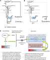
Schematic diagram of hypoxia-induced changes in S-nitrosylation during the hypoxic ventilatory response (HVR). (A) S-nitrosylation status during phase I of the HVR. NR2B, nNOS and GSNOR have a low level of basal S-nitrosylation. NMDAR channel is open in response to glutamate, glycine, and/or GSNO, allowing influx of Ca2+, which activates nNOS, and production of GSNO and NO. The GSNO produced is not efficiently catabolized, as GSNOR activity is minimal. Release of NO enhances release of glutamate from presynaptic cells. (B) S-nitrosylation status during ventilatory roll-off. Continued exposure to hypoxia augments S-nitrosylation of NR2B, nNOS, and GSNOR. S-nitrosylation of the NR2B decreases NMDAR channel opening, thus decreasing Ca2+ influx. Decreased Ca2+ influx and increased S-nitrosylation status of nNOS decreases the production of GSNO and NO. GSNOR activity is elevated, resulting in the increased catabolism of GSNO. Low NO production results in a minimal release of glutamate from presynaptic cells. All processes would thereby promote ventilatory roll-off during hypoxic challenge. (C) Schematic diagram of the potential mechanisms and sites of action of S-nitrosothiols in the HVR. Hb, hemoglobin.

Similar articles

Cited by

References

    1. Lahiri S, Roy A, Baby SM, Hoshi T, Semenza GL, Prabhakar NR. Oxygen sensing in the body. Prog Biophys Mol Biol. 2006;91:249–286. - PubMed
    1. Teppema LJ, Dahan A. The ventilatory response to hypoxia in mammals: mechanisms, measurement, and analysis. Physiol Rev. 2010;90:675–754. - PubMed
    1. Gozal D, Torres JE, Gozal YM, Littwin SM. Effect of nitric oxide synthase inhibition on cardiorespiratory responses in the conscious rat. J Appl Physiol (1985) 1996;81:2068–2077. - PubMed
    1. Gozal D, Gozal E, Simakajornboon N. Signaling pathways of the acute hypoxic ventilatory response in the nucleus tractus solitarius. Respir Physiol. 2000;121:209–221. - PubMed
    1. Gozal D, Simakajornboon N, Czapla MA, Xue YD, Gozal E, Vlasic V, Lasky JA, Liu JY. Brainstem activation of platelet-derived growth factor-beta receptor modulates the late phase of the hypoxic ventilatory response. J Neurochem. 2000;74:310–319. - PubMed

Publication types

MeSH terms

Substances