. 2014 Aug;87(1040):20140184.
doi: 10.1259/bjr.20140184. Epub 2014 Jun 12.

Platelet factor 4 protects bone marrow mesenchymal stem cells from acute radiation injury

Affiliations

Platelet factor 4 protects bone marrow mesenchymal stem cells from acute radiation injury

J-J Chen et al. Br J Radiol. 2014 Aug.

Abstract

Objective: The aim of this study was to find a new radiation protector, platelet factor 4 (PF4) and to identify its effect on haemopoietic microenvironment in vitro and in vivo.

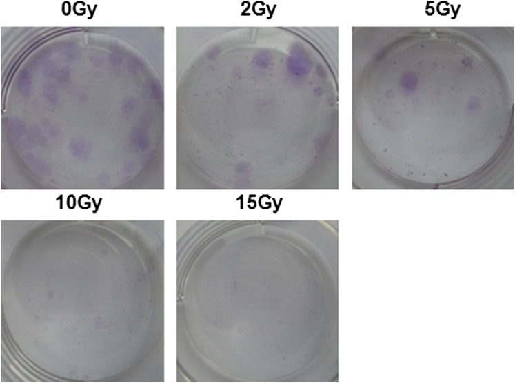
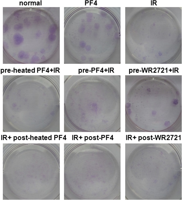
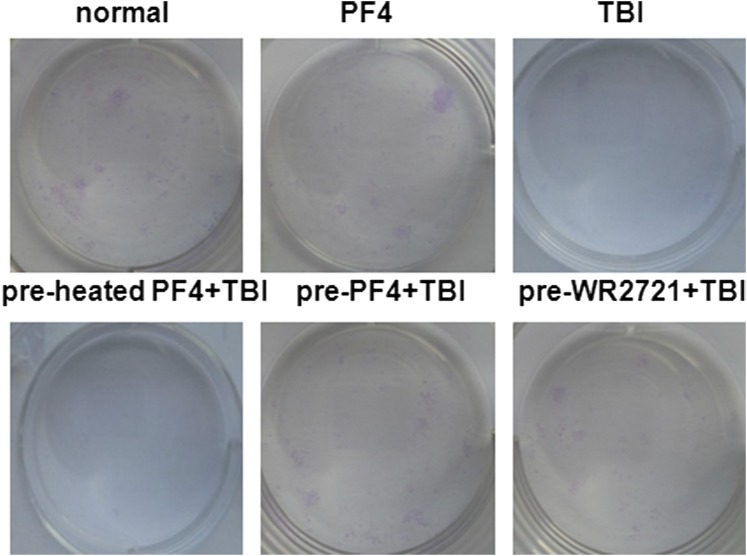
Methods: Radiation damage on bone marrow mesenchymal stem cells ex and in vitro was set up as models. Growth curve analysis, clonogenic survival assay, FACSCalibur™ (BD Immunocytometry Systems, San Jose, CA), 5-ethynyl-2'-deoxyuridine immunofluorescence staining and quantitative reverse transcription-polymerase chain reaction were employed to assess the characterization of bone marrow mesenchymal stem cells (BMSCs), proliferation, apoptosis, cell cycle and gene expression.

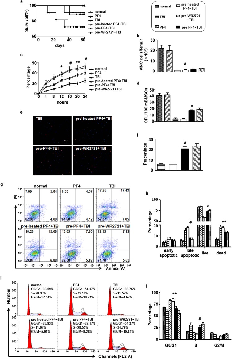
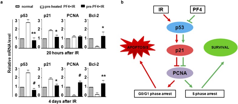
Results: A dose- and time-dependent enhancement of cell viability and survival was observed for PF4 treatment along with 500 cGy γ-radiation in vitro. The same phenomena were noted in vivo, including enhancement of adherence and proliferation ability while inhibition of cell apoptosis, which were associated with a short-term decrease in the G0/G1 ratio owing to S phase arrest. These were accompanied with enhanced Bcl-2 expression and p53/p21 loss.

Conclusion: These results uncover that PF4 might be a novel therapeutic approach, which could reduce DNA damage and increase survival of BMSCs, in part, by inhibiting p53/p21 axis and facilitating DNA damage repair.

Advances in knowledge: This study explores the feasibility of a new radioprotector and hence may be clinically important.


Figures

Figure 1.
Figure 1.
Characterization of bone marrow mesenchymal stem cells (BMSCs). (a) Histogram of cell surface markers determined by flow cytometry, showing that BMSCs were negative for CD45 and CD34 and positive for CD44 and CD29. (b) BMSCs differentiated into adipocytes and osteoblasts for 14 days and subsequently stained with Oil Red and von Kossa staining, respectively. (c) In vitro irradiation survival curves of human BMSCs (hBMSCs). Cells were grown in vitro and irradiated from 0 to 15 Gy alone. (d) The colony-forming unit fibroblasts (CFU-F) in (c) were counted on the 14th day after ionizing irradiation and presented as CFU-F per 100 cultured cells. (e) Effect of platelet factor (PF4) in irradiation killing of hBMSCs in vitro. Cells were irradiated after 12 h incubation in different doses of PF4. (f) Cumulative administration of PF4. Cells were irradiated after different cumulative administrations of PF4 in vitro. Bars, means ± standard deviation. *p < 0.05; **p < 0.01; #p < 0.001; n = 5.
Figure 2.
Figure 2.
Platelet factor 4 (PF4)-promoted human bone marrow mesenchymal stem cells (hBMSCs) proliferation in vitro. (a) Clonogenic survival assay. Cells were incubated with equivalent phosphate-buffered saline, heated PF4, PF4 (1 μg ml−1) or WR-2721 (10 μM) before or after 500 cGy ionizing irradiation and were counted on the 14th day after ionizing irradiation (IR). (b) Representative images of 500-cGy irradiated hBMSCs stained with 5-ethynyl-2′-deoxyuridine (EdU). (c) Quantification of EdU+ cells in (b). (d) Carboxyfluorescein diacetate succinimidyl ester (CFSE) proliferation assay. CFSE-labelled hBMSCs were stimulated with PF4 (1 μg ml−1) or WR-2721 (10 μM) before or after 500 cGy IR. Bars, means ± standard deviation. *p < 0.050; **p < 0.010; #p < 0.001; n = 5. CFU, colony-forming unit.
Figure 3.
Figure 3.
Platelet factor 4 (PF4)–repressed human bone marrow mesenchymal stem cell (hBMSC) apoptosis and induced S phase arrest in vitro. (a–f) hBMSCs were pre-incubated with 1 μg ml−1 of PF4 for 12 h or 10 μM WR-2721 for 30 min before 500- and 0-cGy ionizing irradiation. Cell apoptosis was analysed by FACSCalibur™ (BD Immunocytometry Systems, San Jose, CA) on the second day (a, b); on the fourth day (c, d); and on the sixth day (e, f). (g) Short-term S phase arrest regulation by PF4. Cell cycle was analysed by FACSCalibur at 20 h. (h) Qualitative measurement of (g). Bars, means ± standard deviation. *p < 0.05; **p < 0.01; #p < 0.001; n = 5. IR, ionizing irradiation; PI, propidium iodide.
Figure 4.
Figure 4.
Platelet factor 4 (PF4)–protected bone marrow mesenchymal stem cells (BMSCs) in vivo. Two intraperitoneal injections of PF4 (40 μg kg−1) or equivalent heated PF4 were administered at 6-h intervals, and 20 h after the second injection, the mice received 500-cGy γ-radiation total body irradiation (TBI). WR-2721 (150 mg kg−1) was administered 30 min before TBI for a positive control. The mice were sacrificed by cervical dislocation 3 days after exposure. (a) The survival of the mice was plotted for 60 days. (b) The number of mononuclear cells (MNCs) in different treated mice on the third day after exposure. (c) The percentages of adherent MNC in different culture time. (d) Colony-forming unit fibroblasts (CFU-F) were counted at the 14th day after culture. 5-ethynyl-2′-deoxyuridine (EdU) immunofluorescent staining was performed on the tenth culture day. (e) Representative images of mouse BMSCs stained with EdU. (f) Quantification of EdU+ cells in (e). (g, h) Cell apoptosis was analysed by FACSCalibur™ (BD Immunocytometry Systems, San Jose, CA) on the fifth culture day. (i, j) Cell cycle was performed when isolated bone marrow cells were totally adherent. Bars, means ± standard deviation. *p < 0.05; **p < 0.01; #p < 0.001; n = 8. PI, propidium iodide.
Figure 5.
Figure 5.
Platelet factor 4 (PF4)–modulated cell cycle and apoptosis-related gene expression. (a) Human bone marrow mesenchymal stem cells were pre-incubated with PF4 or equivalent heated PF4 for 12 h before 500 cGy ionizing irradiation, and gene expression was analysed by quantitative reverse transcription–polymerase chain reaction at 20 h and 4 days after ionizing irradiation (IR). The unradiated cells were used as a control. (b) Schematic diagram of potential signaling triggered by PF4. Bars, means ± standard deviation. *p < 0.05; **p < 0.01, #p < 0.001; n = 8. PCNA, proliferating cell nuclear antigen.

Similar articles

Cited by

References

    1. Harfouche G, Martin MT. Response of normal stem cells to ionizing radiation: a balance between homeostasis and genomic stability. Mutat Res 2010; 704: 167–74. doi: 10.1016/j.mrrev.2010.01.007 - DOI - PubMed
    1. Méndez-Ferrer S, Michurina TV, Ferraro F, Mazloom AR, Macarthur BD, Lira SA, et al. . Mesenchymal and haematopoietic stem cells form a unique bone marrow niche. Nature 2010; 466: 829–34. - PMC - PubMed
    1. Greenberger JS, Epperly M. Bone marrow-derived stem cells and radiation response. Semin Radiat Oncol 2009; 19: 133–9. doi: 10.1016/j.semradonc.2008.11.006 - DOI - PMC - PubMed
    1. Singh S, Kloss FR, Brunauer R, Schimke M, Jamnig A, Greiderer-Kleinlercher B, et al. . Mesenchymal stem cells show radioresistance in vivo. J Cell Mol Med 2012; 16: 877–87. doi: 10.1111/j.1582-4934.2011.01383.x - DOI - PMC - PubMed
    1. Nicolay NH, Sommer E, Lopez R, Wirkner U, Trinh T, Sisombath S, et al. . Mesenchymal stem cells retain their defining stem cell characteristics after exposure to ionizing radiation. Int J Radiat Oncol Biol Phys 2013; 87: 1171–8. - PubMed

Publication types

MeSH terms

Substances