. 2014 Jun 12;10(6):e1004431.
doi: 10.1371/journal.pgen.1004431. eCollection 2014 Jun.

Muscle structure influences utrophin expression in mdx mice

Affiliations

Muscle structure influences utrophin expression in mdx mice

Glen B Banks et al. PLoS Genet. .

Abstract

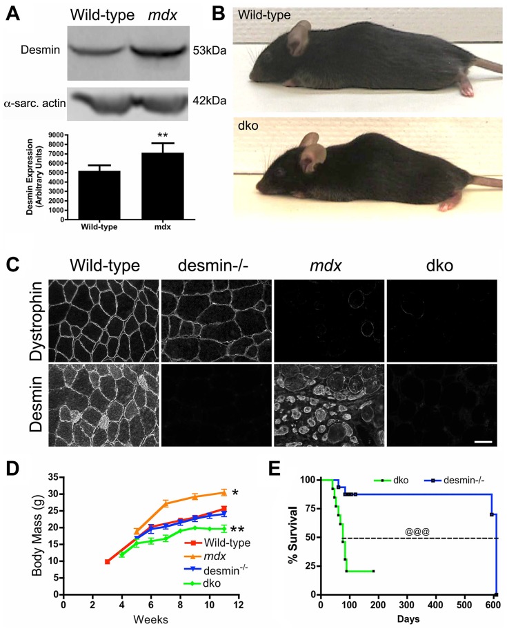
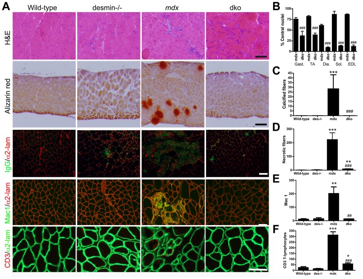
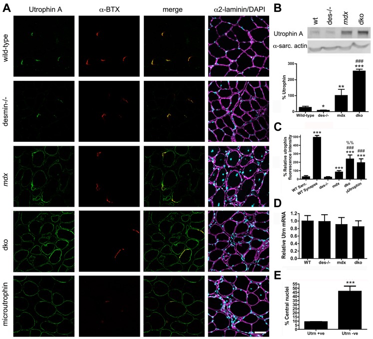
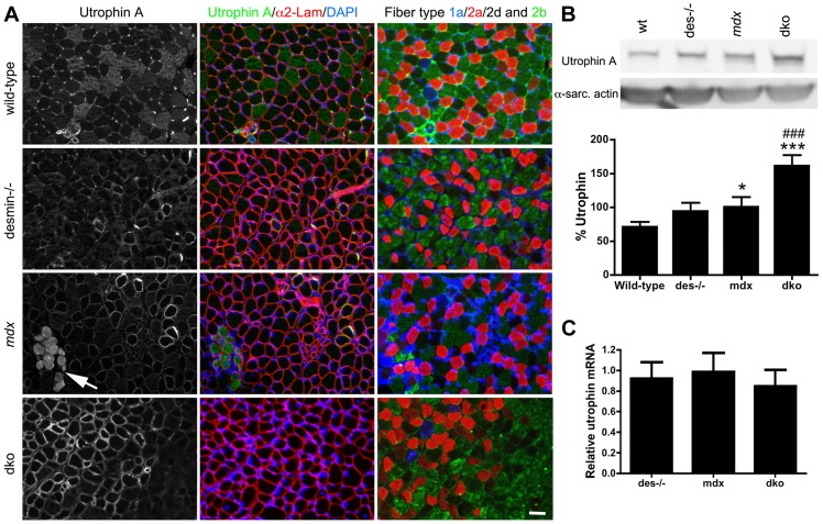
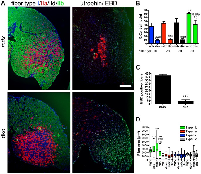
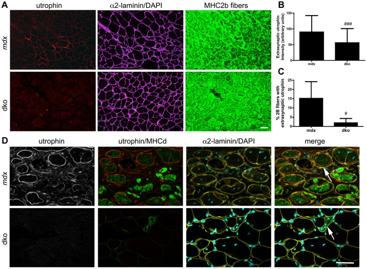
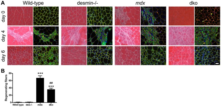
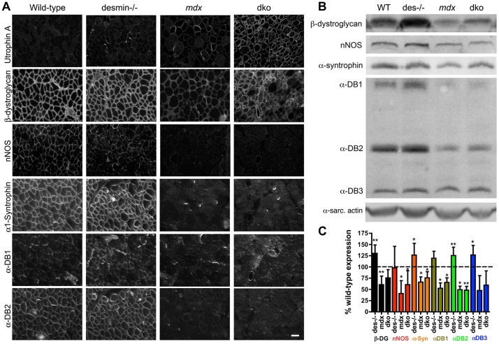
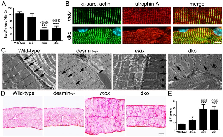
Duchenne muscular dystrophy (DMD) is a severe muscle wasting disorder caused by mutations in the dystrophin gene. To examine the influence of muscle structure on the pathogenesis of DMD we generated mdx4cv:desmin double knockout (dko) mice. The dko male mice died of apparent cardiorespiratory failure at a median age of 76 days compared to 609 days for the desmin-/- mice. An ∼ 2.5 fold increase in utrophin expression in the dko skeletal muscles prevented necrosis in ∼ 91% of 1a, 2a and 2d/x fiber-types. In contrast, utrophin expression was reduced in the extrasynaptic sarcolemma of the dko fast 2b fibers leading to increased membrane fragility and dystrophic pathology. Despite lacking extrasynaptic utrophin, the dko fast 2b fibers were less dystrophic than the mdx4cv fast 2b fibers suggesting utrophin-independent mechanisms were also contributing to the reduced dystrophic pathology. We found no overt change in the regenerative capacity of muscle stem cells when comparing the wild-type, desmin-/-, mdx4cv and dko gastrocnemius muscles injured with notexin. Utrophin could form costameric striations with α-sarcomeric actin in the dko to maintain the integrity of the membrane, but the lack of restoration of the NODS (nNOS, α-dystrobrevin 1 and 2, α1-syntrophin) complex and desmin coincided with profound changes to the sarcomere alignment in the diaphragm, deposition of collagen between the myofibers, and impaired diaphragm function. We conclude that the dko mice may provide new insights into the structural mechanisms that influence endogenous utrophin expression that are pertinent for developing a therapy for DMD.


Conflict of interest statement

The authors have declared that no competing interests exist.

Figures

Figure 1
Figure 1. The genetic deletion of desmin from mdx4cv mice reduces body mass and survival.
A) Desmin expression was significantly increased in the mdx4cv skeletal muscles. Bars represent the mean +/− S.D. densitometry of desmin expression from n = 7 wild-type and n = 6 mdx4cv gastrocnemius muscles. B) Photograph of representative wild-type and dko mice. Note that the dko mice develop a mild kyphosis at 11 weeks of age. C) Confirmation of genotype by immunostaining of frozen gastrocnemius muscle sections with antibodies to dystrophin and desmin. Scale bar  = 50 µm D) Mean +/− S.D. body mass of wild-type (n = 5), mdx4cv (n = 4), desmin−/− (n = 5) and dko mice (n = 9). E) Kaplan-Meyer survival analyses demonstrating the significantly shortened lifespan of dko mice (n = 13) compared with desmin−/− mice (n = 16). *P<0.05, **P<0.01 compared with wild-type. @@@P<0.001 compared to desmin−/−.
Figure 2
Figure 2. The dystrophic histopathology in mdx4cv mice was profoundly improved by the absence of desmin.
A) Frozen sections demonstrating the histopathology of skeletal muscles. Note that the extensive central nucleation and mononuclear cell infiltrate, calcification, necrosis and inflammation in mdx4cv muscles were significantly diminished in the dko skeletal muscles. All panels are representative sections of gastrocnemius muscle except the second row, which are sections of the diaphragm. Scale bars  = 100 µm. B) The number of centrally nucleated fibers was significantly diminished in hind-limb and respiratory muscles in the dko mice when compared with the mdx4cv muscles C) Calcified fibers were found in the mdx4cv diaphragm muscle but not in the dko muscles. D) Quantitation of the total number of necrotic fibers in the gastrocnemius muscles. E) Quantitation of macrophages in the gastrocnemius muscles. F) Quantitation of the CD3 positive T-lymphocytes in the gastrocnemius muscles. N = 4 for all experiments. All bars in the graphs represent mean +/− S.D. *P<0.05, **P<0.01 and ***P<0.001 compared to wild-type; # P<0.05, ## P<0.01 and ### P<0.001 compared to mdx4cv.
Figure 3
Figure 3. Expression and localization of utrophin in wild-type, desmin−/−, mdx4cv and dko muscles at 11 weeks of age.
A) Frozen sections of the gastrocnemius muscles immunolabelled with antibodies to utrophin A and α-bungarotoxin (α-BTX). Utrophin was restricted to the neuromuscular junctions in wild-type and desmin−/− muscles. Utrophin was expressed on the extrasynaptic sarcolemma in the mdx4cv and dko muscles. Note the increased utrophin expression on the sarcolemma of dko fibers compared with the mdx4cv muscles. Scale bar  = 50 µm. B) Western blot analyses of utrophin A expression in whole gastrocnemius muscle lysates from wild-type (n = 3), desmin−/− (n = 3), mdx4cv (n = 7) and dko (n = 6) mice. Quantitation of utrophin expression in whole muscle lysates is shown below the immunoblots. C) Maximal utrophin fluorescence intensity was significantly increased on the sarcolemma of dko fibers compared with mdx4cv fibers. Furthermore, maximal fluorescence intensity was significantly increased in dko fibers compared to mdx:utrophin double knockout fibers expressing microutrophinΔR4–R21. N = 4. D) We found no change in utrophin mRNA when comparing whole gastrocnemius muscle lysates when utrophin mRNA was normalized to the housekeeping gene Ywhaz. N = 4. E) Utrophin prevents muscle degeneration and regeneration in dko gastrocnemius muscles as demonstrated by the reduced proportion of fibers with central nuclei. N = 4. All bar graphs show the mean +/− S.D. *P<0.05 and ***P<0.001 compared to wild-type; # P<0.05 and ### P<0.001 compared to mdx4cv.
Figure 4
Figure 4. Expression and localization of utrophin in wild-type, desmin−/−, mdx4cv and dko muscles at 3 weeks of age.
A) Utrophin expression in gastrocnemius muscles compared to adjacent sections labeled for the different skeletal muscle fiber-types. Note that utrophin expression is restricted to the neuromuscular junctions and non-muscle cells in the wild-type muscles. Utrophin is also restricted to the neuromuscular junctions in most fast 2b fibers in desmin−/− muscles, but remains on the sarcolemma of the type 1a, 2a and 2d/x fiber types. Utrophin is found on the extrasynaptic sarcolemma of mdx4cv muscles, irrespective of fiber-type at 3 weeks of age. Regions where utrophin is lost from the extrasynaptic sarcolemma in mdx4cv muscles have necrotic fibers (arrows). Utrophin expression is lost from most of the fast 2b fibers in the dko by 3 weeks of age, but is retained on the sarcolemma of 1a, 2a and 2d/x fiber types. Scale bar  = 50 µm. B) Western blot analyses of utrophin A expression in whole gastrocnemius muscle lysates from (n = 4), desmin−/− (n = 4), mdx4cv (n = 8) and dko (n = 8) mice. Quantitation of utrophin expression in whole muscle lysates is shown below the immunoblots. C) We found no change in utrophin mRNA when comparing whole gastrocnemius muscle lysates, when utrophin mRNA was normalized to the housekeeping gene Ywhaz. N = 4. *P<0.05, P<0.001 compared to wild-type. ### P<0.001 compared to mdx4cv.
Figure 5
Figure 5. Utrophin maintains the integrity of the dko muscle membrane in a fiber-type specific manner.
A) Shown are frozen sections of the lateral portion of the gastrocnemius muscle immunolabeled with monoclonal antibodies to fiber types 1a (blue), 2a (red), 2d/x (black) and 2b (green; left panel) or utrophin (green; right panel) and Evan's blue dye (EBD; red; right panel). Note that the uneven distribution of utrophin expression in the mdx4cv muscles correlated with patches of adjacent membrane permeable fibers that labeled with EBD. However, an increase in utrophin expression in the dko myofibers excluded EBD from the 1a, 2a and 2d/x fiber types. The dko fast 2b fibers, which lacked utrophin, were permeable to EBD. Scale bar  = 500 µm. B) Bars show the mean +/− S.D. percentage of centrally nucleated fibers in distinct fiber types. Note that all dko muscle fiber types had significantly less myonuclei than the mdx4cv fibers (## P<0.01 and ### P<0.001). The dko fast 2b fibers had more central nuclei than the 1a, 2a and 2d/x fiber types (@@@P<0.001). The mdx4cv fast 2b fibers had more central nuclei than the 1a, 2a and 2d/x fiber types (**P<0.01). C) Bars show the mean +/− S.D. total number of EBD positive fibers in the gastrocnemius muscles. ***P<0.001 compared with mdx4cv myofibers. D) Bars show the mean +/− S.D. area of type 1a, 2a, 2d/x and 2b muscle fiber types. ***P<0.001 compared with wild-type myofibers. ### P<0.001 compared with mdx4cv myofibers. @@@P<0.001 compared with desmin−/− myofibers. All experiments were from n = 4 mice.
Figure 6
Figure 6. Utrophin expression was reduced on the extrasynaptic sarcolemma of dko fast 2b fibers.
A) Utrophin expression in transverse sections of the near pure population of fast 2b fibers in the most superficial region of the gastrocnemius muscles. Shown are single sections from mdx4cv or dko gastrocnemius fast 2b fibers labeled with utrophin in red, α2-laminin in magenta, DAPI in cyan and fast 2b fibers in green. B) Quantitation of maximal extrasynaptic utrophin fluorescence intensity in fast 2b fibers and C) the proportion of fast 2b fibers expressing extrasynaptic utrophin in the mdx4cv and dko. All bars represent the mean +/− S.D. from n = 4 mice. # P<0.05 and ### P<0.001 compared with mdx4cv fibers. D) Regenerating fibers expressed utrophin in the mdx4cv fibers, but not in the dko. Shown are single sections from mdx4cv or dko superficial gastrocnemius muscles labeled with utrophin in red, developmental myosin heavy chain in green, α2-laminin in yellow and DAPI in cyan. Note that utrophin is on the expanding sarcolemma in mdx4cv muscles, but not in the dko (arrows). Scale bars  = 50 µm.
Figure 7
Figure 7. Regenerative capacity of muscle.
A) Transverse sections of uninjured (Day 0) or injured gastrocnemius muscles 4 days and 6 days post notexin administration. Sections stained with hematoxylin and eosin are shown in the left columns and developmental myosin heavy chain (green), α2-laminin (red) and DAPI (blue) are shown in the right columns. Scale bar  = 50 µm. B) Bars represent the mean +/− S.D. of the total number of regenerating fibers in the uninjured gastrocnemius muscles (Day 0). ***P<0.001 compared with wild-type muscles and ## P<0.01 compared with mdx4cv muscles. N = 4 mice for all experiments.
Figure 8
Figure 8. Localization and expression of β-dystroglycan and the NODS complex in skeletal muscles.
A) Adjacent sections of gastrocnemius muscles showing the localization of β-dystroglycan and the NODS complex at the sarcolemma of wild-type, desmin-/-, mdx4cv and dko skeletal muscles. Scale bar  = 50 µm. B) Western analysis of quadriceps muscles reveals an increase in expression of DGC proteins in the desmin-/- and a reduction in the mdx4cv and dko muscles. α-DB is α-dystrobrevin and α-sarc. actin is the α-sarcomeric actin loading control. C) Bars show mean +/− S.D. densitometric quantitation of protein expression graphed as a percentage of wild-type. *P<0.05, **P<0.01 compared to wild-type. N = 4–8 for all experiments.
Figure 9
Figure 9. Impaired diaphragm function in the dko correlates with loss of sarcomere alignment and deposition of collagen.
A) Mean +/− S.D. specific force of diaphragm strips in vitro from wild-type (n = 6), desmin−/− (n = 5), mdx4cv (n = 5) and dko (n = 5) mice. B) Utrophin A colocalizes with α-sarcomeric actin in longitudinal sections. Note the misalignment of α-sarcomeric actin and utrophin A in the dko myofiber. Scale bar  = 6 µm. C) Electron microscopy of longitudinal sections of diaphragm muscle demonstrating the alignment of sarcomeres in wild-type and mdx mice (arrows). Note the alignment of sarcomeres is perturbed in desmin−/− muscles (arrows) and severely impaired in the dko muscles (arrows point to misalignment of sarcomeres while the arrow head points toward a hyper-contracted myofiber). Scale bars  = 2 µm. D) Sirius red staining of collagen in transverse frozen sections of the wild-type (n = 8), desmin−/− (n = 9), mdx4cv (n = 5) and dko (n = 5) diaphragms. Scale bar  = 100 µm. E) Mean +/− S.D. of Sirius red staining as a proportion of the muscle area. *P<0.05, ***P<0.001 compared to wild-type, @@@P<0.001 compared to desmin−/−.

References

    1. Mendell JR, Shilling C, Leslie ND, Flanigan KM, al-Dahhak R, et al. (2012) Evidence-based path to newborn screening for Duchenne muscular dystrophy. Ann Neurol 71: 304–313. - PubMed
    1. Hoffman EP, Brown RH Jr, Kunkel LM (1987) Dystrophin: the protein product of the Duchenne muscular dystrophy locus. Cell 51: 919–928. - PubMed
    1. Koenig M, Hoffman EP, Bertelson CJ, Monaco AP, Feener C, et al. (1987) Complete cloning of the Duchenne muscular dystrophy (DMD) cDNA and preliminary genomic organization of the DMD gene in normal and affected individuals. Cell 50: 509–517. - PubMed
    1. Bhasin N, Law R, Liao G, Safer D, Ellmer J, et al. (2005) Molecular extensibility of mini-dystrophins and a dystrophin rod construct. J Mol Biol 352: 795–806. - PubMed
    1. Mirza A, Sagathevan M, Sahni N, Choi L, Menhart N (2010) A biophysical map of the dystrophin rod. Biochim Biophys Acta 1804: 1796–1809. - PubMed

Publication types

MeSH terms