. 2014 Jun 11;15(6):681-91.
doi: 10.1016/j.chom.2014.05.010.

The Nogo receptor NgR1 mediates infection by mammalian reovirus

Affiliations

The Nogo receptor NgR1 mediates infection by mammalian reovirus

Jennifer L Konopka-Anstadt et al. Cell Host Microbe. .

Abstract

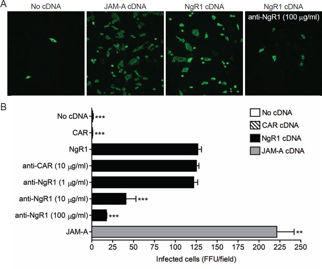
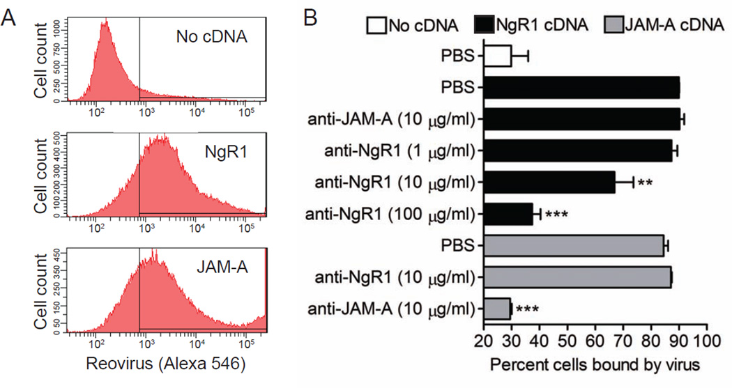
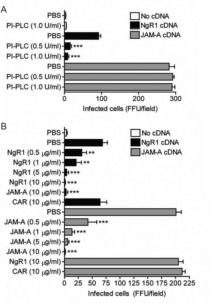
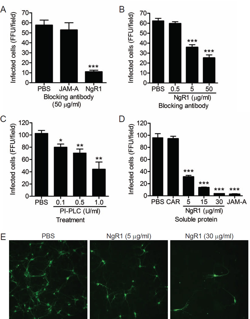
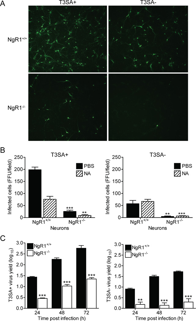
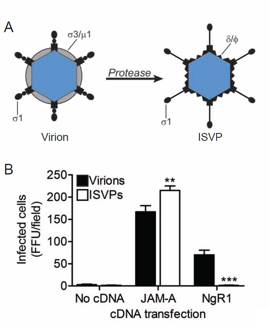
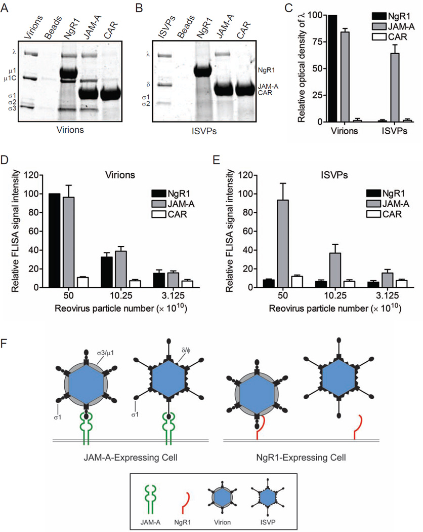
Neurotropic viruses, including mammalian reovirus, must disseminate from an initial site of replication to the central nervous system (CNS), often binding multiple receptors to facilitate systemic spread. Reovirus engages junctional adhesion molecule A (JAM-A) to disseminate hematogenously. However, JAM-A is dispensable for reovirus replication in the CNS. We demonstrate that reovirus binds Nogo receptor NgR1, a leucine-rich repeat protein expressed in the CNS, to infect neurons. Expression of NgR1 confers reovirus binding and infection of nonsusceptible cells. Incubating reovirus virions with soluble NgR1 neutralizes infectivity. Blocking NgR1 on transfected cells or primary cortical neurons abrogates reovirus infection. Concordantly, reovirus infection is ablated in primary cortical neurons derived from NgR1 null mice. Reovirus virions bind to soluble JAM-A and NgR1, while infectious disassembly intermediates (ISVPs) bind only to JAM-A. These results suggest that reovirus uses different capsid components to bind distinct cell-surface molecules, engaging independent receptors to facilitate spread and tropism.


Figures

Figure 1
Figure 1. Expression of NgR1 confers reovirus infection of non-susceptible cells
CHO cells were either mock transfected or transiently transfected with plasmid encoding NgR1, JAM-A, or CAR. After 48 h, cells were pretreated with PBS, a CAR-specific antibody, or increasing concentrations of NgR1-specific antibody prior to being adsorbed with reovirus T3SA- at an MOI of 10 PFU/cell. Cells were fixed at 20 h and scored for reovirus antigen (green) using indirect immunofluorescence. (A) Representative images are shown. (B) Results are expressed as the mean FFU/field for three fields of view in triplicate samples. Error bars indicate SD. **, P < 0.005; ***, P < 0.0005 (as determined by Student’s t test in comparison to NgR1-transfected cells). See also Figures S2, S3, and S6.
Figure 2
Figure 2. Expression of NgR1 allows reovirus binding to non-susceptible cells
CHO cells were either mock transfected or transiently transfected with plasmid encoding NgR1 or JAM-A. After 48 h, cells were treated with PBS or antibodies specific for JAM-A or NgR1 prior to being incubated with 105 particles/cell of Alexa Fluor 546-labeled reovirus T3SA- on ice for 1 h. The percentage of cells bound by virus was quantified by flow cytometry. (A) Representative flow cytometric profiles are shown. (B) Results are expressed as the percent cells bound by virus for triplicate samples. Error bars indicate SD. **, P < 0.005; ***, P < 0.0005 (as determined by Student’s t test in comparison to NgR1- or JAM-A-transfected cells).
Figure 3
Figure 3. Reovirus infectivity is abrogated when NgR1 is removed from the cell surface or by treatment of virions with soluble NgR1 protein
CHO cells were either mock transfected or transiently transfected with plasmid encoding NgR1 or JAM-A for 48 h. (A) Transfected cells were treated with PI-PLC, which cleaves GPI-anchored proteins, for 1 h prior to adsorption with reovirus T3SA- at an MOI of 10 PFU/cell. (B) Reovirus T3SA- was incubated with soluble Fc-tagged NgR1, JAM-A, or CAR at the indicated concentrations prior to inoculation of transfected cells at an MOI of 10 PFU/cell. (A and B) Cells were fixed at 20 h and scored for reovirus antigen using indirect immunofluorescence. Results are expressed as the mean FFU/field for three fields of view in triplicate samples. Error bars indicate SD. **, P < 0.005; ***, P < 0.0005 (as determined by Student’s t test in comparison to NgR1- or JAM-A-transfected cells). See also Figure S4.
Figure 4
Figure 4. Blocking access to cell-surface NgR1 diminishes reovirus infection of neurons
Primary murine cortical neuron cultures prepared from (A) wildtype JAM-A+/+ or (B) isogenic JAM-A−/− embryos were pretreated with either PBS or antibodies specific for JAM-A or NgR1 prior to adsorption with reovirus T3SA+ at an MOI of 500 PFU/cell. (C) Neuron cultures prepared from wildtype cortices were treated with PI-PLC at increasing concentrations for 1 h prior to adsorption with reovirus T3SA+ at an MOI of 500 PFU/cell. (D) Reovirus T3SA+ was preincubated with increasing amounts of soluble Fc-tagged NgR1, JAM-A (30 µg/ml), or CAR (30 µg/ml) prior to inoculation of wildtype neuron cultures at an MOI of 500 PFU/cell. (A – D) Cells were fixed at 20 h and scored for reovirus antigen (green) using indirect immunofluorescence. Results are expressed as the mean FFU/field for three fields of view in triplicate samples. Representative images of an experiment in (D) are shown in (E). Error bars indicate SD. *, P < 0.05; **, P < 0.005; ***, P < 0.0005 (as determined by Student’s t test in comparison to PBS-treated infected cells). See also Figures S1 and S5.
Figure 5
Figure 5. NgR1 is required for efficient reovirus infection of primary cortical neurons
Primary murine cortical neuron cultures prepared from wildtype NgR1+/+ or isogenic NgR1−/− embryos were pretreated with either PBS or NA prior to adsorption with reovirus T3SA+ or T3SA- at an MOI of 500 PFU/cell. Cells were fixed at 20 h and scored for reovirus antigen (green) using indirect immunofluorescence. (A) Representative images are shown. (B) Results are expressed as the mean FFU/field for three fields of view in triplicate samples. (C) Primary murine cortical neuron cultures prepared from wildtype NgR1+/+ or isogenic NgR1−/− embryos were adsorbed with reovirus T3SA+ or T3SA- at an MOI of 10 PFU/cell. Titers of virus in cell lysates at the indicated intervals were determined by plaque. Results are expressed as mean viral yields for triplicate samples. Error bars indicate SD. **, P < 0.005; ***, P < 0.0005 (as determined by Student’s t test in comparison to NgR1+/+ neurons). See also Figure S7.
Figure 6
Figure 6. NgR1 mediates infection of reovirus virions but not ISVPs
(A) Reovirus virions undergo proteolytic disassembly leading to formation of ISVPs. ISVPs are characterized by loss of outer-capsid protein σ3, an extended conformer of σ1, and cleavage of outer-capsid protein µ1 to form δ and Φ. (B) CHO cells were transiently transfected with plasm ids encoding NgR1 or JAM-A and adsorbed with equal particle numbers of reovirus T3SA- virions or ISVPs. Cells were fixed at 20 h scored for reovirus antigen using indirect immunofluorescence. Results are expressed as the mean FFU/field for three fields of view in triplicate samples. Error bars indicate SD. **, P < 0.005; ***, P < 0.0005 (as determined by Student’s t test in comparison to cells infected with virus).
Figure 7
Figure 7. Reovirus virions but not ISVPs bind to soluble NgR1
Soluble Fc-tagged NgR1, JAM-A, or CAR was immobilized onto protein G beads. Receptor-conjugated beads were incubated with 1011 particles of either reovirus T3SA+ (A) virions or (B) ISVPs, washed extensively, resuspended and boiled in protein-dissociation buffer to release bound material. Precipitated proteins were resolved by SDS-polyacrylamide gel electrophoresis and visualized by colloidal blue staining. (C) Reovirus λ band intensity was quantified by the Odyssey imaging system for three independent experiments. Results are expressed as the mean optical density relative to that of NgR1-precipated λ bands. Error bars indicate SD. (D, E) Plates were coated with soluble Fc-tagged NgR1, JAM-A, or CAR and adsorbed with serial dilutions of reovirus T3SA+ virions (D) or ISVPs (E) in a FLISA binding assay. Wells were washed and incubated with reovirus-specific antibodies and fluorescent secondary antibodies to detect virions or ISVPs bound to receptor protein. Signal intensity was quantified using the Odyssey imaging system. Results are shown as the mean relative FLISA signal for three independent experiments. Error bars indicate SD. (F) Both reovirus virions and ISVPs efficiently bind JAM-A. However, ISVPs are incapable of engaging NgR1, suggesting that the reovirus ligand for NgR1 is either σ3 or the virion-associated conformer of σ1.

Comment in

References

    1. Akbik F, Cafferty WB, Strittmatter SM. Myelin associated inhibitors: a link between injury-induced and experience-dependent plasticity. Exp. Neurol. 2012;235:43–52. - PMC - PubMed
    1. Antar AAR, Konopka JL, Campbell JA, Henry RA, Perdigoto AL, Carter BD, Pozzi A, Abel TW, Dermody TS. Junctional adhesion molecule-A is required for hematogenous dissemination of reovirus. Cell Host Microbe. 2009;5:59–71. - PMC - PubMed
    1. Baer GS, Dermody TS. Mutations in reovirus outer-capsid protein σ3 selected during persistent infections of L cells confer resistance to protease inhibitor E64. J. Virol. 1997;71:4921–4928. - PMC - PubMed
    1. Barton ES, Connolly JL, Forrest JC, Chappell JD, Dermody TS. Utilization of sialic acid as a coreceptor enhances reovirus attachment by multistep adhesion strengthening. J. Biol. Chem. 2001a;276:2200–2211. - PubMed
    1. Barton ES, Forrest JC, Connolly JL, Chappell JD, Liu Y, Schnell F, Nusrat A, Parkos CA, Dermody TS. Junction adhesion molecule is a receptor for reovirus. Cell. 2001b;104:441–451. - PubMed

Publication types

MeSH terms