. 2014 Jun 11;15(6):692-705.
doi: 10.1016/j.chom.2014.05.006.

Circulating avian influenza viruses closely related to the 1918 virus have pandemic potential

Affiliations

Circulating avian influenza viruses closely related to the 1918 virus have pandemic potential

Tokiko Watanabe et al. Cell Host Microbe. .

Abstract

Wild birds harbor a large gene pool of influenza A viruses that have the potential to cause influenza pandemics. Foreseeing and understanding this potential is important for effective surveillance. Our phylogenetic and geographic analyses revealed the global prevalence of avian influenza virus genes whose proteins differ only a few amino acids from the 1918 pandemic influenza virus, suggesting that 1918-like pandemic viruses may emerge in the future. To assess this risk, we generated and characterized a virus composed of avian influenza viral segments with high homology to the 1918 virus. This virus exhibited pathogenicity in mice and ferrets higher than that in an authentic avian influenza virus. Further, acquisition of seven amino acid substitutions in the viral polymerases and the hemagglutinin surface glycoprotein conferred respiratory droplet transmission to the 1918-like avian virus in ferrets, demonstrating that contemporary avian influenza viruses with 1918 virus-like proteins may have pandemic potential.


Figures

Fig. 1
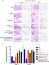
Fig. 1. Replicative ability of 1918 virus, an authentic avian virus, 1918-like avian virus, and 1918-like avian mutant viruses
Virus replication in respiratory organs of ferrets. Ferrets were intranasally infected with 106 PFU of virus. Six animals per group were euthanized on days 3 and 6 post-infection for virus titration. Virus titers in nasal turbinates, tracheae, and lungs were determined by plaque assay in MDCK cells. Horizontal bars indicate the mean virus titers. Asterisks indicate virus titers significantly different from those of the 1918-like avian virus (, p < 0.05; **, p < 0.01). See also Table S2.
Fig. 2
Fig. 2. Pathological analyses of 1918 virus, an authentic avian virus, 1918-like avian virus, and 1918-like avian mutant viruses
(A) Histopathological findings in virus-infected ferrets. Shown are representative pathological changes in tracheae and lungs of ferrets infected with 106 PFU of the indicated viruses on day 6 post-infection. Three ferrets per group were infected intranasally with 106 PFU of virus, and tissues were collected on day 6 after infection for pathological examination. No virus was detected from the lungs of the DK/ALB-infected ferrets. Left panel, hematoxylin-eosin (HE) staining. Right panel, immunohistochemical staining for influenza viral antigen (NP). Scale bars indicate 100 µm. (B) Pathological severity scores for infected ferrets. To represent comprehensive histological changes, respiratory tissue slides were evaluated by scoring pathological changes as described in Supplemental Experimental Procedures. The sum of the pathologic scores for all 5 lung lobes was calculated for each ferret. The means ± standard deviations from three ferrets are shown. Asterisks indicate virus pathological scores significantly different from that of the 1918-like avian virus (Dunnett’s test; p < 0.05). See also Fig. S2.
Fig. 3
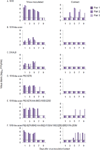
Fig. 3. Respiratory droplet transmission in ferrets
Groups of three ferrets were infected intranasally with 106 PFU of the indicated viruses. One day later, a naïve ferret (contact ferret) was placed in a cage adjacent to each infected ferret. Nasal washes were collected from infected ferrets on day 1 after inoculation and from contact ferrets on day 1 after co-housing, and then every other day (for up to 9 days) for virus titration. The lower limit of detection is indicated by the horizontal dashed line. See also Fig. S4 and Table S3.
Fig. 4
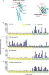
Fig. 4. HA structural analysis and glycan microarray analysis
(A) Localization of amino acid changes identified in viruses recovered from ferrets in the transmission study. Shown is the three-dimensional structure of the monomer of A/Brevig Mission/1/18 (H1N1) HA in complex with human receptor analogues [Protein Data Bank (PDB) code, 2WRG]. A close-up view of the globular head is also shown to the right. Mutations known to increase affinity to human-type receptors are shown in red (E190D and G225D). Mutations that emerged in HA during replication and/or transmission in ferrets are shown in green (E89D, S113N, I187T, and D265V). The amino acid changes at positions 89 and 113 are located close to an amino acid at position 110 (103 with H5 numbering) that was previously found to be associated with the transmissibility of an H5 virus (Herfst et al., 2012). Images were created with MacPymol [http://www.pymol.org/]. (B–E) The receptor specificities of viruses possessing 1918 HA (B), 1918-like avian HA (C), 1918-like avian HA-190D/225D (D), and 1918-like avian HA-89D/113N/190D/225D (E) were assessed by using a glycan microarray containing a diverse library of α2–3 and α2–6 sialosides (Xu et al., 2013). Viruses, directly labeled with biotin, were applied at 128 hemagglutination units/ml for 1 h, and, after washing, were incubated with Streptavidin-AlexaFluor647 (1 µg/ml) for 1 h to detect bound virus. Error bars represent the standard deviation calculated from 6 replicate spots of each glycan. A complete list of glycans is found in Table S6. See also Fig. S5.
Fig. 5
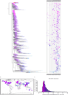
Fig. 5. Global patterns of PB2 proteins derived from avian influenza viruses
(A) Phylogenetic tree of 1,022 randomly selected amino acid sequences of PB2 genes derived from avian influenza viruses. The tree was rooted to the PB2 sequence of the 1918 virus, A/Brevig Mission/1/18 (H1N1) (red arrow). The PB2 from A/blue-winged teal/Ohio/926/2002 (H3N8), which is expressed by the 1918-like avian virus, is indicated by the blue arrow. The year in which the strains were isolated is indicated by horizontal bars to the right of the tree drawn at the same vertical position as the position of the strain in the tree. The tree and time-series are color-coded according to the number of amino acid differences from the 1918 virus defined in the histogram shown in (C). (B) Geographic map indicating locations where the respective viruses shown in (A) were isolated. The map is color-coded according to the number of amino acid differences defined in the histogram shown in (C). (C) Histogram showing the distribution of the amino acid differences of all avian influenza PB2 proteins from the 1918 PB2 protein and color scheme for panels (A) and (B) (red = 0–5 amino acid substitutions; magenta = 6–10; purple =11–15; etc). See also Fig. S6 and Tables S1 and S7.

Comment in

References

    1. Beare AS, Webster RG. Replication of avian influenza viruses in humans. Arch Virol. 1991;119:37–42. - PubMed
    1. Belser JA, Gustin KM, Pearce MB, Maines TR, Zeng H, Pappas C, Sun X, Carney PJ, Villanueva JM, Stevens J, et al. Pathogenesis and transmission of avian influenza A (H7N9) virus in ferrets and mice. Nature. 2013;501:556–559. - PMC - PubMed
    1. Chutinimitkul S, Herfst S, Steel J, Lowen AC, Ye J, van Riel D, Schrauwen EJ, Bestebroer TM, Koel B, Burke DF, et al. Virulence-associated substitution D222G in the hemagglutinin of 2009 pandemic influenza A(H1N1) virus affects receptor binding. J Virol. 2010;84:11802–11813. - PMC - PubMed
    1. de Vries RP, Zhu X, McBride R, Rigter A, Hanson A, Zhong G, Hatta M, Xu R, Yu W, Kawaoka Y, et al. Hemagglutinin Receptor Specificity and Structural Analyses of Respiratory Droplet-Transmissible H5N1 Viruses. J Virol. 2014;88:768–773. - PMC - PubMed
    1. Gabriel G, Herwig A, Klenk HD. Interaction of polymerase subunit PB2 and NP with importin alpha1 is a determinant of host range of influenza A virus. PLoS Pathog. 2008;4:e11. - PMC - PubMed

Publication types

MeSH terms