. 2014 Jun 11;15(6):768-78.
doi: 10.1016/j.chom.2014.05.012.

Porphyromonas gingivalis manipulates complement and TLR signaling to uncouple bacterial clearance from inflammation and promote dysbiosis

Affiliations

Porphyromonas gingivalis manipulates complement and TLR signaling to uncouple bacterial clearance from inflammation and promote dysbiosis

Tomoki Maekawa et al. Cell Host Microbe. .

Abstract

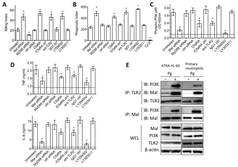
Certain low-abundance bacterial species, such as the periodontitis-associated oral bacterium Porphyromonas gingivalis, can subvert host immunity to remodel a normally symbiotic microbiota into a dysbiotic, disease-provoking state. However, such pathogens also exploit inflammation to thrive in dysbiotic conditions. How these bacteria evade immunity while maintaining inflammation is unclear. As previously reported, P. gingivalis remodels the oral microbiota into a dysbiotic state by exploiting complement. Now we show that in neutrophils P. gingivalis disarms a host-protective TLR2-MyD88 pathway via proteasomal degradation of MyD88, whereas it activates an alternate TLR2-Mal-PI3K pathway. This alternate TLR2-Mal-PI3K pathway blocks phagocytosis, provides "bystander" protection to otherwise susceptible bacteria, and promotes dysbiotic inflammation in vivo. This mechanism to disengage bacterial clearance from inflammation required an intimate crosstalk between TLR2 and the complement receptor C5aR and can contribute to the persistence of microbial communities that drive dysbiotic diseases.


Figures

Figure 1
Figure 1. Pg exploits C5aR and TLR2 in vivo to inhibit neutrophil killing but not inflammation
(A) Pg (109 CFU) was injected into implanted chambers in mice and recruited cells were phenotypically characterized by flow cytometry. The majority (>97%) were neutrophils (Ly6G+) whereas macrophages (F4/80+) or T cells (CD3+) were essentially absent. (B and C) Pg (109 CFU) was injected into implanted chambers in WT mice or in the indicated knockout mice (with or without 10 μg C5aRA, iC5aRA control, anti-TLR2, or isotype control). Fluid was aspirated from the chambers 2h post-inoculation and was used to determine viable Pg CFU (B) and measure the indicated cytokine responses by ELISA (C). Baseline levels (prior to Pg injection) for TNF and IL-1β were undetectable; baseline levels of IL-6 were <10% of the Pg-induced response in WT. (D) Pg and F. nucleatum (Fn) were injected either alone (109 CFU) or together (5×10⁸ CFU each) into implanted chambers in WT mice or in the indicated knockout mice. At 24h post-inoculation, chamber fluid was aspirated to determine viable CFU. In B and D, each symbol represents an individual mouse and horizontal lines indicate means. In C, data are means ± SD (n=5-6 mice per group except for WT; n=10 mice). The experiments were performed twice yielding consistent results. *P<0.05 and **P<0.01 compared with untreated WT control (B and C) or between the indicated groups (D). NS, not significant. See also Figure S1.
Figure 2
Figure 2. Interactions of Pg with human neutrophils
(A-C) In vitro killing (A, B) and oxidative burst (C) assays using purified human neutrophils and Pg in the presence of the indicated compounds. In A, mAbs to TLR2, CR3, CXCR4, and isotype controls were used at 10 μg/ml; AMD3100, 1 μg/ml; C5aRA and iC5aRA control, each at 1 μM; SQ22536, 200 μM; PKI 6-22, 1 μM; and H-89, 10 μM. In B and C exogenous C5a was added at 10 nM, whereas C5aRA and iC5aRA control were each added at 1 μM. (D and E) Neutrophil activation by Pg induces C5aR-TLR2 co-association. (D) Human neutrophils were challenged with Syto9-labeled Pg (green) for 30 min (MOI=10:1) followed by fixation and staining for C5aR (red) and TLR2 (blue). Bottom right, merged image. (E) Human neutrophils were challenged with Pg (MOI=10:1) for 10 min. FRET between the indicated donors and acceptors was measured from the increase in donor (Cy3) fluorescence after acceptor (Cy5) photobleaching. All data are means ± SD (n =3) from one of at least two experiments yielding similar results. The horizontal dashed line indicates the maximum (max) energy transfer efficiency in the experimental system determined as the energy transfer between two different epitopes on the same molecule (C5aR). *P<0.01 compared with untreated control (medium only) or between the indicated groups.
Figure 3
Figure 3. Pg causes a reduction in the levels of MyD88, which promotes bacterial killing
(A) Pg (109 CFU) was injected into implanted chambers in WT mice or in the indicated knockout mice (with or without 10 μg C5aRA, iC5aRA control, anti-TLR2, or isotype control). Fluid was aspirated from the chambers 24h post-inoculation and was used to determine viable Pg CFU. (B-E) Pg was incubated in vitro with human neutrophils (MOI=10:1) for the indicated times and immunoblotting of neutrophil lysates was used to monitor the levels of MyD88 protein (or β-actin; control). In B, Pg was compared with Pam3CSK4 (10 ng/ml) and their effects on MyD88 were monitored also at the mRNA level using real-time PCR. In C, neutrophils were stimulated with Pg in the presence of C5aRA or iC5aRA control (each at 1 μM) or anti-TLR2 mAb or isotype control (10 μg/ml). In D, cell lysates immunoprecipitated using anti-MyD88 were probed with antibodies to MyD88 or to ubiquitin. In E, neutrophils were stimulated with Pg in the presence of increasing concentrations of epoxomycin. (F) ATRA–HL-60 neutrophils were transfected with siRNA to Smurf1 and treated with Pg (MOI=10:1) for 8h. Whole cell-lysate immunoblotting with specific antibodies was used to monitor the levels of Smurf1 and MyD88 protein (β-actin was used as control). Cell lysates immunoprecipitated using anti-MyD88 were immunoblotted with anti-ubiquitin. (G) Human neutrophils were treated with Pg and assayed for MyD88 degradation and ubiquitination as in F in the absence or presence of anti-TGF-β mAb. (H) The indicated bacteria or compounds were injected (Pg, 109 CFU; Pam3SK4, 10 μg; F. nucleatum, 109 CFU) into implanted chambers in mice for the indicated times and immunoblotting of neutrophil lysates was used to monitor the levels of MyD88 protein. (I) Assay for in vitro killing of Pg by purified mouse neutrophils of the indicated genotypes. All experiments were performed two (A) or three times (B-I) yielding similar findings. In A, each symbol represents an individual mouse and horizontal lines indicate means. In I, data are means ± SD (n=4). *P<0.05 and **P<0.01 compared with WT control or between the indicated groups. IB, immunoblotting; IP, immunoprecipitation; WCL, whole cell lysates. See also Figure S2.
Figure 4
Figure 4. PI3K promotes inflammation and the survival of Pg by inhibiting its phagocytosis in vivo
(A-C) Pg (109 CFU) was injected into implanted chambers in WT mice, with or without 10 μg LY294002 (PI3K inhibitor) or LY303511 (control). Fluid aspirated from the chambers 2h post-inoculation was used to determine viable Pg CFU (A), neutrophil recruitment (B), and Pg phagocytosis (C). In C, the experiment additionally included treatments with C5aRA, anti-TLR2, and respective controls (all at 10 μg). (D) Activation of PI3K in mouse neutrophils after 15-min-stimulation with Pg (left) or with 10 nM C5a and/or 100 ng/ml Pam3CSK4 (right) using FACE PI3Kp85 ELISA. In the left panel, neutrophils were from WT or indicated knockout mice. (E) Pg and F. nucleatum (Fn) were injected either alone (109 CFU) or together (5×10⁸ CFU each) into implanted chambers in WT mice with or without 10 μg LY294002 or LY303511. At 24h post-inoculation, chamber fluid was aspirated to determine viable CFU. (F) Fluid aspirated from chambers 2h post-inoculation with Pg was used to measure the indicated cytokine responses by ELISA. (G) Changes in polymerized F-actin over time in human neutrophils exposed to Pg (MOI=10:1) and treated with or without C5aRA or iC5aRA control (1 μM), anti-TLR2 mAb or isotype control (10μg/ml), LY294002 or LY303511 (25 μM). Results are expressed as fold change relative to F-actin at baseline set as 1. (H) Human neutrophils were treated as in G and after 20-min stimulation with Pg the activation of RhoA was determined using the G-LISA RhoA assay kit. The dashed line indicates baseline RhoA activation (in cells not exposed to Pg). (I) Human neutrophils were treated as in G and after 6h-stimulation with Pg the indicated cytokine responses were measured by ELISA. In A and E, each symbol represents an individual mouse and horizontal lines indicate means. In bar graphs, data are means ± SD (B, C, and F, n=5; D and G-I, n=3). The experiments were performed two (A-C and E-H) or three times (D,I) yielding consistent results. *P <0.01 compared with control (WT or untreated) or between the indicated groups. NS, not significant. See also Figure S3.
Figure 5
Figure 5. PI3K promotes dysbiotic inflammation in the periodontal tissue
Mice were orally inoculated or not with Pg (109 CFU; three times at 2-day intervals) and seven days after the last inoculation were injected with the indicated experimental or control compounds into the palatal gingiva, on the mesial of the first molar and in the papillae between first and second and third molars on both sides of the maxilla (1 μl of 1 μg per site; total of 6 μg in six sites). Two days later, the mice were euthanized and maxillary periodontal tissue was harvested to determine Pg and total bacterial numbers using real-time PCR (A) or was used to measure the indicated cytokine responses at the mRNA (B) or protein (C) level. The mRNA expression levels were normalized against GAPDH mRNA and expressed as fold induction relative to the transcript levels of sham-infected mice, which were assigned an average value of 1. Data are means ± SD (n=5 mice) from one of two experiments yielding consistent results. *P<0.01 compared with untreated control (A) or with sham control (B, C); P<0.01 compared with Pg alone. No Pg could be detected in sham-infected mice. See also Figure S4.
Figure 6
Figure 6. Mal is a component of the C5aR-TLR2 subversive pathway acting upstream of PI3K
(A-D) ATRA-differentiated HL-60 neutrophils were transiently transfected with control siRNA or siRNA to Mal or MyD88 (all at 40 nM), or were treated with the indicated compounds (LY294002 or LY303511, 25 μM; C5aRA or iC5aRA control, 1 μM; anti-TLR2 mAb or isotype control, 10μg/ml). The cells were used in assays of Pg killing (A) and phagocytosis (B) or in assays of PI3K activation (C) and cytokine release (D). In B, cytochalasin D (CytD) was used as negative control for phagocytosis. In C and D, the horizontal dashed lines indicate baseline levels of PI3K activation and cytokines determined on unstimulated cells. (E) Pg-stimulated lysates of ATRA-differentiated HL-60 or primary neutrophils were immunoprecipitated with antibodies to TLR2 or Mal and were probed with antibodies to PI3K, Mal, and TLR2. Pg stimulation was carried out at a MOI equal to 10:1 and for 15 min. Data are means ± SD (A and D, n=5; B and C, n =3). All experiments were performed two times or more yielding similar results. *P<0.01 compared with untreated control. IB, immunoblotting; IP, immunoprecipitation; WCL, whole cell lysates. See also Figure S5.
Figure 7
Figure 7. Model of Pg subversion of neutrophils leading to dysbiotic inflammation
Pg co-activates TLR2 and C5aR in neutrophils and the resulting crosstalk leads to ubiquitination and proteasomal degradation of MyD88, thereby inhibiting a host-protective antimicrobial response. This activity requires C5aR/TLR2-dependent release of TGF-β1 (Figure S2J), which mediates ubiquitin-proteasome degradation of MyD88 via the E3 ubiquitin ligase Smurf1. Moreover, the C5aR-TLR2 crosstalk activates PI3K, which prevents phagocytosis through inhibition of RhoA activation and actin polymerization, while stimulating an inflammatory response. In contrast to MyD88, Mal is a component of the subversive pathway acting upstream of PI3K. The integrated mechanism provides ‘bystander’ protection to otherwise susceptible bacterial species and promotes polymicrobial dysbiotic inflammation in vivo.

Comment in

References

    1. Abe T, Hosur KB, Hajishengallis E, Reis ES, Ricklin D, Lambris JD, Hajishengallis G. Local complement-targeted intervention in periodontitis: proof-of-concept using a C5a receptor (CD88) antagonist. J. Immunol. 2012;189:5442–5448. - PMC - PubMed
    1. Abusleme L, Dupuy AK, Dutzan N, Silva N, Burleson JA, Strausbaugh LD, Gamonal J, Diaz PI. The subgingival microbiome in health and periodontitis and its relationship with community biomass and inflammation. ISME J. 2013;7:1016–1025. - PMC - PubMed
    1. Brzezinska AA, Johnson JL, Munafo DB, Ellis BA, Catz SD. Signalling mechanisms for Toll-like receptor-activated neutrophil exocytosis: key roles for interleukin-1-receptor-associated kinase-4 and phosphatidylinositol 3-kinase but not Toll/IL-1 receptor (TIR) domain-containing adaptor inducing IFNβ (TRIF) Immunology. 2009;127:386–397. - PMC - PubMed
    1. Burns E, Eliyahu T, Uematsu S, Akira S, Nussbaum G. TLR2-dependent inflammatory response to Porphyromonas gingivalis is MyD88 independent, whereas MyD88 is required to clear infection. J. Immunol. 2010;184:1455–1462. - PubMed
    1. Choi CH, Spooner R, DeGuzman J, Koutouzis T, Ojcius DM, Yilmaz O. Porphyromonas gingivalis-nucleoside-diphosphate-kinase inhibits ATP-induced reactive-oxygen-species via P2X7 receptor/NADPH-oxidase signalling and contributes to persistence. Cell Microbiol. 2013;15:961–976. - PMC - PubMed

Publication types

MeSH terms