. 2014 Jul 10;57(13):5579-601.
doi: 10.1021/jm5002502. Epub 2014 Jun 25.

Discovery and structure-activity relationship of novel 2,3-dihydrobenzofuran-7-carboxamide and 2,3-dihydrobenzofuran-3(2H)-one-7-carboxamide derivatives as poly(ADP-ribose)polymerase-1 inhibitors

Affiliations

Discovery and structure-activity relationship of novel 2,3-dihydrobenzofuran-7-carboxamide and 2,3-dihydrobenzofuran-3(2H)-one-7-carboxamide derivatives as poly(ADP-ribose)polymerase-1 inhibitors

Maulik R Patel et al. J Med Chem. .

Abstract

Novel substituted 2,3-dihydrobenzofuran-7-carboxamide (DHBF-7-carboxamide) and 2,3-dihydrobenzofuran-3(2H)-one-7-carboxamide (DHBF-3-one-7-carboxamide) derivatives were synthesized and evaluated as inhibitors of poly(ADP-ribose)polymerase-1 (PARP-1). A structure-based design strategy resulted in lead compound 3 (DHBF-7-carboxamide; IC50 = 9.45 μM). To facilitate synthetically feasible derivatives, an alternative core was designed, DHBF-3-one-7-carboxamide (36, IC50 = 16.2 μM). The electrophilic 2-position of this scaffold was accessible for extended modifications. Substituted benzylidene derivatives at the 2-position were found to be the most potent, with 3',4'-dihydroxybenzylidene 58 (IC50 = 0.531 μM) showing a 30-fold improvement in potency. Various heterocycles attached at the 4'-hydroxyl/4'-amino of the benzylidene moiety resulted in significant improvement in inhibition of PARP-1 activity (e.g., compounds 66-68, 70, 72, and 73; IC50 values from 0.718 to 0.079 μM). Compound 66 showed selective cytotoxicity in BRCA2-deficient DT40 cells. Crystal structures of three inhibitors (compounds (-)-13c, 59, and 65) bound to a multidomain PARP-1 structure were obtained, providing insights into further development of these inhibitors.


Figures

Chart 1
Chart 1. PARP Inhibitors Currently Being Evaluated in the Clinic
Figure 1
Figure 1
XP-Glide predicted binding mode of compound 3 within the active site of PARP-1, represented in ribbon form, with the interacting amino acids represented as sticks and atoms colored according to the following: carbon, beige; hydrogen, white; nitrogen, blue; oxygen, red. The inhibitor is shown as ball and stick with the same color scheme depicted above except that carbons are represented as orange. Dotted red lines indicate intra- and intermolecular hydrogen bonding interaction, whereas the dotted yellow lines indicate the distance between the two atoms/groups/centroids in angstroms. The centroids are marked as green stars. The image was generated using PyMOL, version 1.6.0.
Scheme 1
Scheme 1. Synthesis of 5-Substituted 2,3-Dihydrobenzofuran-7-carboxamide Derivatives
Reagents and conditions: (a) (i) n-BuLi, TEMED, hexane, rt, 4 h; (ii) dry ice, overnight; conc HCl; (b) (i) i-BuOCOCl, NMM, THF, −20 °C, 4 h; (ii) 30% NH4OH, rt, 1 h; (c) Br2, CH3COONa, CH3COOH, 80 °C, 3 h; (d) TFA, HNO3, 0 °C to rt, 3 h; (e) SnCl2·2H2O, EtOAc, reflux, 4 h; (f) H2SO4, MeOH, reflux, 3 h.
Scheme 2
Scheme 2. Synthesis of 2,5-Disubstituted 2,3-Dihydrobenzofuran-7-carboxamides
Reagents and conditions: (a) SOCl2, MeOH, reflux, 12 h; (b) allyl bromide, K2CO3, NaI, DMF, rt, 12 h; (c) neat, 160–190 °C, 2 h; (d) ZrCl4, DCM, rt, 10 h; (e) NaOH, MeOH, reflux, 2 h; (f) (i) i-BuOCOCl, NMM, THF, −20 °C, 4 h; (ii) 30% NH4OH, rt, 1 h; (g) when R = H, TFA, HNO3, 0 °C to rt, 3 h; (h) SnCl2·2H2O, EtOAc, reflux, 4 h.
Figure 2
Figure 2
ORTEP diagram of salt of (R)-(−)-12a with (S)-(−)-α-methylbenzylamine.
Scheme 3
Scheme 3. Synthesis of 5-Fluoro-2,3-dihydrobenzofuran-7-carboxamide
Reagents and conditions: (a) TFA, HNO3, 0 °C to rt, 3 h; (b) H2/Pd/C, EtOH, 60 psi, rt, 2–18 h; (c) (i) THF, HCl, HBF4, rt to −15 °C, NaNO2, 30 min; (ii) xylene, reflux, 2 h; (d) R=H, (i) n-BuLi, TEMED, hexane, rt, 4 h; (ii) dry ice, overnight; (e) when R = -COOCH3; NaOH, MeOH, reflux, 3 h; (f) (i) i-BuOCOCl, NMM, THF, −20 °C, 4 h; (ii) 30% NH4OH, rt, 1 h.
Scheme 4
Scheme 4. Synthesis of 4-Amino-2-methyl-2,3-dihydrobenzofuran-7-carboxamide
Reagents and conditions: (a) SOCl2, MeOH, reflux, 12 h; (b) allyl bromide, K2CO3, NaI, DMF, rt, 12 h; (c) carbitol, 170–180 °C, 2 h; (d) ZrCl4, DCM, rt, 10 h; (e) NaOH, MeOH, reflux; (f) H2/Pd/C, EtOH, 60 psi, rt, 4 h; (g) NaOH, MeOH, reflux, 18 h; (h) (i) NMM, i-BuOCOCl, THF, −20 °C, 20 min; (ii) dry NH3, rt, 45 min.
Scheme 5
Scheme 5. Synthesis of 3-Oxo-2,3-dihydrobenzofuran-7-carboxamide
Reagents and conditions: (a) SOCl2, MeOH, reflux, 12 h; (b) BrCH2COOEt, K2CO3, NaI, DMF, rt, 12 h; (c) KOH, MeOH, reflux, 4 h; (d) KMnO4, water, reflux, 2 h; (e) NaOAc, (CH3CO)2O, CH3COOH, reflux, 5 h; (f) HCl (11 N)/H2O/MeOH, reflux, 1 h; (g) (i) i-BuOCOCl, NMM, THF, −20 °C, 4 h; (ii) 30% NH4OH, rt, 1 h.
Scheme 6
Scheme 6. Synthesis of Substituted Benzaldehydes 3740 and 42
Reagents and conditions: (a) K2CO3, 4-(2-chloroethyl)morpholine (for compounds 37 and 39) and 4-(2-chloroethyl)piperidine (for compounds 38 and 40), CH3CN, reflux, 6 h; (b) K2CO3, KI, bromochloroethane, CH3CN, reflux, 12 h; (c) K2CO3, KI, N-methylpiperazine, CH3CN, reflux, 2–6 h.
Scheme 7
Scheme 7. Synthesis of Substituted Benzaldehydes 44 and 4649
Reagents and conditions: (a) K2CO3, bromochloropropane, CH3CN, reflux, 3 h; (b) K2CO3, morpholine, CH3CN, reflux, 6 h; (c) chloroacetyl chloride, DCM, triethylamine, 0 °C to rt, 3 h; (d) K2CO3, 4-hydroxybenzaldehyde, CH3CN, reflux, 6 h; (e) epibromhydrin, K2CO3, CH3CN, reflux, 6 h; (f) morpholine, K2CO3, CH3CN, reflux, 6 h; (g) 4-fluorophenylpiperazine, K2CO3, CH3CN, reflux, 48 h.
Scheme 8
Scheme 8. Synthesis of Target Compounds 5073
Reagents and conditions: (a) NH4OAc, commercially available/synthesized R-CHO, toluene, reflux, 1–24 h.
Scheme 9
Scheme 9. Synthesis of Substituted Benzaldehyde Intermediates 72d and 72e Required for Respective Synthesis of Target Compounds 72 and 73
Reagents and conditions: (a) SnCl2·2H2O, ethyl acetate, reflux, 4 h; (b) chloroethanesulfonyl chloride (for compound 72b) or chloropropanesulfonyl chloride (for compound 72c), DCM, triethylamine, 0 °C to rt, 6 h; (c) N-methylpiperazine, K2CO3, CH3CN, reflux, 6 h.
Figure 3
Figure 3
Crystal structure of inhibitors in the active site of PARP-1. (A) Structural representation of the PARP-1 activated complex, which highlights the NAD+ binding site (active site) where most PARP inhibitors bind. (B–D) FoFc electron density difference map (blue mesh) of PARP-1 crystals soaked with 1–5 mM (−)-13c, 59, or 65 (PBD code 4OPX, 4OQA, or 4OQB, respectively), contoured to 2.5σ in which density is calculated in the absence of ligand. (E–G) Compounds bound to the NAD+ site of PARP-1 make π–π stacking interactions between Tyr907 and Tyr896 and H-bond interactions with the backbone of Gly863 and Ser904 consistent with the traditional benzamide pharmacophore of most PARP inhibitors. Compounds 59 and 65 extend out of the nicotinamide pocket and make further interactions in the ADP-ribose pocket of the NAD+ binding site.
Figure 4
Figure 4
Predicted binding mode of compounds 66 (A) and 72 (B) within the active site of PARP-1. The color scheme for the active site amino acid residues and the inhibitor is the same as in Figure 1. The centroids are generated as green stars. The yellow dotted lines indicate the distance between the two atoms/groups/centroids (in angstroms). The image was generated in PyMOL, version 1.6.0.
Figure 5
Figure 5
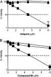
Cell viability of DT40 cells after continuous exposure to (A) veliparib or (B) compound 66 for 72 h. Cellular ATP concentration was used to measure cellular viability. The viability of untreated cells was set as 100%. Error bars represent standard deviation (n = 3). Circle: wild-type cell. Triangle: PARP1-deficient cells. Square: BRCA2-deficient cells. Invisible error bars are encompassed within the symbol sizes.

References

    1. Gibson B. A.; Kraus W. L. New insights into the molecular and cellular functions of poly(ADPribose) and PARPs. Nat. Rev. Mol. Cell Biol. 2012, 13, 411–424. - PubMed
    1. Luo X.; Kraus W. L. On PAR with PARP: cellular stress signaling through poly(ADP-ribose) and PARP-1. Genes Dev. 2012, 26, 417–432. - PMC - PubMed
    1. Jankevicius G.; Hassler M.; Golia B.; Rybin V.; Zacharias M.; Timinszky G.; Ladurner A. G. A family of macrodomain proteins reverses cellular mono-ADP-ribosylation. Nat. Struct. Mol. Biol. 2013, 20, 508–514. - PMC - PubMed
    1. Hottiger M. O.; Hassa P. O.; Luscher B.; Schuler H.; Koch-Nolte F. Toward a unified nomenclature for mammalian ADP ribosyl transferases. Trends Biochem. Sci. 2010, 35, 208–219. - PubMed
    1. Rouleau M.; Patel A.; Hendzel M. J.; Kaufmann S. H.; Poirier G. G. PARP inhibition: PARP1 and beyond. Nat. Rev. Cancer 2010, 10, 293–301. - PMC - PubMed

Publication types