Review
. 2014 Dec;20(6):683-96.
doi: 10.1089/ten.TEB.2014.0086. Epub 2014 Jul 22.

Review of collagen I hydrogels for bioengineered tissue microenvironments: characterization of mechanics, structure, and transport

Affiliations
Review

Review of collagen I hydrogels for bioengineered tissue microenvironments: characterization of mechanics, structure, and transport

Elizabeth E Antoine et al. Tissue Eng Part B Rev. 2014 Dec.

Abstract

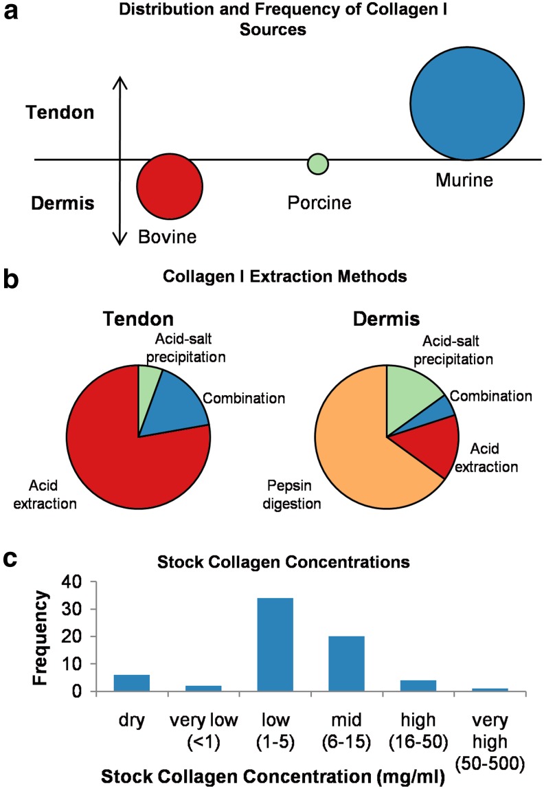
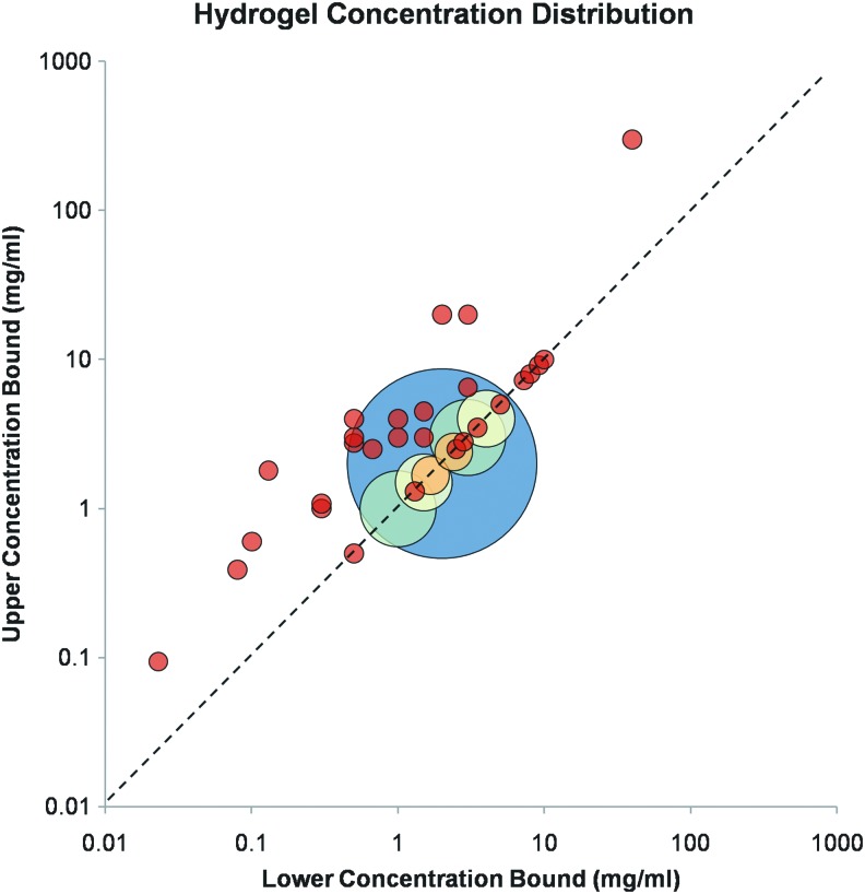
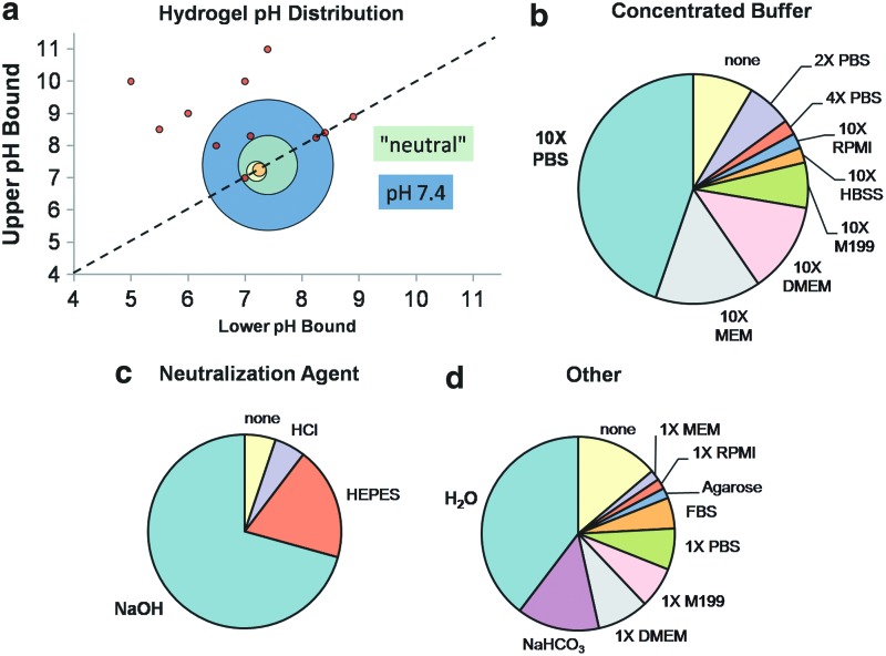
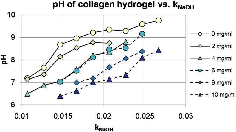
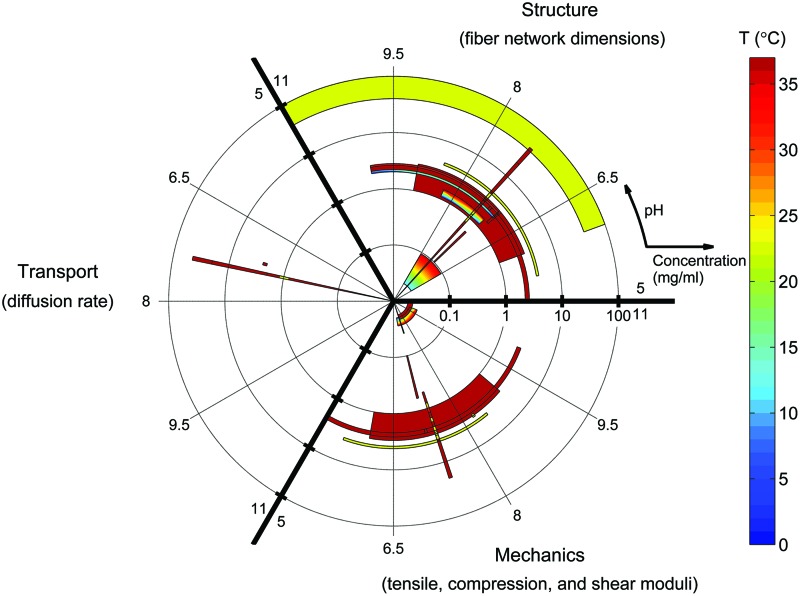
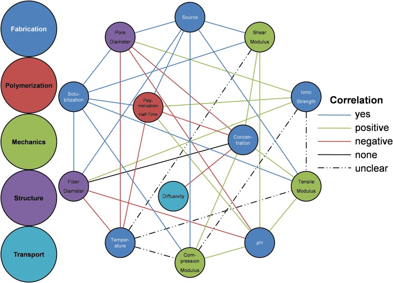
Type I collagen hydrogels have been used successfully as three-dimensional substrates for cell culture and have shown promise as scaffolds for engineered tissues and tumors. A critical step in the development of collagen hydrogels as viable tissue mimics is quantitative characterization of hydrogel properties and their correlation with fabrication parameters, which enables hydrogels to be tuned to match specific tissues or fulfill engineering requirements. A significant body of work has been devoted to characterization of collagen I hydrogels; however, due to the breadth of materials and techniques used for characterization, published data are often disjoint and hence their utility to the community is reduced. This review aims to determine the parameter space covered by existing data and identify key gaps in the literature so that future characterization and use of collagen I hydrogels for research can be most efficiently conducted. This review is divided into three sections: (1) relevant fabrication parameters are introduced and several of the most popular methods of controlling and regulating them are described, (2) hydrogel properties most relevant for tissue engineering are presented and discussed along with their characterization techniques, (3) the state of collagen I hydrogel characterization is recapitulated and future directions are proposed. Ultimately, this review can serve as a resource for selection of fabrication parameters and material characterization methodologies in order to increase the usefulness of future collagen-hydrogel-based characterization studies and tissue engineering experiments.


Figures

<b>FIG. 1.</b>
FIG. 1.
Overview of collagen I sources and solubilization methods. (a) Source animal compared with source tissue; (b) comparison of extraction methods by source tissue; and (c) collagen content of stock solutions. Source data can be found in Supplementary Table S1. Color images available online at www.liebertpub.com/teb
<b>FIG. 2.</b>
FIG. 2.
Collagen concentration ranges used in the tissue engineering literature. Source data can be found in Supplementary Table S2. 1 mg/mL=0.1% wt. Color images available online at www.liebertpub.com/teb
<b>FIG. 3.</b>
FIG. 3.
Collagen hydrogel configurations used in the tissue engineering literature. (a) pH ranges. (b–d) Distribution of noncollagen hydrogel components. Components in bold occur most frequently and match the commercial BD/Vitrogen hydrogel preparation protocols. Source data can be found in Supplementary Table S3. Color images available online at www.liebertpub.com/teb
<b>FIG. 4.</b>
FIG. 4.
Dependence of hydrogel pH on NaOH fraction and absolute collagen concentration. Color images available online at www.liebertpub.com/teb
<b>FIG. 5.</b>
FIG. 5.
Diagram of a typical spectrophotometric measurement for quantification of polymerization kinetics. The polymerization half-time t1/2 is defined as the time at which half of the total change in absorbance ΔAbs/2 is attained. The rate of change of absorbance at the half-time is defined as the polymerization rate dAbs/dt, while the lag time is defined as the zero-absorbance intercept of the line with slope dAbs/dt and intercepting (t1/2, ΔAbs/2). Color images available online at www.liebertpub.com/teb
<b>FIG. 6.</b>
FIG. 6.
Modes and physical scales of collagen hydrogel mechanical characterization. Data points corresponding to studies that investigate a range of scales are centered at the mean value and plotted with a bar extending to the upper bound of the range investigated. Because of the log-log scale, lower bounds are not plotted. Source data and application notes can be found in Supplementary Table S4. Color images available online at www.liebertpub.com/teb
<b>FIG. 7.</b>
FIG. 7.
Representative scanning electron microscopy (SEM) image of collagen hydrogel annotated to indicate commonly quantified fiber structure parameters P (pore size), D (fiber diameter), and Ø (orientation). Scale bar=500 nm. Figure reproduced with permission of the publisher.
<b>FIG. 8.</b>
FIG. 8.
Representative images of collagen hydrogels using various modalities. (a) Transmission electron microscopy (scale bar=500 nm), (b) SEM (scale bar=500 nm), (c) second-harmonic generation (scale bar=50 μm), and (d) confocal reflectance microscopy (scale bar=50 μm). Figures reproduced with permission of the publishers. Color images available online at www.liebertpub.com/teb
<b>FIG. 9.</b>
FIG. 9.
State of collagen hydrogel characterization. Each object represents the fabrication parameter ranges covered by a single experimental study from the literature. Log(collagen concentration) and polymerization pH are plotted as radius and angle, respectively. Polymerization temperature is plotted as a color contour. Studies outside the ranges (0–100) mg/mL, (5–11) pH units, and (0–37) °C are not shown. Diffusing-molecule hydrodynamic radius and deformation mode for mechanical characterization are not represented in this figure. Color images available online at www.liebertpub.com/teb
<b>FIG. 10.</b>
FIG. 10.
Correlations between collagen hydrogel fabrication parameters and material properties. Color images available online at www.liebertpub.com/teb

References

    1. Kumar V., Brewster L., Caves J., and Chaikof E.Tissue engineering of blood vessels: functional requirements, progress, and future challenges. Cardiovasc Eng Technol 2,137, 2011 - PMC - PubMed
    1. Ingram M., Techy G.B., Ward B.R., Imam S.A., Atkinson R., Ho H., et al. Tissue engineered tumor models. Biotech Histochem 85,213, 2010 - PubMed
    1. Sung J.H., and Shuler M.L.Microtechnology for mimicking in vivo tissue environment. Ann Biomed Eng 40,1289, 2012 - PubMed
    1. Buchanan C., and Rylander M.N.Microfluidic culture models to study the hydrodynamics of tumor progression and therapeutic response. Biotechnol Bioeng 110,2063, 2013 - PubMed
    1. Tibbitt M.W., and Anseth K.S.Hydrogels as extracellular matrix mimics for 3D cell culture. Biotechnol Bioeng 103,655, 2009 - PMC - PubMed

Publication types