. 2014 Jul 22;8(7):6620-32.
doi: 10.1021/nn501652j.

Targeted iron-oxide nanoparticle for photodynamic therapy and imaging of head and neck cancer

Targeted iron-oxide nanoparticle for photodynamic therapy and imaging of head and neck cancer

Dongsheng Wang et al. ACS Nano. .

Abstract

Photodynamic therapy (PDT) is a highly specific anticancer treatment modality for various cancers, particularly for recurrent cancers that no longer respond to conventional anticancer therapies. PDT has been under development for decades, but light-associated toxicity limits its clinical applications. To reduce the toxicity of PDT, we recently developed a targeted nanoparticle (NP) platform that combines a second-generation PDT drug, Pc 4, with a cancer targeting ligand, and iron oxide (IO) NPs. Carboxyl functionalized IO NPs were first conjugated with a fibronectin-mimetic peptide (Fmp), which binds integrin β1. Then the PDT drug Pc 4 was successfully encapsulated into the ligand-conjugated IO NPs to generate Fmp-IO-Pc 4. Our study indicated that both nontargeted IO-Pc 4 and targeted Fmp-IO-Pc 4 NPs accumulated in xenograft tumors with higher concentrations than nonformulated Pc 4. As expected, both IO-Pc 4 and Fmp-IO-Pc 4 reduced the size of HNSCC xenograft tumors more effectively than free Pc 4. Using a 10-fold lower dose of Pc 4 than that reported in the literature, the targeted Fmp-IO-Pc 4 NPs demonstrated significantly greater inhibition of tumor growth than nontargeted IO-Pc 4 NPs. These results suggest that the delivery of a PDT agent Pc 4 by IO NPs can enhance treatment efficacy and reduce PDT drug dose. The targeted IO-Pc 4 NPs have great potential to serve as both a magnetic resonance imaging (MRI) agent and PDT drug in the clinic.


Figures

Figure 1
Figure 1
Construction of targeted IO nanoparticles carrying Pc 4. (A) Schematic illustration of synthesis of water-soluble Fmp-IO-Pc 4 using Ocean’s amphiphilic polymer-coated IO nanoparticles. (B) TEM image of IO NPs, which are highly homogeneous with an average size of 10 nm. (C) Size-dependent migration of different sized IO, Fmp-IO and Fmp-IO-Pc 4 NPs in 2% agarose gel. Since Fmp-IO-Pc 4 has the largest molecular weight and size, it migrated slowest in the gel. (D) The absorption spectra of synthesized Fmp-IO-Pc 4. As shown, Fmp-IO-Pc 4 (purple) has a similar absorption at around 675 nm as the free Pc 4 (blue) while IO (red) and solvent (green) show no specific absorption at 675 nm.
Figure 2
Figure 2
In vitro inhibition of HNSCC cell growth. SRB assay shows that both free Pc 4 and NP-based Pc 4 have good drug efficacy in several HNSCC cancer cell lines, M4E, M4E-15, 686LN, and TU212. The cancer cells were grown in medium with 50 nM or 100 nM Pc 4, IO-Pc 4 or Fmp-IO-Pc 4 at equivalent concentrations of Pc 4 for 24 h. Cells were then subjected to laser treatment. SRB assay was performed 24 and 48 h post laser treatment. (A) 24 h after laser treatment, both Fmp-IO-Pc 4 and IO-Pc 4 showed dose dependent efficacy equal to that of free Pc 4. (B) 48 h after laser treatment, all treatments caused death of the majority of cells at the same concentration of 50 nM, indicating that the encapsulated Pc4 in Fmp-IO-Pc 4 or IO-Pc 4 is as biologically active as free Pc 4.
Figure 3
Figure 3
In vitro binding assays to compare targeted Fmp-IO-Pc 4 with nontargeted IO-Pc 4 NPs in HNSCC cells. M4E, an integrin β1-positive cell line, and M4E-15, an integrin β1 knock-down derivative of M4E, were seeded on chamber slides at 3000 cells per well. IO-Pc 4 and Fmp-IO-Pc 4 at 100 nM were added 24 h later. Cells were kept at 4 or 37 °C for 2 h. (A) At 4 °C only Fmp-IO-Pc4 bound to integrin β1-positive M4E cells, while very low positive signals were detected for nontargeted IO-Pc4. Very low Fmp-IO-Pc 4 binding was detected in M4E-15 cells. (B) At 37 °C, binding of both Fmp-IO-Pc 4 and IO-Pc 4 was detected to M4E and M4E-15 cells, but Fmp-IO-Pc 4 had a significantly higher signal than IO-Pc 4. Quantified Pc 4 signals on the cells as indicated at 4 °C (C) and 37 °C (D) were obtained using the Nuance Multispectral Image System Nuance 3.1.
Figure 4
Figure 4
Inhibition of xenograft tumor formation by Pc 4 PDT delivered by IO nanoparticles. (A–D) Tumor growth and representative images of tumors on both sides of the mice in the PBS control, free Pc 4, IO-Pc 4, and Fmp-IO-Pc4 groups, respectively. Pc 4 was given at a concentration of 0.4 mg/kg. Laser treatment was performed 48 h after the drug administration. Three out of six mice from each group are shown as representatives. Statistical analysis indicated a significant difference in the longitudinal tumor volume across the 5 groups within the right side (laser treated), (p < 0.0013). Both IO-Pc 4 and Fmp-IO-Pc 4 groups had a significantly lower tumor growth volume than the PBS control group (p < 0.022 for IO-Pc 4 and 0.0038 for Fmp-IO-Pc 4). The Pc 4 group had a marginally significantly lower tumor growth volume than the control group (p < 0.071). The Pc 4 group had a significantly higher tumor growth volume than both the IO-Pc 4 and Fmp-IO-Pc 4 groups (p < 0.049 for IO-Pc 4 group and 0.040 for Fmp-IO-Pc 4). No tumor growth difference was found between IO-Pc 4 and Fmp-IO-Pc 4 groups (p = 0.98). There was no significant difference in the longitudinal tumor volume across the 4 groups on the left side tumor (no laser treatment, p = 0.4987). None of the pairwise comparisons in tumor volume between any two groups with untreated left tumors was significantly different (results are omitted). (E) Tumor growth curve using a lower dose (0.06 mg/kg) and shorter period of time between drug administration and laser treatment than used in (A–D). Tumors in the Fmp-IO-Pc 4 (targeted) group grew significantly slower than those in the IO-Pc 4 group (nontargeted) (p < 0.025).
Figure 5
Figure 5
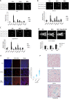
Tissue biodistribution of free Pc 4 and both targeted and nontargeted IO-Pc 4 NPs. Drug distribution studies show that Fmp-IO-Pc 4 has a more prolonged existence in xenografted tumors than free Pc 4 and nontargeted IO-Pc 4. Mice were given Pc 4, IO-Pc 4 or Fmp-IO-Pc 4. Mouse whole-body images and organ images were taken 4, 24, and 48 h after drug administration. (A–C) Images of different organs, including the xenograft tumors, and levels of Pc 4 delivered as free Pc 4, IO-Pc 4 and Fmp-IO-Pc 4 at different time points, respectively (images represent 1 out of 3 mice). (D) Levels of Pc 4 delivered as free Pc 4, IO-Pc 4, and Fmp-IO-Pc 4 in tumors at different time points by whole-body imaging. As shown, the targeted nanoparticle Fmp-IO-Pc 4 has a more prolonged retention in tumors than either free Pc4 or the nontargeted nanoparticle IO-Pc 4. (E) Pc4 staining in fixed tumor tissue from the free Pc 4, IO-Pc 4, and Fmp-IO-Pc 4 groups. 4′,6-Diamidino-2-phenylindole (DAPI)was used for nuclear labeling. Greater Pc 4 presence was observed in tumor tissues in Fmp-IO-Pc 4 treated mice than in those treated with IO-Pc 4 or free Pc 4 (images represent 1 out of 3 mice). (F) Tumor sections from 3 mice injected with free Pc 4, IO-Pc 4 and Fmp-IO-Pc 4, respectively. No blue staining was found in tumor cells from free Pc 4 treated mice. Higher numbers of tumor cells with blue staining were observed in tumors from Fmp-IO-Pc4 treated mice than from IO-Pc 4 treated mice.
Figure 6
Figure 6
In vitro MRI imaging experiment of Fmp-IO-Pc 4. MR imaging and T2 maps of tumor cells incubated with Fmp-IO or IO nanoparticles were collected. (A) shows significant T2 signal decrease in the cells incubated with Fmp-IO as compared to IO nanoparticles or the control without IO. The first well of the upper panel of M4E cells shows a decrease in T2 contrast (darker) for Fmp-IO. The lower panels display the level of T2 values measured by T2 relaxometry mapping method. A low T2 value (green color) correlates with a higher iron concentration, indicating higher level of specific binding of Fmp-IO nanoparticles to tumor cells (image represents 1 out of 3 experiments). Quantitative measures of T2 values show that the cells with Fmp-IO had lower T2 values (107.6 ± 0.47 ms) as compared to those with IO (118.5 ± 1.94 ms) as shown in (B).

Similar articles

Cited by

References

    1. Siegel R.; Naishadham D.; Jemal A. Cancer Statistics. Ca—Cancer J. Clin. 2013, 63, 11–30. - PubMed
    1. Jermal A.; Bray F.; Center M. M.; Ferlay J.; Forman D. Global Cancer Statistics. Ca—Cancer J. Clin. 2011, 61, 69–90. - PubMed
    1. Molin Y.; Fayette J. Current Chemotherapies for Recurrent/Metastatic Head and Neck Cancer. Anticancer Drugs 2011, 22, 621–625. - PubMed
    1. Shin D. M.; Khuri F. R. Advances in the Management of Recurrent or Metastatic Squamous Cell Carcinoma of the Head and Neck. Head Neck 2013, 35, 443–453. - PubMed
    1. Dolmans D. E.; Fukumura D.; Jain R. K. Photodynamic Therapy for Cancer. Nat. Rev. Cancer 2003, 3, 380–387. - PubMed

Publication types

MeSH terms