Review
. 2014 Jun;10(3 Suppl):S242-53.
doi: 10.1016/j.jalz.2014.04.003.

Military-related traumatic brain injury and neurodegeneration

Affiliations
Review

Military-related traumatic brain injury and neurodegeneration

Ann C McKee et al. Alzheimers Dement. 2014 Jun.

Abstract

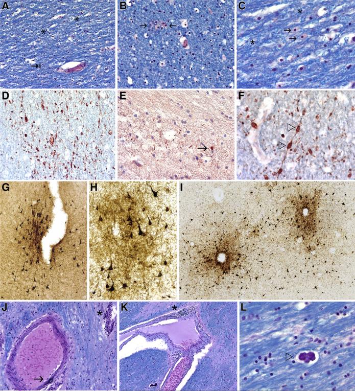
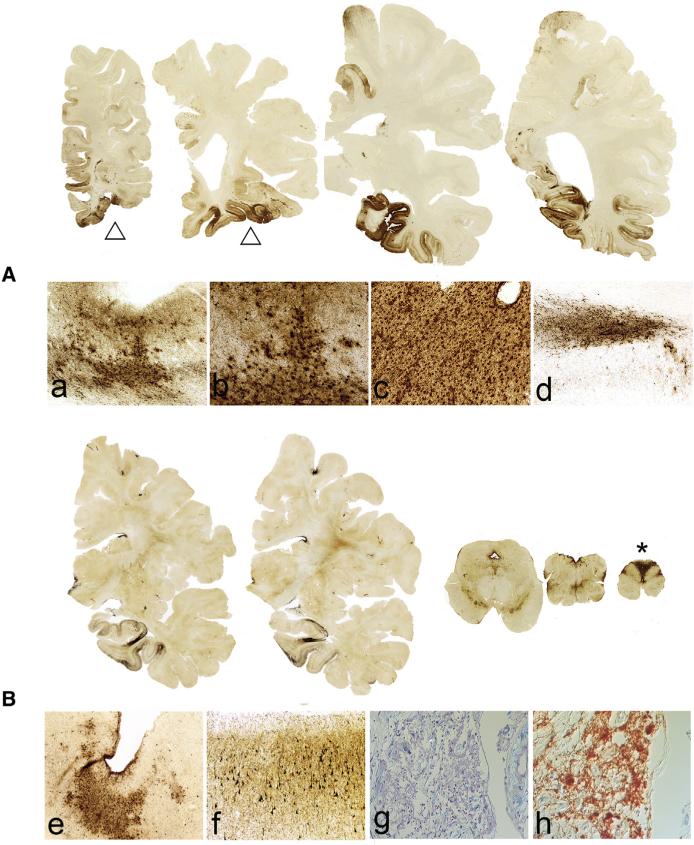
Mild traumatic brain injury (mTBI) includes concussion, subconcussion, and most exposures to explosive blast from improvised explosive devices. mTBI is the most common traumatic brain injury affecting military personnel; however, it is the most difficult to diagnose and the least well understood. It is also recognized that some mTBIs have persistent, and sometimes progressive, long-term debilitating effects. Increasing evidence suggests that a single traumatic brain injury can produce long-term gray and white matter atrophy, precipitate or accelerate age-related neurodegeneration, and increase the risk of developing Alzheimer's disease, Parkinson's disease, and motor neuron disease. In addition, repetitive mTBIs can provoke the development of a tauopathy, chronic traumatic encephalopathy. We found early changes of chronic traumatic encephalopathy in four young veterans of the Iraq and Afghanistan conflict who were exposed to explosive blast and in another young veteran who was repetitively concussed. Four of the five veterans with early-stage chronic traumatic encephalopathy were also diagnosed with posttraumatic stress disorder. Advanced chronic traumatic encephalopathy has been found in veterans who experienced repetitive neurotrauma while in service and in others who were accomplished athletes. Clinically, chronic traumatic encephalopathy is associated with behavioral changes, executive dysfunction, memory loss, and cognitive impairments that begin insidiously and progress slowly over decades. Pathologically, chronic traumatic encephalopathy produces atrophy of the frontal and temporal lobes, thalamus, and hypothalamus; septal abnormalities; and abnormal deposits of hyperphosphorylated tau as neurofibrillary tangles and disordered neurites throughout the brain. The incidence and prevalence of chronic traumatic encephalopathy and the genetic risk factors critical to its development are currently unknown. Chronic traumatic encephalopathy has clinical and pathological features that overlap with postconcussion syndrome and posttraumatic stress disorder, suggesting that the three disorders might share some biological underpinnings.

Keywords: Alzheimer's disease; Chronic traumatic encephalopathy; Neurodegeneration; TDP-43; Tauopathy; Traumatic brain injury; Veterans.


Figures

Fig. 1
Fig. 1
Neuropathological changes associated with blast injury. (A–C). Subcortical frontal white matter shows loss of myelinated fibers, abnormal myelin clumps (asterisks), and astrocytosis (arrows), case 1, luxol fast blue-hematoxylin and eosin stain, original magnification (A) ×200, (B) ×400, and (C) ×600. (D) Axons in the frontal white matter are severely depleted with many irregular axonal swellings, case 1, SMI-34 immunostaining, original magnification ×400. (E) APP immunostaining shows axon retraction bulbs and other axonal irregularities (arrow), APP immunostaining, case 1, original magnification ×400. (F) Axonal swellings, case 1, SMI-34 immunostaining, original magnification ×600. (G) A single focus of perivascular NFTs and neurites at the depth of sulcus in inferior parietal cortex consistent with Stage I/IV CTE, case 1, AT8 immunostaining for hyperphosphorylated tau (p-tau), original magnification ×100. (H) Perivascular NFTs at the sulcal depths of frontal cortex, case 3, AT8 immunostaining, original magnification ×200. (I) Multiple areas of perivascular NFTs, case 2, AT8 immunostaining, original magnification ×100. (J) Small blood vessels in the thalamus show prominent perivascular lymphocytic cuffing (asterisk) whereas a neighboring medium-sized artery shows focal degenerative calcification of the blood vessel wall (arrow), case 4, luxol fast blue-hematoxylin and eosin stain, original magnification ×200. (K) A medium-sized vessel in the thalamus shows lymphocytic infiltration of the vascular wall with hemosiderin-laden macrophages (asterisk), case 4, luxol fast blue-hematoxylin and eosin stain, original magnification ×200. (L) Focus of dystrophic calcification in the white matter (open triangles), case 4, luxol fast blue-hematoxylin and eosin stain, original magnification ×400. APP, amyloid precursor protein; NFTs, neurofibrillary tangles; CTE, chronic traumatic encephalopathy.
Fig. 2
Fig. 2
CTE in two nonathlete veterans. (A) A 77-year-old veteran experienced a severe TBI with LOC during an assault while in service, resulting in intraparenchymal cystic cavities with calcification in the left inferior frontal cortex, left hippocampus, and left calcarine cortex (open triangles). Afterward, he developed poorly controlled posttraumatic grand mal epilepsy that resulted in multiple visits to the emergency department for mTBI. At autopsy, he was found to have a marked tauopathy, most severe in the areas surrounding the traumatic lesions, which was strikingly perivascular, most severe at the depths of the sulci, and affected the white matter and subpial regions. (A) Whole-mount, AT8-immunostained, free-floating coronal sections, original magnifications (a) ×100, (b) ×200, (c) × 200, and (d) ×100. (B) An 82-year-old veteran suffered a TBI and cervical spinal injury in a motor-vehicle accident while in service. Upon discharge, he was involved in multiple assaults as a police officer that eventually resulted in permanent disability and quadriplegia. At autopsy, his brain showed p-tau-immunoreactive NFTs and neurites in an irregular, multifocal distribution in the frontal and temporal cortices, with a predilection for sulcal depths and perivascular, periventricular, and subpial regions diagnostic of Stage III/IV CTE. The posterior columns in the medulla showed intense p-tau immunoreactivity (asterisk). In addition, his cervical spinal cord from C2 and below was replaced by loose neuroglial tissue that showed intense immunoreactivity for p-tau. (B) Whole mount, AT8-immunostained, free-floating coronal sections: (e) irregular clusters of p-tau NFTs at depths of sulcus, original magnification !40; (f) p-tau NFTs in the superficial frontal cortex, original magnification ×100; (g) loose neuroglial tissue at C2 level of the spinal cord, luxol fast blue-hematoxylin and eosin stain, original magnification ×200; (h) neuroglial tissue at C2 level of the spinal cord, AT8 immunostaining, original magnification ×200. CTE, chronic traumatic encephalopathy; TBI, traumatic brain injury; LOC, loss of consciousness; p-tau, hyperphosphorylated tau.

References

    1. Wojcik BE, Stein CR, Bagg K, Humphrey RJ, Orosco J. Traumatic brain injury hospitalizations of U.S. army soldiers deployed to Afghanistan and Iraq. Am J Prev Med. 2010;38(1 suppl):S108–16. - PubMed
    1. Hoge CW, McGurk D, Thomas JL, Cox AL, Engel CC, Castro CA. Mild traumatic brain injury in U.S. Soldiers returning from Iraq. N Engl J Med. 2008;358:453–63. - PubMed
    1. Bell RS, Vo AH, Neal CJ, Tigno J, Roberts R, Mossop C, et al. Military traumatic brain and spinal column injury: a 5-year study of the impact blast and other military grade weaponry on the central nervous system. J Trauma. 2009;66(4 suppl):S104–11. - PubMed
    1. Terrio H, Brenner LA, Ivins BJ, Cho JM, Helmick K, Schwab K, et al. Traumatic brain injury screening: Preliminary findings in a US Army Brigade Combat Team. J Head Trauma Rehabil. 2009;24:14–23. - PubMed
    1. Mott FW. The effects of high explosives upon the central nervous system, lecture I. Lancet. 1916;48:331–8.

Publication types

MeSH terms