. 2014 Jun 13:13:149.
doi: 10.1186/1476-4598-13-149.

Repression of phosphoinositide-dependent protein kinase 1 expression by ciglitazone via Egr-1 represents a new approach for inhibition of lung cancer cell growth

Affiliations

Repression of phosphoinositide-dependent protein kinase 1 expression by ciglitazone via Egr-1 represents a new approach for inhibition of lung cancer cell growth

Swei Sunny Hann et al. Mol Cancer. .

Abstract

Background: Peroxisome proliferator-activated receptors gamma (PPARγ) ligands have been shown to inhibit the growth of non-small cell lung cancer (NSCLC) cells. However, the mechanisms underlying this effect remain incompletely elucidated.

Methods: Cell proliferation and apoptosis were measured by cell viability, MTT and caspase3/7 activity assays. Phosphorylation/protein expression and gene silence/overexpression of AMPKα, phosphoinositide-dependent protein kinase 1 (PDK1), Egr-1 and PPARγ were performed by Western blot and siRNA/transfection assays. Dual-Luciferase Reporter Kit was used to measure the PPAR response elements (PPRE) reporter and PDK1 promoter activities, and ChIP assay was used to detect the Egr-1 protein binding to the DNA site in the PDK1 gene promoter.

Results: We found that ciglitazone, one synthetic PPARγ ligand, inhibited growth and induced apoptosis of NSCLC cells through decreased expression of PDK1, which was not blocked by GW9662 (a specific PPARγ antagonist). Overexpression of PDK1 overcame the effect of ciglitazone on cell growth and caspase 3/7 activity. Ciglitazone increased the phosphorylation of AMPKα and c-Jun N-terminal kinase (JNK), and the inhibitor of AMPK (compound C), but not JNK (SP600125), reversed the effect of ciglitazone on PDK1 protein expression. Ciglitazone reduced PDK1 gene promoter activity, which was not observed in cells exposed to compound C, but not silenced of PPARγ siRNA. Combination of ciglitazone and metformin further reduced PDK1 expression and promoter activity. Furthermore, we showed that ciglitazone induced the protein expression of Egr-1, which was not observed in cells silencing of AMPKα. Moreover, silencing of Egr-1 abrogated the effect of ciglitazone on PDK1 promoter activity and cell growth. On the contrary, overexpression of Egr-1 enhanced the effect of ciglitazone on PDK1 gene promoter activity. ChIP assays demonstrated that ciglitazone induced Egr-1 protein bind to the specific DNA site in the PDK1 gene promoter.

Conclusion: Collectively, our results demonstrate that ciglitazone inhibits PDK1 expression through AMPKα-mediated induction of Egr-1 and Egr-1 binding to the specific DNA site in the PDK1 gene promoter, which is independent of PPARγ. Activation of AMPKα by metformin enhances the effect of ciglitazone. In turn, this leads to inhibition of NSCLC cell proliferation.


Figures
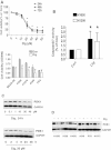
Figure 1
Figure 1
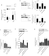
Ciglitazone decreased growth and induced apoptosis in lung cancer cells. A, H1299 cells were treated with increased concentrations of ciglitazone for up to 72 h (upper panel). NSCLC cells indicated were treated with ciglitazone (20 μM) or up to 48 h (lower panel). The cell viability was determined using the MTT assay as described in the Materials and Methods section in three separate experiments. B, Caspase 3/7 activity assay was performed on H1299 cells treated with or without ciglitazone for 48 h. Relative caspase 3/7 activity is indicated as percentage of untreated control cells. Results represent those obtained in three experiments. *indicates significant difference as compared to the untreated control group (P < 0.05). C, Cellular protein was isolated from H1299 cells that were cultured with increased concentrations of ciglitazone for up to 24 h (upper), or with ciglitazone (20 μM) for indicated period of time (lower), followed by Western blot. D, Cellular protein were isolated from NSCLC cells (H1299, PC9, A549, H1957, H358 and H1650) that were cultured with ciglitazone (20 μM) for up to 24, followed by Western Blot.
Figure 2
Figure 2
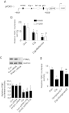
Ciglitazone inhibited PDK1 protein expression independent of PPARγ. A, H1299 and H1650 cells were transfected with control or PPRE X3-TK-luc reporter (from Addgene) for 24 h, followed by treating with ciglitazone for an additional 24 h. Afterwards, the Luciferase reporter activity was measured using Luciferase Assay System (Promega) according to manufacturer's instructions. The bars represent the mean ± SD of at least three independent experiments for each condition. *indicates significant difference as compared to the untreated control group (P < 0.05). B, Cellular protein was isolated from H1299 and H1650 cells cultured for 1 h in the presence or absence of GW9662 (20 μM) before exposing the cells to ciglitazone (20 μM) for an additional 24 h, then subjected to Western blot analysis. C, H1299 cells were transfected with control or PDK1 siRNA (80 nM) for 40 h, followed by exposing the cells to ciglitazone (20 μM) for an additional 24 h. Afterwards, the luminescence of viable cells was detected using Cell Titer-Glo Luminescent Cell Viability Assay kit. D-E, H1299 cells were transfected with the control and PDK1 expression vectors using the oligofectamine reagent according to the manufacturer’s instructions. After 24 h of incubation, cells were treated with or without ciglitazone for an additional 24 h. Afterwards, the luminescence of viable cells was detected using Cell Titer-Glo Luminescent Cell Viability Assay kit (D). In separate experiment, the relative caspase 3/7 activity (E) is indicated as percentage of untreated control cells. The bars represent the mean ± SD of at least four independent experiments for each condition. Insert on the top panel shows a Western blot for PDK1 protein. *indicates significant difference as compared to the untreated control group (P < 0.05). **Indicates significance of combination treatment as compared with ciglitazone alone (P < 0.05).
Figure 3
Figure 3
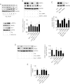
The role of AMPK and SAPK/JNK in mediating the effect of ciglitazone on PDK1 protein expression. A-B, Cellular protein were isolated from H1299 cells that were cultured with ciglitazone for up to 24, followed by Western Blot for phosphor-AMPKα, SAPK/JNK and total AMPKα, SAPK/JNK. C, Cellular protein was isolated from H1299 and H1650 cells treated with SP600125 (10 μM) or compound C (20 μM) for 1 h before exposure of the cells to ciglitazone for an additional 24 h. Afterwards, Western blot was performed. D, Cellular protein was isolated from H1299 cells transfected with control or AMPKα siRNA (80 nM) for 40 h, followed by exposing the cells to ciglitazone for an additional 24 h. Afterwards, Western blot was performed. E, Cellular protein was isolated from H1299 cells treated with ciglitazone and metformin (5 mM) for 24 h, followed by Western Blot. GAPDH served as internal controls for normalization purposes. The bar graph represents the mean ± SD of PDK1/GAPDH of at least three independent experiments. *Indicates significant difference from untreated control. **Indicates significance of combination treatment as compared with ciglitazone alone (P < 0.05).
Figure 4
Figure 4
Ciglitazone decreased PDK1 promoter activity. A, The human PDK1 wild type reporter construct schematics are presented. These regions contain several transcription factor binding sites including PPRE, Egr-1, p53 and NF-κB. B, H1299 and H1650 lung cancer cells (1 × 10⁵ cells) were transfected with a wild type human PDK1 promoter reporter construct ligated to luciferase reporter gene and an internal control Renilla Luciferase Reporter Vector for 24 h. Afterward, cells were treated with ciglitazone for an additional 24 h. C, H1299 cells were transfected with control or PPARγ siRNAs (80 nM) together with a wild type ILK promoter construct for 30 h, then cells were exposed to ciglitazone for an additional 24 h. Insert on the top shows Western blot result for PPARγ protein. GAPDH served as internal control for normalization purposes. D, H1299 cells (1 × 10⁵ cells) were transfected with a wild type human PDK1 promoter reporter construct ligated to luciferase reporter gene and an internal control Renilla Luciferase Reporter Vector as described in Materials and Methods for 24 h. Afterwards, cells were treated with SP600125 (10 μM) for 1 h before exposure of the cells to ciglitazone for an additional 24 h. The ratio of firefly luciferase to renilla luciferase activity was quantified as described in Material and Methods. The bars represent the mean ± SD of at least four independent experiments for each condition. *Indicates significant increase of activity as compared to controls. **Indicates significance of combination treatment as compared with ciglitazone alone (P < 0.05).
Figure 5
Figure 5
Ciglitazone induces Egr-1 protein expression; silencing of Egr-1 abrogates the effect of ciglitazone on PDK1 promoter activity, protein expression and cell proliferation. A, Cellular proteins were isolated from H1299 cells treated with ciglitazone (20 μM) for the indicated time period (upper panel) or with metformin (5 mM) for 24 h (lower panel). Afterwards, Western blot analyses were performed for detecting Egr-1, p65 and p53 proteins. B, H1299 cells were transfected with control or AMPKα siRNA (80 nM) for 30 h before exposing the cells to ciglitazone (20 μM) for an additional 24 h followed by Western blot. C, H1299 cells were transfected with control or Egr-1 siRNA (80 nM) together with a wild type PDK1 promoter construct for 30 h, then cells were exposed to ciglitazone (20 μM) for an additional 24 h. Insert shows the Western blot result for Egr-1 protein. D, H1299 cells were transfected with control or Egr-1 siRNA (80 nM) for 30 h before exposing the cells to ciglitazone (20 μM) for an additional 24 h, followed by Western blot. E, H1299 cells were transfected with control or Egr-1 siRNA (80 nM) for 30 h before exposure of the cells to ciglitazone (20 μM) for an additional 24 h. Afterwards, the luminescence of viable cells was detected using Cell Titer-Glo Luminescent Cell Viability Assay kit. The bars represent the mean ± SD of at least four independent experiments for each condition. *Indicates significant difference as compared to the control. **Indicates significance of combination treatment as compared with ciglitazone alone (P < 0.05).
Figure 6
Figure 6
Overexpression of Egr-1 reduces PDK1 promoter activity. A, H1299 cells (1x10⁵ cells) were transfected with control and Egr-1 expression reporter constructs, and together with a wild type PDK1 promoter construct and an internal control Renilla Luciferase Reporter Vector as described in Material and Methods section for 24 h, then treated with ciglitazone (20 μM) for an additional 24 h. The insert in upper panel represents Western blot results for Egr-1 protein. B, H1299 cells were lysed after exposure of ciglitazone (20 μM) for 24 h, and nuclei were isolated and then sonicated. Chromatin from H1650 cells was immunoprecipitated using antibodies against Egr-1 protein or preimmune serum (pre-immune). PCR analysis using primers surrounding the Egr-1 site shows that this DNA sequence (−4392 to −4402 bp) is specifically immunoprecipitated indicating that Egr-1 binds to endogenous DNA sites in the PDK1 promoter. A non- Egr-1 sequence was used as control. Aliquots of the chromatin were also analyzed before immunoprecipitation (input). C, Diagram demonstrates that ciglitazone inhibits PDK1 expression through AMPKα-mediated induction of Egr-1 protein expression and Egr-1 protein binding to the DNA sequence in the PDK1 gene promoter independent of PPARγ. Activation of AMPKα enhances the effect of ciglitazone on Egr-1 and PDK1 protein expression. In turn, this results in inhibition of NSCLC cell proliferation.

Similar articles

Cited by

References

    1. Lee CB, Stinchcombe TE, Rosenman JG, Socinski MA. Therapeutic advances in local-regional therapy for stage III non-small-cell lung cancer: evolving role of dose-escalated conformal (3-dimensional) radiation therapy. Clin Lung Cancer. 2006;8:195–202. doi: 10.3816/CLC.2006.n.047. - DOI - PubMed
    1. Dempke WC, Suto T, Reck M. Targeted therapies for non-small cell lung cancer. Lung Cancer. 2010;67:257–274. doi: 10.1016/j.lungcan.2009.10.012. - DOI - PubMed
    1. Siegel R, Naishadham D, Jemal A. Cancer statistics, 2013. CA Cancer J Clin. 2013;63:11–30. doi: 10.3322/caac.21166. - DOI - PubMed
    1. Tan NS, Michalik L, Desvergne B, Wahli W. Multiple expression control mechanisms of peroxisome proliferator-activated receptors and their target genes. J Steroid Biochem Mol Biol. 2005;93:99–105. doi: 10.1016/j.jsbmb.2004.12.025. - DOI - PubMed
    1. Choi JH, Banks AS, Kamenecka TM, Busby SA, Chalmers MJ, Kumar N, Kuruvilla DS, Shin Y, He Y, Bruning JB, Marciano DP, Cameron MD, Laznik D, Jurczak MJ, Schürer SC, Vidović D, Shulman GI, Spiegelman BM, Griffin PR. Antidiabetic actions of a non-agonist PPARgamma ligand blocking Cdk5-mediated phosphorylation. Nature. 2011;477:477–481. doi: 10.1038/nature10383. - DOI - PMC - PubMed

Publication types

MeSH terms