. 2014 Nov 1;23(21):5793-804.
doi: 10.1093/hmg/ddu297. Epub 2014 Jun 11.

TNNI3K mutation in familial syndrome of conduction system disease, atrial tachyarrhythmia and dilated cardiomyopathy

Affiliations

TNNI3K mutation in familial syndrome of conduction system disease, atrial tachyarrhythmia and dilated cardiomyopathy

Jeanne L Theis et al. Hum Mol Genet. .

Abstract

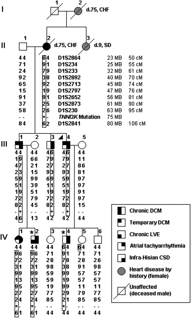
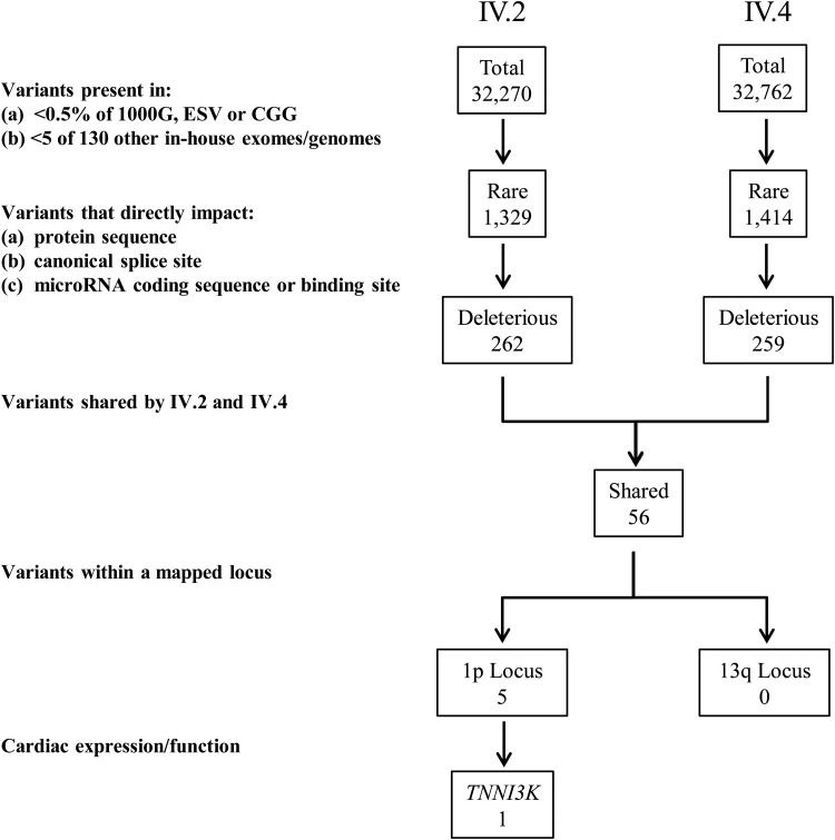
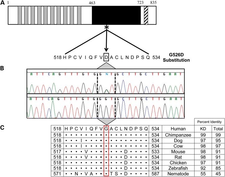
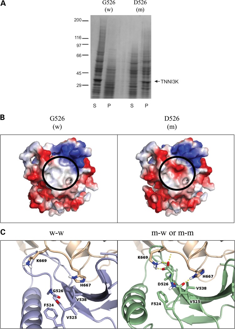
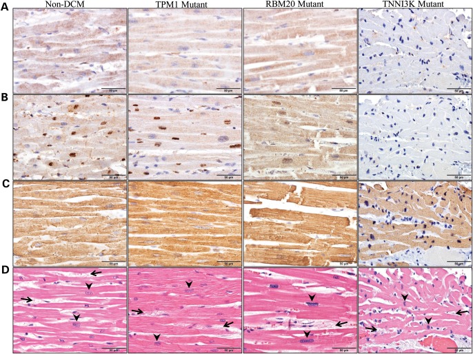
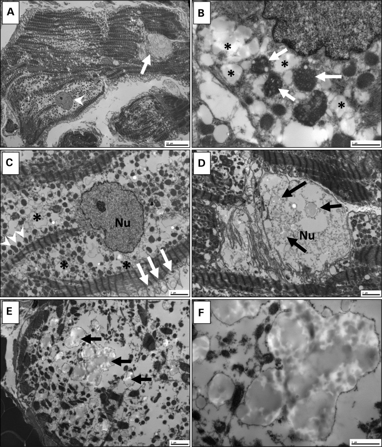
Locus mapping has uncovered diverse etiologies for familial atrial fibrillation (AF), dilated cardiomyopathy (DCM), and mixed cardiac phenotype syndromes, yet the molecular basis for these disorders remains idiopathic in most cases. Whole-exome sequencing (WES) provides a powerful new tool for familial disease gene discovery. Here, synergistic application of these genomic strategies identified the pathogenic mutation in a familial syndrome of atrial tachyarrhythmia, conduction system disease (CSD), and DCM vulnerability. Seven members of a three-generation family exhibited the variably expressed phenotype, three of whom manifested CSD and clinically significant arrhythmia in childhood. Genome-wide linkage analysis mapped two equally plausible loci to chromosomes 1p3 and 13q12. Variants from WES of two affected cousins were filtered for rare, predicted-deleterious, positional variants, revealing an unreported heterozygous missense mutation disrupting the highly conserved kinase domain in TNNI3K. The G526D substitution in troponin I interacting kinase, with the most deleterious SIFT and Polyphen2 scores possible, resulted in abnormal peptide aggregation in vitro and in silico docking models predicted altered yet energetically favorable wild-type mutant dimerization. Ventricular tissue from a mutation carrier displayed histopathological hallmarks of DCM and reduced TNNI3K protein staining with unique amorphous nuclear and sarcoplasmic inclusions. In conclusion, mutation of TNNI3K, encoding a heart-specific kinase previously shown to modulate cardiac conduction and myocardial function in mice, underlies a familial syndrome of electrical and myopathic heart disease. The identified substitution causes a TNNI3K aggregation defect and protein deficiency, implicating a dominant-negative loss of function disease mechanism.


Figures

Figure 1.
Figure 1.
Pedigree structure and haplotypes at 1p31-p36.1 locus for family with autosomal dominant electrical and myopathic heart disease. Phenotypic traits are indicated by shaded quadrants within the pedigree symbol. A triangle (▸) designates the proband whereas an asterisk (*) indicates individuals who underwent WES. Short tandem repeat DNA markers are listed from p telomere to centromere, with map locations according to the National Center for Biotechnology Information Web site (hg19 human reference genome) and given in megabases (MB) and centimorgans (cM). Haplotypes for marker genotypes are shown in columns below pedigree symbols with identical disease-associated haplotypes (boxed) inherited by all seven affected family members. A recombination event in III.1 defines D1S2864 as the upper-flanking marker whereas the lower-flanking marker, D1S2841, is defined by recombination events in III.3 and III.4. TNNI3K maps within this 57 MB locus. Presence of the identified TNNI3K-G526D mutant allele is indicated by a plus symbol (+) and its absence by a minus (−) symbol. CHF, congestive heart failure; CSD, conduction system disease; d., age at death; DCM, dilated cardiomyopathy; LVE, left ventricular enlargement; SD, sudden unexpected death.
Figure 2.
Figure 2.
Variant filtering scheme for WES data. An iterative filtering approach was applied to SNVs and insertion/deletions identified in individuals IV.2 and IV.4 by WES. The population frequency of individual variants and their impact on protein structure or expression were assessed as inclusion criteria. While there were no variants at the 13q locus, 5 variants resided within the 1p locus. One of these occurred in TNNI3K, a gene that is highly and specifically expressed in the heart and has an established role in cardiac physiology.
Figure 3.
Figure 3.
Location and conservation of TNNI3K-G526D mutation. (A) The protein topology is shown for TNNI3K, including 10 functional ankyrin repeat domains (gray), a serine-rich domain (diagonal hatch lines) and a single functional kinase domain (black) where the heterozygous c.1577 G > A resides (asterisk). (B) Sanger sequencing verified the mutation, which resulted in a G526D substitution. (C) Conservation of this residue is illustrated whereby • indicates identical residues. Overall, the amino acid sequence of TNNI3K is highly conserved with the amino acid identity to the human TNNI3K ortholog indicated for both the kinase domain (KD) and the entire protein.
Figure 4.
Figure 4.
In vitro expression and in silico modeling of mutant peptide. (A) Kinase domains of wild-type (w; G526) and mutant (m; D526) TNNI3K were expressed and the resulting pellet (P) and supernatant (S) fractions were analyzed by SDS-Page and Coomassie Blue, demonstrating poor solubility of the mutant peptide. The arrow indicates the 32 kilodalton kinase domain of TNNI3K protein and the molecular weight standards (×10−3) are shown on the left. (B) Electrostatic protein modeling (21) highlights a large hydrophobic patch on the surface of the kinase domain (white), which becomes negatively charged (red) in the presence of the G526D mutation. (C) The left panel depicts docking of wild-type (blue) to another wild-type TNNI3K (tan) (w–w) whereas the right panel shows the docking pose for mutant TNNI3K (green) to either wild-type or mutant TNNI3K (both represented in tan) (m-w and m–m). Residues within 3.5 Å of residue 526 in either pose are shown in atomic detail. Hydrogen bonds involving these indicated residues are drawn as dashed yellow lines. When mutated, the loop containing residue 526 shifts toward the binding partner to make more contacts including an electrostatic lysine-aspartic acid interaction (depicted as h-bonds).
Figure 5.
Figure 5.
Detection of TNNI3K protein in ventricular tissue by immunohistochemistry. Immunohistochemistry was performed using three antibodies on ventricular tissue from four individuals. Column 1 illustrates cardiac tissue from an individual without DCM (non-DCM), Columns 2 and 3 illustrate cardiac tissue from individuals with genetically defined familial DCM due to mutations in TPM1 (TPM1 Mutant) or RBM20 (RBM20 Mutant), respectively. The fourth column illustrates cardiac tissue from individual III.4 who carries the G526D mutation in TNNI3K (TNNI3K Mutant). Antibodies that recognize (A) the N-terminus of TNNI3K or (B) the C-terminus of TNNI3K reveal markedly reduced TNNI3K protein expression in the sarcoplasm (A and B) and the nuclei (B) of cardiomyocytes in the individual with the G526D mutation as compared with control tissue. (C) In contrast, actin expression was retained in all individuals. (D) H&E staining demonstrated non-specific histopathologic changes, including cardiomyocyte hypertrophy (evidenced by enlargement of cardiomyocyte nuclei, arrowheads) and mild interstitial fibrosis (increased fibrous connective tissue between individual cardiomyocytes, arrows), although the degree of cardiomyocyte hypertrophy was only mild in the TNNI3K mutant, in contrast with the moderate-to-severe hypertrophy in the other three patients.
Figure 6.
Figure 6.
Transmission electron microscopy of ventricular tissue. Ultrastructure of ventricular myocardium was determined for individual III.4. (A) At scanning magnification, non-specific mitochondriosis (increased number of mitochondria) is apparent with variable loss of myofilaments. Some nuclei are normal (arrowhead); others are abnormal (arrow). (B) Some mitochondria (arrows) show architectural disorganization of cristae with vacuolization and degenerative changes, and are surrounded by sarcoplasmic inclusions (*). (C) The sarcoplasm contains many severely swollen mitochondria (arrows), as well as scattered preserved mitochondria (arrowheads) within extensive zones (*) of complete mitochondrial disruption. In this cardiomyocyte, the nucleus (Nu) is intact. (D) Other nuclei are abnormal, containing numerous amorphous intranuclear inclusions (arrows). (E) Similar inclusions (arrows) are present in the sarcoplasm, particularly in areas of marked myofilament loss. (F) At higher magnification, inclusions are amorphous, globular and electron-opaque. Some appear to be membrane-bound, possibly representing distended mitochondria or lysozomes. Scale bar: 10 µm (A); 2 µm (C–E); 1 µm (B and F).

References

    1. Lloyd-Jones D.M., Wang T.J., Leip E.P., Larson M.G., Levy D., Vasan R.S., D'Agostino R.B., Massaro J.M., Beiser A., Wolf P.A., et al. Lifetime risk for development of atrial fibrillation: the Framingham Heart Study. Circulation. 2004;110:1042–1046. - PubMed
    1. Darbar D., Herron K.J., Ballew J.D., Jahangir A., Gersh B.J., Shen W.-K., Hammill S.C., Packer D.L., Olson T.M. Familial atrial fibrillation is a genetically heterogeneous disorder. J. Am. Coll. Cardiol. 2003;41:2185–2192. - PubMed
    1. Chen Y.H., Xu S.J., Bendahhou S., Wang X.L., Wang Y., Xu W.Y., Jin H.W., Sun H., Su X.Y., Zhuang Q.N., et al. KCNQ1 gain-of-function mutation in familial atrial fibrillation. Science. 2003;299:251–254. - PubMed
    1. Hodgson-Zingman D.M., Karst M.L., Zingman L.V., Heublein D.M., Darbar D., Herron K.J., Ballew J.B., de Andrade M., Burnett J.C., Olson T.M. Atrial natriuretic peptide frameshift mutation in familial atrial fibrillation. New Engl. J. Med. 2008;359:158–165. - PMC - PubMed
    1. Zhang X., Chen S., Yoo S., Chakrabarti S., Zhang T., Ke T., Oberti C., Yong S.L., Fang F., Li L., et al. Mutation in nuclear pore component NUP155 leads to atrial fibrillation and early sudden cardiac death. Cell. 2008;135:1017–1027. - PubMed

Publication types

MeSH terms