. 2014 Nov 1;23(21):5781-92.
doi: 10.1093/hmg/ddu296. Epub 2014 Jun 11.

POMK mutations disrupt muscle development leading to a spectrum of neuromuscular presentations

Affiliations

POMK mutations disrupt muscle development leading to a spectrum of neuromuscular presentations

Stefania Di Costanzo et al. Hum Mol Genet. .

Abstract

Dystroglycan is a transmembrane glycoprotein whose interactions with the extracellular matrix (ECM) are necessary for normal muscle and brain development, and disruptions of its function lead to dystroglycanopathies, a group of congenital muscular dystrophies showing extreme genetic and clinical heterogeneity. Specific glycans bound to the extracellular portion of dystroglycan, α-dystroglycan, mediate ECM interactions and most known dystroglycanopathy genes encode glycosyltransferases involved in glycan synthesis. POMK, which was found mutated in two dystroglycanopathy cases, is instead involved in a glycan phosphorylation reaction critical for ECM binding, but little is known about the clinical presentation of POMK mutations or of the function of this protein in the muscle. Here, we describe two families carrying different truncating alleles, both removing the kinase domain in POMK, with different clinical manifestations ranging from Walker-Warburg syndrome, the most severe form of dystroglycanopathy, to limb-girdle muscular dystrophy with cognitive defects. We explored POMK expression in fetal and adult human muscle and identified widespread expression primarily during fetal development in myocytes and interstitial cells suggesting a role for this protein during early muscle differentiation. Analysis of loss of function in the zebrafish embryo and larva showed that pomk function is necessary for normal muscle development, leading to locomotor dysfuction in the embryo and signs of muscular dystrophy in the larva. In summary, we defined diverse clinical presentations following POMK mutations and showed that this gene is necessary for early muscle development.


Figures

Figure 1.
Figure 1.
A nonsense mutation in POMK causes LGMD and cognitive deficits. (A) Two affected individuals, a female (P1) and a male (P2), were born to consanguineous parents (1–6 and 1–7) in Family 1. DNA was collected from all numbered individuals. Calf hypertrophy was observed in P1 (B) and P2 (Supplementary Material, Fig.S1B), and an arachnoid cyst was also identified on the left hemisphere in P2 via MRI imaging (C). (D) Both individuals carried a nonsense mutation truncating the POMK protein at glutamine 109 (c.325C>T). Parents were confirmed to be heterozygous for the variant.
Figure 2.
Figure 2.
Severe compound heterozygous changes in POMK cause WWS. (A) Family 2 was an Italian trio including a boy affected by WWS (P3). (B) One of the diagnostic features of WWS, cobblestone lissencephaly with hydrocephalus, was noted upon MRI imaging. (C) Exome sequencing identified two heterozygous mutations in POMK, which were confirmed by Sanger sequencing. One change was a 1 bp deletion leading to a frameshift (c.286delT) and the second change was a missense affecting a highly conserved amino acid (c.905T > A) (D). (E) The truncating mutations in Families 1 and 2 are found very early in the protein and are both predicted to completely remove the putative kinase domain in POMK.
Figure 3.
Figure 3.
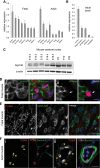
POMK is highly expressed during muscle and brain development. (A and B) Quantitative PCR analysis of POMK expression in human tissues shows highest expression in brain, skeletal muscle, kidney and heart in fetal and adult tissues. Highest expression in the brain is observed in the cerebral cortex. (C) Cortical expression was studied in the mouse and found to be dynamic increasing after E16.5. (D) The expression analysis of POMK in the developing human muscle was also revealed to be dynamic. In fetal muscle cells, POMK is found in both laminin-positive (i) and laminin-negative cells (arrow in ii). Scale bar: 10 µm (E) Lamin A/C staining outlines the nuclear envelope and POMK decorates perinuclear and cytoplasmic structures in all cells in the fetal muscle. Scale bar: 50 µm (F) In the adult human muscle, POMK expression is restricted to interstitial cells between the myofibers and in cells closely associated with the blood vessels (yellow arrowheads). Scale bar: 50 µm.
Figure 4.
Figure 4.
POMK knockdown reduces dystroglycan glycosylation but does not affect myotube formation. (A) POMK is highly expressed in both MCAM-positive and MCAM-negative cells (arrow) purified from human fetal muscle. MCAM-positive cells are myogenic cells and we wondered whether POMK knockdown would affect the early stages of myocyte development. Scale bars: 10 µm. (B) Purified myocytes will fuse to form myotubes (arrows) over the first week in vitro. Scale bar: 100 µm. (C) POMK is highly expressed starting at Day 1 after myogenic cell purification. (D) A highly efficient siRNA pool was tested and shown to generate consistent knockdown starting at a concentration of 25 nm. (E) No effect was observed in myocyte fusion during the first week in vitro, but α-DG glycosylation was reduced following POMK knockdown. Day 4 samples are shown.
Figure 5.
Figure 5.
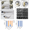
pomk knockdown disrupts muscle development in the zebrafish embryo. (A) pomk MO injection affects brain, eye and muscle development in the zebrafish embryo. The tail is thickened and the muscle appears disorganized, the head is smaller and retinal fusion is delayed (compare distance between small arrows in WT and MO right panels). Scale bar: 500 µm. (B) Locomotor activity in the developing zebrafish embryo is disrupted at 20 hpf. Embryo body movement is tracked in a video (Supplementary Material, videos): head movement is tracked in blue and tip of the tail movement in yellow. Both head and tail change position dramatically in the wild type due to spontaneous tail coiling. In the pomk morphants, the head and tail always remain in the same relative situation because the embryo is unable to shift position within the chorion. (B) Morphant larvae at 3 dpf show a shorter and bent tail. Scale bar: 500 µm. (C) pomk morphants muscle at 3 dpf shows signs of muscular dystrophy, laminin and dystrophin (dystr) staining are reduced and glycosylated α-DG is almost completely missing. Disorganization of the muscle fibers can also be noted by the disruption in nuclear orientation revealed by Hoechst staining. Scale bar: 100 µm. (E) Injection of human full-length POMK mRNA in the pomk morphant embryo causes significant phenotypic improvement, while injection of truncated (Q109*) POMK mRNA does not (left graph). Overexpression of mRNA up to 1 ng has no effect on the embryos (right graph). Results are from at least three independent experiment and are quantified at 1 dpf scoring the phenotypes shown in Figure 5A. Embryos scored as severe are greatly underdeveloped with barely distinguishable head and tail.

References

    1. Muntoni F., Torelli S., Wells D.J., Brown S.C. Muscular dystrophies due to glycosylation defects. Curr. Opin. Neurol. 2011;24:437–442. - PubMed
    1. Wells L. The o-mannosylation pathway: glycosyltransferases and proteins implicated in congenital muscular dystrophy. J. Biol. Chem. 2013;288:6930–6935. - PMC - PubMed
    1. Cormand B., Pihko H., Bayés M., Valanne L., Santavuori P., Talim B., Gershoni-Baruch R., Ahmad A., Van Bokhoven H., Brunner H.G., et al. Clinical and genetic distinction between Walker-Warburg syndrome and muscle-eye-brain disease. Neurology. 2001;56:1059–1069. - PubMed
    1. Dobyns W.B., Pagon R.A., Armstrong D., Curry C.J., Greenberg F., Grix A., Holmes L.B., Laxova R., Michels V.V., Robinow M. Diagnostic criteria for Walker-Warburg syndrome. Am. J. Med. Genet. 1989;32:195–210. - PubMed
    1. Balci B., Uyanik G., Dincer P., Gross C., Willer T., Talim B., Haliloglu G., Kale G., Hehr U., Winkler J., et al. An autosomal recessive limb girdle muscular dystrophy (LGMD2) with mild mental retardation is allelic to Walker-Warburg syndrome (WWS) caused by a mutation in the POMT1 gene. Neuromuscular Disord. 2005;15:271–275. - PubMed

Publication types

MeSH terms