. 2014 Jun 12;9(6):e100108.
doi: 10.1371/journal.pone.0100108. eCollection 2014.

ABA renewal involves enhancements in both GluA2-lacking AMPA receptor activity and GluA1 phosphorylation in the lateral amygdala

Affiliations

ABA renewal involves enhancements in both GluA2-lacking AMPA receptor activity and GluA1 phosphorylation in the lateral amygdala

Kyungjoon Park et al. PLoS One. .

Abstract

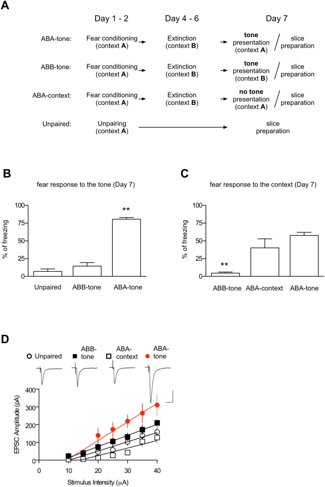
Fear renewal, the context-specific relapse of fear following fear extinction, is a leading animal model of post-traumatic stress disorders (PTSD) and fear-related disorders. Although fear extinction can diminish fear responses, this effect is restricted to the context where the extinction is carried out, and the extinguished fear strongly relapses when assessed in the original acquisition context (ABA renewal) or in a context distinct from the conditioning and extinction contexts (ABC renewal). We have previously identified Ser831 phosphorylation of GluA1 subunit in the lateral amygdala (LA) as a key molecular mechanism for ABC renewal. However, molecular mechanisms underlying ABA renewal remain to be elucidated. Here, we found that both the excitatory synaptic efficacy and GluA2-lacking AMPAR activity at thalamic input synapses onto the LA (T-LA synapses) were enhanced upon ABA renewal. GluA2-lacking AMPAR activity was also increased during low-threshold potentiation, a potential cellular substrate of renewal, at T-LA synapses. The microinjection of 1-naphtylacetyl-spermine (NASPM), a selective blocker of GluA2-lacking AMPARs, into the LA attenuated ABA renewal, suggesting a critical role of GluA2-lacking AMPARs in ABA renewal. We also found that Ser831 phosphorylation of GluA1 in the LA was increased upon ABA renewal. We developed a short peptide mimicking the Ser831-containing C-tail region of GluA1, which can be phosphorylated upon renewal (GluA1S); thus, the phosphorylated GluA1S may compete with Ser831-phosphorylated GluA1. This GluA1S peptide blocked the low-threshold potentiation when dialyzed into a recorded neuron. The microinjection of a cell-permeable form of GluA1S peptide into the LA attenuated ABA renewal. In support of the GluA1S experiments, a GluA1D peptide (in which the serine at 831 is replaced with a phosphomimetic amino acid, aspartate) attenuated ABA renewal when microinjected into the LA. These findings suggest that enhancements in both the GluA2-lacking AMPAR activity and GluA1 phosphorylation at Ser831 are required for ABA renewal.


Conflict of interest statement

Competing Interests: The authors have declared that no competing interests exist.

Figures

Figure 1
Figure 1. ABA renewal-inducing stimuli produce a context-dependent enhancement of synaptic efficacy at T-LA synapses.
A. The behavioral procedure. On Day 7, brain slices were prepared immediately after a tone test (ABB-tone and ABA-tone group) or context exposure (ABC-context group). In unpaired controls, one set of rats was killed for the preparation of brain slices, while another set was monitored for conditioned freezing to a CS on Day 7. B and C. Pooled behavioral results. **, p<0.01. D. Input-output curves for EPSCs in unpaired controls (n = 7), ABC-context (n = 7), ABB-tone (n = 16) and ABA-tone groups (n = 9). The representative traces are the averages of four responses evoked by input stimulations of 35 µA. Scale bars, 20 ms and 100 pA.
Figure 2
Figure 2. ABA renewal-inducing stimuli enhance the amplitude of AMPAR-mediated mEPSCs at T-LA synapses.
A. The behavioral procedure. On Day 7, brain slices were prepared immediately after a tone test (ABA-tone group). In the extinction group, one set of rats was killed for the preparation of brain slices, while another set was monitored for conditioned freezing to CS on Day 7. B. Upper left: Sample traces of evoked EPSCs in the presence of Ca2+ or Sr2+. Scale bars, 50ms and 10 pA. Note that the representative traces were further processed with a digital Gaussian filter for better display. Lower left: Cumulative amplitude distributions of evoked mEPSCs in the presence of Sr2+ (n = 300 events per cell). Upper right: Mean amplitude of mEPSCs evoked in the presence of Sr2+ (F2,16 = 5.896, p = 0.0121; naïve, 14.3±0.9 pA, n = 7; extinction,14.0±0.7 pA, n = 7; ABA-tone, 17.5±0.4 pA, n = 5; *, p<0.05). Lower right: Mean frequency of mEPSCs evoked in the presence of Sr2+ (naïve, 8.0±1.1Hz, n = 7; extinction, 7.4±1.2Hz, n = 7; ABA-tone, 9.4±1.7Hz, n = 5).
Figure 3
Figure 3. ABA renewal-inducing stimuli enhance GluR2-lacking AMPAR activity at T-LA synapses.
A. ABA renewal-inducing stimuli increased the RI of synaptic AMPA currents compared with the extinction group (extinction, n = 13; ABA-tone, n = 12). Superimposed averages of AMPAR-mediated EPSCs recorded at −60 mV and +40 mV are shown in the inset (see Methods for additional details). B. PhTx, a selective blocker for GluA2-lacking AMPARs, inhibited AMPAR-mediated EPSCs in the renewal group, but not in the extinction group. D-AP5 (100 µM) was included in the recording solution. EPSCs were elicited at a frequency of 0.33 Hz. *, p<0.05 (paired t-test).
Figure 4
Figure 4. The GluR2-lacking AMPAR activity is also enhanced during low-threshold potentiation.
A. The pairing protocol induced low-threshold potentiation, and ABA renewal occluded the low-threshold potentiation. Left, Pairing of four stimulus pulses (100 Hz) with a postsynaptic depolarization (40 ms) induced significantly less potentiation in the renewal group than in the extinction controls. The baseline responses for the first 3-min period were omitted because these responses tended to show a spontaneous increase after starting the whole-cell recordings. Instead, the baseline responses for 2 min before the delivery of the pairing protocol are shown (the pairing protocol was given 5 min after start of the whole-cell recordings). Right, The baseline responses were stable at least for 30 min in slices prepared from the extinction group. Representative paired traces are the averages of four traces within 2 min before and 25 min after pairing, respectively. Scale bars, 10 ms and 50 pA. B. The low-threshold potentiation was associated with an enhancement in the RI of EPSCs recorded at −70 mV and +40 mV (see Methods for additional details). B1 Left: A representative experiment in which the RI was estimated during low-threshold potentiation using an internal solution with spermine. B1 Right: Superimposed averages of EPSCs for a neuron shown in the left graph. B2 Left: The magnitude of the low-threshold potentiation and the rectification index were significantly correlated (r2 = 0.6473). B2 Right: Summary data (n = 13) for the RI changes after the induction of low-threshold potentiation. C. Left, Summary data (n = 12) for the RI changes after the induction of low-threshold potentiation in the experiments without intracellular spermine. Right, Example traces. In this experiment, we observed a significant increase (>130% of baseline responses) in NMDA EPSCs after pairing in 25% of cells recorded and ruled out these cells from further analyses. In B and C, D-AP5 was not included in the recording solution. *, p<0.05; **, p<0.01 (paired t-test). Scale bars in B and C, 50 ms and 100 pA. D. PhTx inhibited enhanced EPSCs after the induction of the low-threshold potentiation. EPSCs were elicited at a frequency of 0.33 Hz.
Figure 5
Figure 5. Inhibition of GluR2-lacking AMPAR activity in the LA is required for ABA renewal.
A. The behavioral procedure for the experiments. NASPM, a selective blocker of GluA2-lacking AMPARs, was microinjected 15 min before tone presentation in context C on Day 6. In this figure, a weaker conditioning protocol was used (see Methods for additional details). B. Microinjection of NASPM (40 µg) into the LA impaired ABA renewal relative to the microinjection of saline (F2,23 = 10.44, p = 0.0006, one-way ANOVA; Saline, 66.9±7.0%, n = 12; 10 µg NASPM, 44.1±11.6%, n = 7; 40 µg NASPM, 16.0±3.5%, n = 7; p<0.001, Newman-Keuls post-test). C. Schematic representation of the injector cannula tips. Histological plates illustrating the injection site in the LA were adopted from the rat brain atlas .
Figure 6
Figure 6. ABA renewal-inducing stimuli enhance Ser-831 phosphorylation of surface GluR1 in the LA synaptosomes.
A. The behavioral procedure. On Day 7, the LA synaptosomal membranes were prepared immediately after a tone test (ABA-tone group). In the unpaired and extinction groups, one set of rats was killed for the preparation of LA synaptosomal membranes, while another set was monitored for conditioned freezing to CS on Day 7. B. Representative immunoblot. C. Pooled results showing that Ser-831 phosphorylation was enhanced upon ABA renewal. *, p<0.05, one-way ANOVA (F2,12 = 5.850, p = 0.0169) followed by Newman-Keuls post-test. D. There was no significant difference across the three groups in terms of the amount of surface GluR1 from the LA synaptosomes. The number of rats used in each group was as follows: unpaired = 20, extinction = 18, renewal = 19.
Figure 7
Figure 7. The GluR1S peptide inhibits both low-threshold potentiation and its associated enhancement in the RI.
A. Left: Inclusion of GluA1S in the internal solution inhibited the low-threshold potentiation relative to GluA1A (GluR1A, 135.9±7.8% of baseline, n = 12; GluA1S, 113.3±6.6% of baseline, n = 16). Representative traces are superimposed averages of the EPSCs before and 20 min after pairing. Scale bars, 20 ms and 50 pA. Right: The inclusion of GluA1S or GluA1A in the internal solution did not have a significant effect on basal synaptic transmission (GluA1s, 104.1±6.7% of baseline, n = 5, p>0.5; GluA1A, 109.7±5.8 of baseline, n = 5, p>0.1; paired t-test). B1. The pairing protocol produced an enhancement in the RI and the AMPA EPSC amplitudes when cells were dialyzed with GluA1A. Left: Sample traces from an individual experiment. Right: Summary data for the RI changes after the induction of low-threshold potentiation (114.9±5.9% of baseline, n = 13). NMDAR-meditated EPSCs did not change significantly after the induction of low-threshold potentiation (113.4±7.4% of baseline, p = 0.0930, paired t-test). B2. The inclusion of GluA1S in the internal solution attenuated the RI increase associated with the low-threshold potentiation. Left, Sample traces from an individual experiment. Right, Summary data for the RI changes after the induction of low-threshold potentiation (103.9±4.5% of baseline, n = 13). NMDAR-meditated EPSCs did not change (106.9±4.6% of baseline, p = 0.1563, paired t-test). *, p<0.05.
Figure 8
Figure 8. The microinjection of a cell permeable form of the GluA1-derived peptides into the LA attenuates ABA renewal.
A. The behavioral procedure. The tone stimulus used for renewal was 60sec in duration on Day 6. In this figure, a weaker conditioning protocol was used (see Methods for additional details). B1. Upper, The microinjection of GluA1S into the LA impaired ABA renewal relative to GluA1A (GluA1A, 41.43±2.68%, n = 8; GluA1S, 21.24±5.63%, n = 9; p<0.01, unpaired t-test). Lower, Schematic representation of the injector cannula tips. Histological plates illustrating the injection site in the LA were adopted from the rat brain atlas (○, GluA1A; •, GluA1S). B2. Top, GluA1A peptide injection had no effects on ABA renewal relative to vehicle controls (vehicle, 56.09±6.78%, n = 9; GluA1A, 53.70±11.05%, n = 10; p>0.5, unpaired t-test). Bottom, Schematic representation of the injector cannula tips. Histological plates illustrating the injection site in the LA were adopted from the rat brain atlas (○, Vehicle; •, GluA1A). C. Diffusion of the fluorescent dansyl-tat-GluR1S peptide (1nmol) within 1h after the microinjection, as visualized with a multiphoton microscope (top). The white arrow indicates the end of the injector cannula. Peptide transduction in individual LA neurons at high magnification (bottom). Ce, central amygdala; BA, basal amygdala. D1. Top: Microinjection of GluA1D into the LA impaired ABA renewal relative to GluA1A (GluA1A, 66.82±5.62%, n = 6; GluA1D, 36.70±6.77%, n = 6; p<0.01, unpaired t-test). Bottom: Schematic representation of the injector cannula tips (○, GluA1A; •, GluA1D). D2. Top: GluA1A injection had no effects on ABA renewal relative to vehicle controls (vehicle, 74.01±5.65%, n = 6; GluA1A, 77.37±7.24%, n = 4; p>0.05, unpaired t-test). Bottom: Schematic representation of the injector cannula tips (○, Vehicle; •, GluA1A).

Similar articles

Cited by

References

    1. Hermans D, Craske MG, Mineka S, Lovibond PF (2006) Extinction in human fear conditioning. Biol Psychiatry 60: 361–368 10.1016/j.biopsych.2005.10.006 - DOI - PubMed
    1. Vervliet B, Craske MG, Hermans D (2013) Fear extinction and relapse: state of the art. Annu Rev Clin Psychol 9: 215–248 10.1146/annurev-clinpsy-050212-185542 - DOI - PubMed
    1. Bouton ME (2004) Context and behavioral processes in extinction. Learn Mem 11: 485–494 10.1101/lm.78804 - DOI - PubMed
    1. Corcoran KA, Maren S (2001) Hippocampal inactivation disrupts contextual retrieval of fear memory after extinction. J Neurosci 21: 1720–1726. - PMC - PubMed
    1. Herry C, Ciocchi S, Senn V, Demmou L, Müller C, et al. (2008) Switching on and off fear by distinct neuronal circuits. Nature 454: 600–606 10.1038/nature07166 - DOI - PubMed

Publication types

Substances