

What's special about task in dystonia? A voxel-based morphometry and diffusion weighted imaging study
- PMID: 24925463
- PMCID: PMC4139455
- DOI: 10.1002/mds.25934
What's special about task in dystonia? A voxel-based morphometry and diffusion weighted imaging study
Abstract
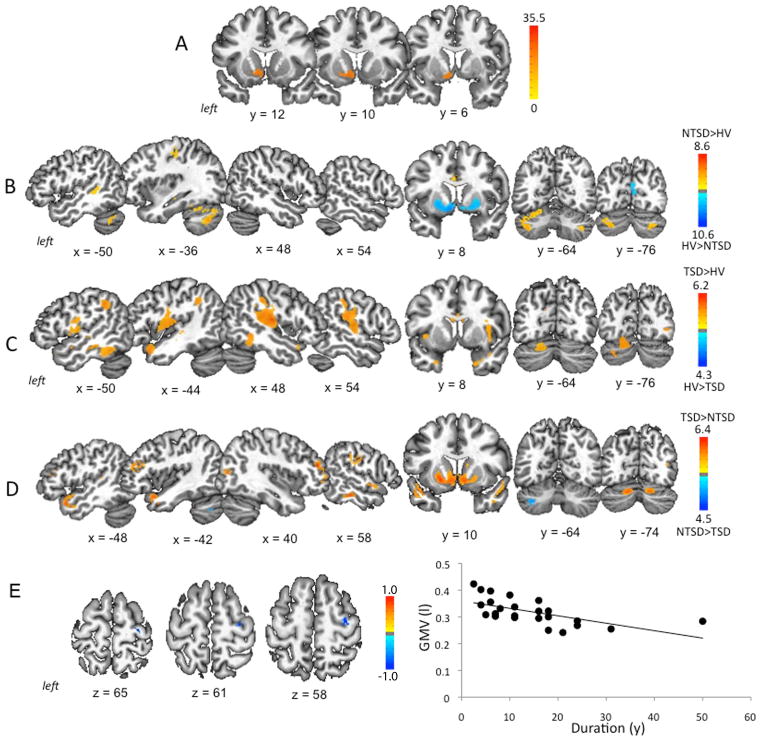
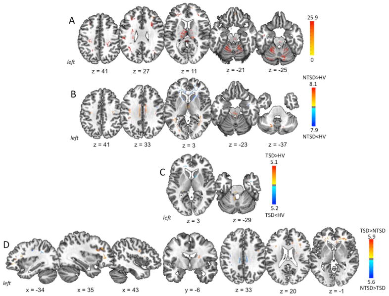
Numerous brain imaging studies have demonstrated structural changes in the basal ganglia, thalamus, sensorimotor cortex, and cerebellum across different forms of primary dystonia. However, our understanding of brain abnormalities contributing to the clinically well-described phenomenon of task specificity in dystonia remained limited. We used high-resolution magnetic resonance imaging (MRI) with voxel-based morphometry and diffusion weighted imaging with tract-based spatial statistics of fractional anisotropy to examine gray and white matter organization in two task-specific dystonia forms, writer's cramp and laryngeal dystonia, and two non-task-specific dystonia forms, cervical dystonia and blepharospasm. A direct comparison between both dystonia forms indicated that characteristic gray matter volumetric changes in task-specific dystonia involve the brain regions responsible for sensorimotor control during writing and speaking, such as primary somatosensory cortex, middle frontal gyrus, superior/inferior temporal gyrus, middle/posterior cingulate cortex, and occipital cortex as well as the striatum and cerebellum (lobules VI-VIIa). These gray matter changes were accompanied by white matter abnormalities in the premotor cortex, middle/inferior frontal gyrus, genu of the corpus callosum, anterior limb/genu of the internal capsule, and putamen. Conversely, gray matter volumetric changes in the non-task-specific group were limited to the left cerebellum (lobule VIIa) only, whereas white matter alterations were found to underlie the primary sensorimotor cortex, inferior parietal lobule, and middle cingulate gyrus. Distinct microstructural patterns in task-specific and non-task-specific dystonias may represent neuroimaging markers and provide evidence that these two dystonia subclasses likely follow divergent pathophysiological mechanisms precipitated by different triggers.
Keywords: brain imaging; focal dystonia; task specificity.
© 2014 International Parkinson and Movement Disorder Society.
Conflict of interest statement
Ritesh A. Ramdhani, MD, has nothing to disclose. Veena Kumar has nothing to disclose. Miodrag Velickovic, MD, has nothing to disclose. Steven J. Frucht, MD, has received consulting fees from Merz and Impax Laboratories, Inc., unrelated to the research in this article. Michele Tagliati, MD, has received speaker honoraria from Medtronic, Inc., and consultation fees from St. Jude Medical, Inc. (formerly Advanced Neuromodulation Systems), Abbvie, Allergan, Boston Scientific and Impax Laboratories, Inc., unrelated to the research in this article. Kristina Simonyan, MD, PhD, has nothing to disclose.
Figures
References
-
- Zoons E, Booij J, Nederveen AJ, Dijk JM, Tijssen MA. Structural, functional and molecular imaging of the brain in primary focal dystonia--a review. Neuroimage. 2011;56(3):1011–1020. - PubMed
-
- Garraux G, Bauer A, Hanakawa T, Wu T, Kansaku K, Hallett M. Changes in brain anatomy in focal hand dystonia. Ann Neurol. 2004;55(5):736–739. - PubMed
-
- Delmaire C, Vidailhet M, Elbaz A, et al. Structural abnormalities in the cerebellum and sensorimotor circuit in writer’s cramp. Neurology. 2007;69(4):376–380. - PubMed
Publication types
MeSH terms
Supplementary concepts
Grants and funding
LinkOut - more resources
Full Text Sources
Other Literature Sources
