. 2014 Dec 20;21(18):2515-30.
doi: 10.1089/ars.2013.5391. Epub 2014 Jul 30.

p62/SQSTM1 plays a protective role in oxidative injury of steatotic liver in a mouse hepatectomy model

Affiliations

p62/SQSTM1 plays a protective role in oxidative injury of steatotic liver in a mouse hepatectomy model

Sanae Haga et al. Antioxid Redox Signal. .

Abstract

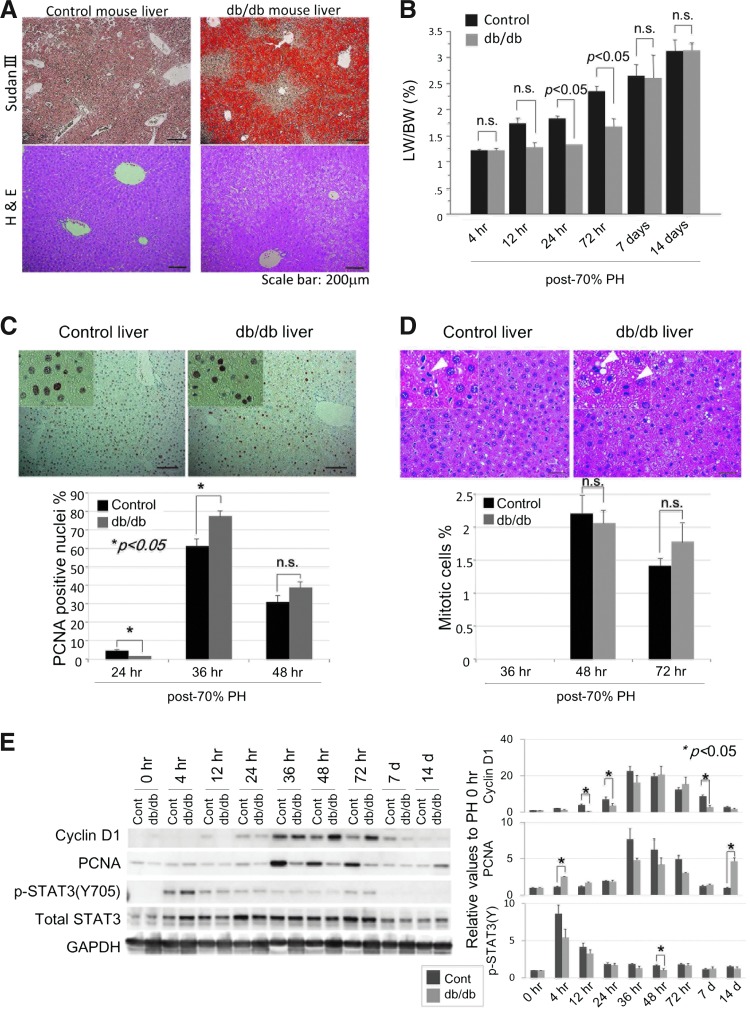
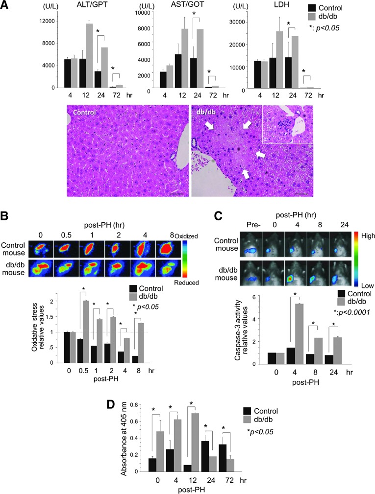
Aims: Liver injury and regeneration involve complicated processes and are affected by various physio-pathological factors. We investigated the mechanisms of steatosis-associated liver injury and delayed regeneration in a mouse model of partial hepatectomy.

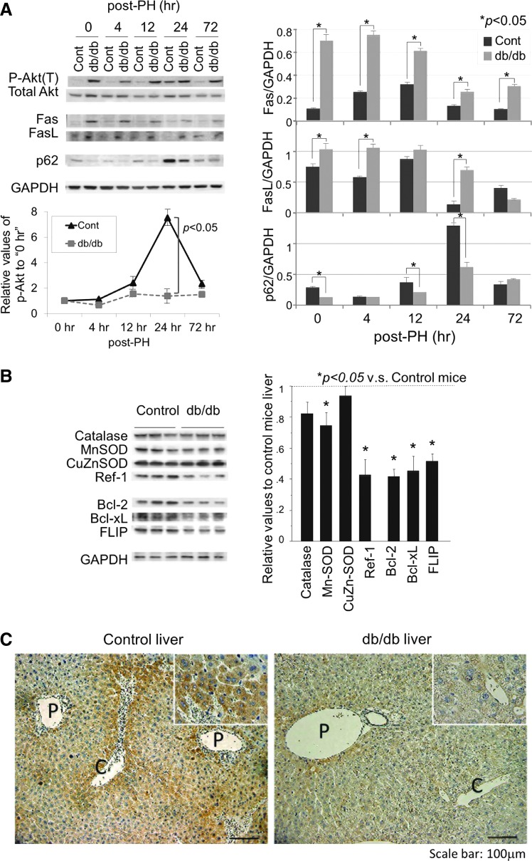
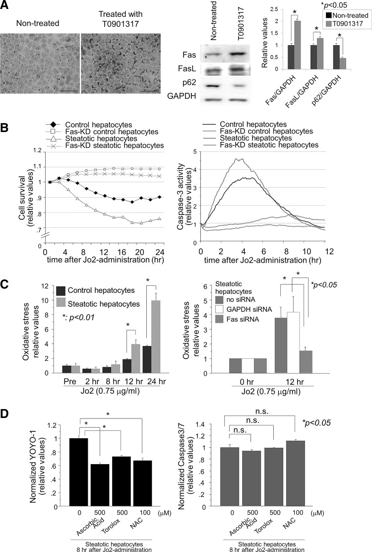
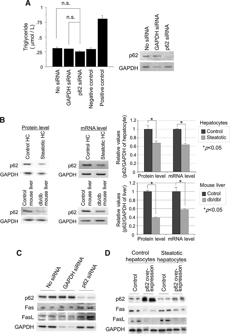
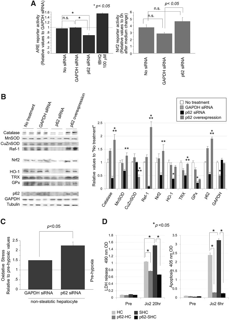
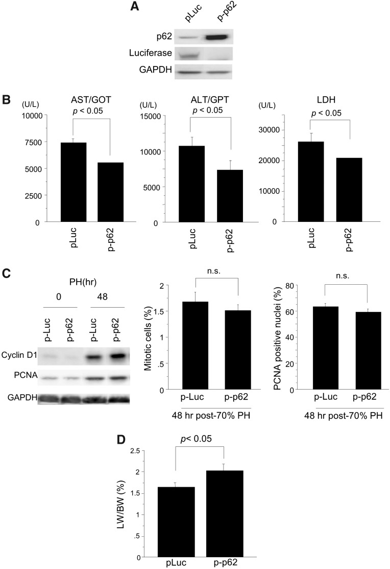
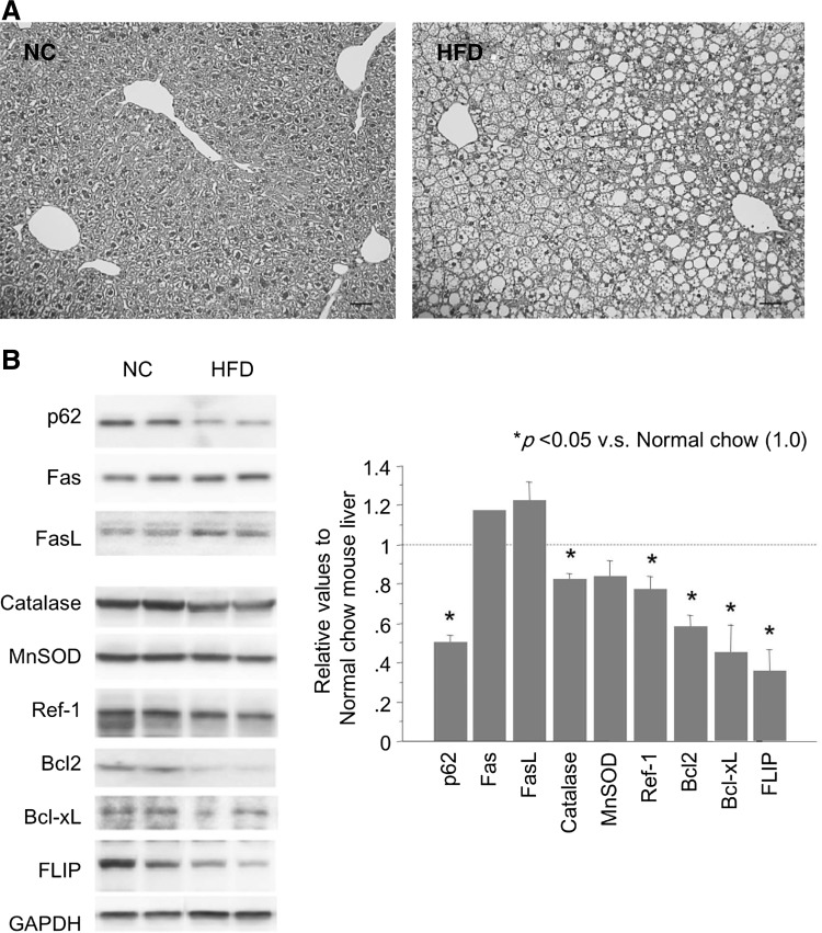
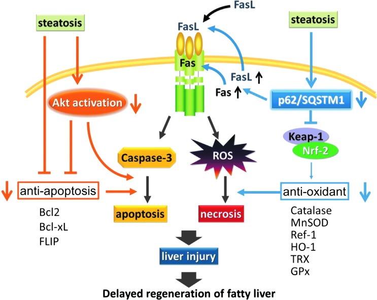
Results: Initial regeneration of the steatotic liver was significantly delayed after hepatectomy. Although hepatocyte proliferation was not significantly suppressed, severe liver injury with oxidative stress (OS) occurred immediately after hepatectomy in the steatotic liver. Fas-ligand (FasL)/Fas expression was upregulated in the steatotic liver, whereas the expression of antioxidant and anti-apoptotic molecules (catalase/MnSOD/Ref-1 and Bcl-2/Bcl-xL/FLIP, respectively) and p62/SQSTM1, a steatosis-associated protein, was downregulated. Interestingly, pro-survival Akt was not activated in response to hepatectomy, although it was sufficiently expressed even before hepatectomy. Suppression of p62/SQSTM1 increased FasL/Fas expression and reduced nuclear factor erythroid 2-related factor-2 (Nrf-2)-dependent antioxidant response elements activity and antioxidant responses in steatotic and nonsteatotic hepatocytes. Exogenously added FasL induced severe cellular OS and necrosis/apoptosis in steatotic hepatocytes, with only the necrosis being inhibited by pretreatment with antioxidants, suggesting that FasL/Fas-induced OS mainly leads to necrosis. Furthermore, p62/SQSTM1 re-expression in the steatotic liver markedly reduced liver injury and improved liver regeneration.

Innovation: This study is the first which demonstrates that reduced expression of p62/SQSTM1 plays a crucial role in posthepatectomy acute injury and delayed regeneration of steatotic liver, mainly via redox-dependent mechanisms.

Conclusion: In the steatotic liver, reduced expression of p62/SQSTM1 induced FasL/Fas overexpression and suppressed antioxidant genes, mainly through Nrf-2 inactivation, which, along with the hypo-responsiveness of Akt, caused posthepatectomy necrotic/apoptotic liver injury and delayed regeneration, both mainly via a redox-dependent mechanism.


Figures

<b>FIG. 1.</b>
FIG. 1.
Although post-PH initial liver regeneration is disturbed in db/db mice, mitotic responses occur equally in lean control and db/db steatotic mice. (A) Lipid content was confirmed by Sudan III stain in the liver of control and db/db mice before PH. Lipid accumulation was more evident in the central area than in the portal area of the db/db mouse liver. (B) Initial liver regeneration was disturbed in the db/db mice after PH. (C) Counts of PCNA-positive hepatocytes in the regenerating liver showed that the mitotic response peaked at 36 h post-PH in lean control and db/db mouse liver. (D) Histological examination (H&E staining) revealed similar mitotic responses at 36, 48, and 72 h post-PH in the liver of lean control and db/db steatotic mice. Scale bar: 50 μm. At least five mice were used for each experiment (B–D). Data are expressed as mean±SEM. (E) Expression of cell cycle-associated proteins was examined in the liver. Each blot represents at least three independent experiments. The intensity of each band was quantified by densitometry, showing the chronological relative changes for each protein, and is plotted on the right. The results are expressed as mean±SEM of at least three independent experiments; p<0.05 was considered statistically significant (B–E). Groups without an asterisk (*) were not statistically different (E). H&E, hematoxylin and eosin; PCNA, proliferating cell nuclear antigen; PH, partial hepatectomy; SEM, standard error of the mean. To see this illustration in color, the reader is referred to the web version of this article at www.liebertpub.com/ars
<b>FIG. 2.</b>
FIG. 2.
Steatotic liver shows acute OS and injury immediately after PH. (A) Blood biochemistry shows immediate liver injury after PH in the db/db steatotic mouse liver. Histologically, sporadic necrosis in the peri-portal areas (area surrounded by arrows) and neutrophil infiltration (inset) are observed at 24 h after PH. (B) Bio-imaging of liver OS after PH. Emission from the roGFP fluorescent probe was measured directly at the exposed liver surface, imaged, and quantified. Photographs are representative images of the dynamic changes in hepatic redox states (oxidation: green to blue; reduction: orange to red). Early post-PH, OS is observed in the db/db steatotic liver. For each experiment, the intensities of the hepatic roGFP signals are plotted relative to the pre-PH values. (C) Bio-imaging of liver caspase-3 activity after PH. The pcFluc-DEVD probe emitted signals at 4–24 h post-PH in the remnant liver of db/db mice, with no evident signal in the control liver. Data in the graph are expressed as mean±SEM and are expressed relative to the pre-PH control. (D) Apoptotic cell death is markedly increased at 4 h post-PH in the steatotic liver. Results are expressed as mean±SEM of five independent experiments; p<0.05 was considered statistically significant (AD). Each experiment was performed five times, and representative photographs are shown (B, C). OS, oxidative stress; roGFP, reduction-oxidation–sensitive green fluorescent protein. To see this illustration in color, the reader is referred to the web version of this article at www.liebertpub.com/ars
<b>FIG. 3.</b>
FIG. 3.
Analysis of signals associated with cell proliferation and survival, apoptosis, and OS in hepatocytes. (A) Protein expression of phospho-Akt, Akt, Fas, FasL, and p62/SQSTM1 was analyzed by Western blotting. (B) Anti-oxidant/anti-apoptotic proteins were analyzed in the liver of the control and db/db mice. Each blot represents at least three independent experiments. The intensity of each band was quantified by densitometry and is plotted on the right. The data are expressed as mean±SEM relative to the lean control values; p<0.05 was considered statistically significant (A, B). (C) Immunohistochemical study of the p62/SQSTM1 expression in mouse liver, demonstrating the reduced expression of p62/SQSTM1 in the db/db steatotic liver. (P: portal vein; C: central vein). FasL, Fas-ligand. To see this illustration in color, the reader is referred to the web version of this article at www.liebertpub.com/ars
<b>FIG. 4.</b>
FIG. 4.
Fas/FasL expression, ROS generation, and cell death (necrosis/apoptosis) in steatotic hepatocytes. (A) Steatosis was induced in AML12 liver cells by treatment with 1 μM T0901317 for 5 days. Oil Red O stain showed a marked and homogenous accumulation of lipid in the treated hepatocytes. Fas and FasL expression was upregulated in the T0901317-treated steatotic hepatocytes. Scale bar: 50 μm. Photographs are representative of three independent experiments. The intensities of each band are expressed relative to the intensities of the “Nontreated” band; p<0.05 was considered significant. (B) Cell survival and caspase-3 activity were monitored in the control and steatotic hepatocytes with or without Fas after anti-Fas antibody challenge (0.75 μg/ml of Jo2). (C) Anti-Fas-induced OS in the steatotic hepatocytes was evaluated using a redox-sensitive roGFP probe. For each group, the experiment was performed thrice and the data are expressed relative to values before anti-Fas treatment. (D) The effect of antioxidants on necrosis and apoptosis was examined in the steatotic hepatocytes. Antioxidants (ascorbic acid, Trolox, and NAC) suppressed anti-Fas-induced necrotic death but not apoptotic death in steatotic hepatocytes. Cell apoptosis and necrosis were determined using the fluorogenic probes Caspase-Glo 3/7 and YOYO-1, respectively. For each group, the experiment was performed thrice and the data are expressed relative to the values of the untreated cells. NAC, N-acetyl cysteine; ROS, reactive oxygen species.
<b>FIG. 5.</b>
FIG. 5.
Cellular steatosis reduces the expression of p62/SQSTM1, and the expression of FasL and Fas is negatively regulated by p62/SQSTM1, regardless of steatosis. (A) Adipogenesis assay of p62/SQSTM1-ablated AML12 cells. Ablation of p62/SQSTM1 alone did not induce steatosis in AML12 liver cells. Untreated AML12 cells were used as a negative control, and T0901317-treated AML12 cells were used as a positive control. (B) mRNA and protein expression levels of p62/SQSTM1 are reduced in steatotic hepatocytes and steatotic liver. The intensity of each band was quantified and normalized to control hepatocytes by densitometry and is plotted on the right; p<0.05 was considered statistically significant. (C) Ablation of p62/SQSTM1 alone induced FasL and Fas expression in AML12 hepatocytes, even without steatosis. (D) Overexpression of p62/SQSTM1 in both nonsteatotic and steatotic hepatocytes led to a reduction of FasL and Fas. Each experiment was performed at least thrice independently, and representative data are shown.
<b>FIG. 6.</b>
FIG. 6.
Reduced p62/SQSTM1 leads to a decrease in cellular antioxidant properties by suppressing ARE activity. (A) Ablation of p62/SQSTM1-suppressed ARE reporter activity in AML12 cells (left panel), but not Nrf-2 reporter activity (right panel). ARE reporter activity is expressed relative to the ARE activity against GAPDH siRNA. tBHQ was used as a positive control of the ARE reporter probe. (B) Ablation of p62/SQSTM1 reduced the expression of antioxidant molecules, whereas its overexpression increased the levels of these molecules. Photographs are representative of three independent experiments. The intensity of each band was quantified, normalized against tubulin, and expressed relative to the “no treatment” band (*p<0.05 vs. GAPDH-siRNA; **p<0.05 vs. GAPDH-siRNA and vs. p62-siRNA). (C) A roGFP redox-sensitive probe showed that ablation of p62/SQSTM1 enhanced hypoxia/reoxygenation-induced ROS generation in AML12 liver cells (4-h hypoxia, 10-min reoxygenation). (D) Induction of p62/SQSTM1 suppressed Jo2-induced cell death (LDH release and apoptosis) in both control and steatotic hepatocytes (0.75 μg/ml of Jo2, anti-Fas antibody). In each group, the data are expressed as mean±SEM values relative to the corresponding prehypoxic cells; p<0.05 was considered significant. ARE, antioxidant response elements; LDH, lactate dehydrogenase; Nrf-2, nuclear factor erythroid 2-related factor-2; siRNA, small-interfering RNA; tBHQ, tert-butylhydroquinone.
<b>FIG. 7.</b>
FIG. 7.
Hepatic transduction of the p62/SQSTM1 gene markedly reduces post-PH liver injury and improves regeneration of the steatotic liver of db/db mice. Post-PH liver injury and regeneration were evaluated in db/db mouse liver transduced with the p62/SQSTM1 gene. (A) Western blot analysis shows the robust induction of p62/SQSTM1 protein in the liver of db/db mice by hydrodynamic injection of the p62/SQSTM1 gene (p-p62). Plasmid DNA of luciferase gene was injected as a control (p-Luc). (B) Serum levels of AST, ALT, and LDH were reduced at 12 h post-PH in the p62/SQSTM1-transduced steatotic liver, as compared with the luciferase-transduced control liver. (C) Expression of PCNA and cyclinD1 protein peaked at 48 h post-PH similarly in both the control and p62/SQSTM1-expressing liver; in addition, there was no significant difference in the mitotic index and the counts of PCNA-positive hepatocytes between the two types of liver. (D) Liver regeneration recovered partially, but not completely, after transduction of p62/SQSTM1 into the steatotic liver. At least five mice were used for each experiment. Data are expressed as mean±SEM; p<0.05 was considered statistically significant. ALT, alanine aminotransferase; AST, aspartate aminotransferase.
<b>FIG. 8.</b>
FIG. 8.
HFD induces liver steatosis and p62/SQSTM1 reduction in mouse liver, leading to an increase in FasL/Fas and a decrease in antioxidant/-apoptotic molecules. (A) HFD feeding made hepatocytes sufficiently steatotic in the liver tissue (H&E staining). Photographs are representative of three independent experiments. NC: Normal chow. (B) Western blot analysis showed reduced expression of p62/SQSTM1, increased FasL/Fas, and decreased antioxidant (catalase, MnSOD, and Ref-1) and anti-apoptotic (Bcl-2, Bcl-xL, and FLIP) molecules. Each blot represents at least three independent experiments. The intensity of each band was quantified, normalized against GAPDH, and expressed relative to the “NC mouse liver” band. FLIP, FILCE-like inhibitory protein; HFD, high fat diet; MnSOD, manganese superoxide dismutase; Ref-1, redox factor-1.
<b>FIG. 9.</b>
FIG. 9.
Schematic showing the pivotal role of p62/SQSTM1 in injury of the steatotic liver. Steatosis-associated reduction of p62/SQSTM1 induces FasL/Fas in the steatotic liver. Along with exogenous FasL, this directly causes post-PH necrotic and apoptotic acute liver injury in, respectively, a redox-dependent and redox-independent manner. Reduction of p62/SQSTM1 reduces Keap-1/Nrf-2 binding, which suppresses the expression of antioxidant molecules (catalase, MnSOD, Ref-1, HO-1, TRX, and GPx) and, therefore, makes the liver susceptible to OS. Furthermore, the hypo-responsiveness of Akt enhances necrotic and apoptotic injury, along with the reduced expression of antioxidant molecules and anti-apoptotic molecules (Bcl-2, Bcl-xL, and FLIP), respectively. These mechanisms may collectively underlie steatosis-associated liver injury in the mouse. GPx, glutathione peroxidase; HO-1, heme oxygenase 1; Keap-1, Kelch-like ECH-associated protein 1; TRX, thioredoxin. To see this illustration in color, the reader is referred to the web version of this article at www.liebertpub.com/ars

References

    1. Aoyama T, Ikejima K, Kon K, Okumura K, Arai K, and Watanabe S. Pioglitazone promotes survival and prevents hepatic regeneration failure after partial hepatectomy in obese and diabetic KK-A(y) mice. Hepatology 49: 1636–1644, 2009 - PubMed
    1. Caraceni P, Domenicali M, Vendemiale G, Grattagliano I, Pertosa A, Nardo B, Morselli-Labate AM, Trevisani F, Palasciano G, Altomare E, and Bernardi M. The reduced tolerance of rat steatotic liver to ischemia reperfusion is associated with mitochondrial oxidative injury. J Surg Res 124: 160–168, 2005 - PubMed
    1. Cardoso AR, Kakimoto PA, and Kowaltowski AJ. Diet-sensitive sources of reactive oxygen species in liver mitochondria: role of very long chain acyl-CoA dehydrogenases. PLoS One 8: e77088, 2013 - PMC - PubMed
    1. Cha JY. and Repa JJ. The liver X receptor (LXR) and hepatic lipogenesis. The carbohydrate-response element-binding protein is a target gene of LXR. J Biol Chem 282: 743–751, 2007 - PubMed
    1. Deretic V. Autophagy in infection. Curr Opin Cell Biol 22: 252–262, 2010 - PMC - PubMed

Publication types

MeSH terms

LinkOut - more resources