

Melanocytoma of the ciliary body misdiagnosed as iridodialysis
- PMID: 24926192
- PMCID: PMC4049885
- DOI: 10.2147/OPTH.S63328
Melanocytoma of the ciliary body misdiagnosed as iridodialysis
Abstract
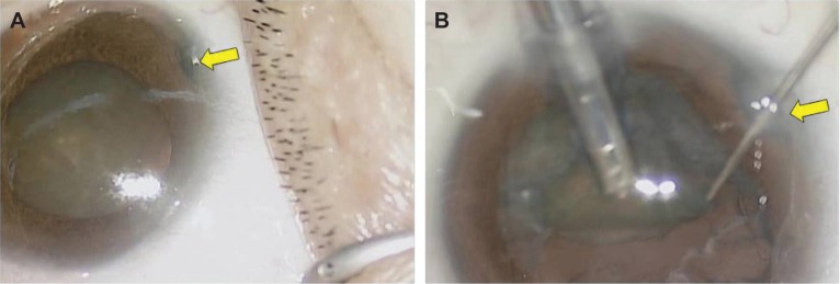
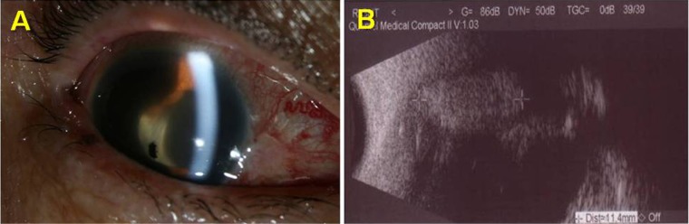
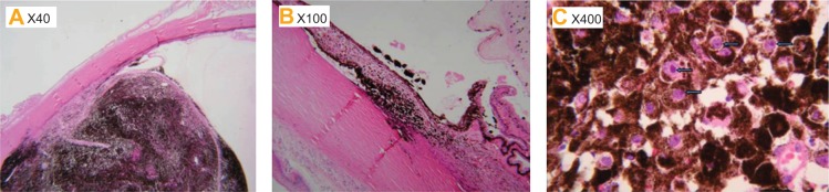
A 62-year-old female presented to our institution with dimness of vision in her right eye. On examination, her best corrected visual acuity was 20/100 in the right eye. The intraocular pressures were 14 mmHg in both eyes. Slit-lamp examination revealed nuclear sclerotic cataracts bilaterally and iridodialysis in her right eye. Seven days after the first visit, cataract surgery was performed without any complications. One year later, she presented to our institution with acute visual loss and ocular pain in the right eye. Best corrected visual acuity of the right eye was light perception and the intraocular pressure was 44 mmHg. Slit-lamp examination revealed a ciliary body mass with widespread pigment dispersion in the anterior segment. Due to no useful vision and uncontrolled pain, enucleation of the right eye was performed. Histopathologic examination revealed a melanocytoma of the ciliary body.
Keywords: ciliary body; iridodialysis; melanocytoma.
Figures
Similar articles
-
Ciliary body melanocytoma with anterior segment pigment dispersion and elevated intraocular pressure.J Glaucoma. 1999 Apr;8(2):129-33. J Glaucoma. 1999. PMID: 10209730
-
Transvitreal endocyclophotocoagulation.Trans Am Ophthalmol Soc. 1996;94:589-676. Trans Am Ophthalmol Soc. 1996. PMID: 8981713 Free PMC article.
-
Large Choroidal Melanocytoma Simulating Choroidal Melanoma: A Difficult Differential Diagnosis and an Inevitable Enucleation.Case Rep Ophthalmol Med. 2020 Nov 21;2020:8890857. doi: 10.1155/2020/8890857. eCollection 2020. Case Rep Ophthalmol Med. 2020. PMID: 33294243 Free PMC article.
-
A Case of Iris Melanocytoma Demonstrating Diffuse Melanocytic Proliferation with Uncontrolled Intraocular Pressure.Case Rep Ophthalmol. 2017 Mar 30;8(1):190-194. doi: 10.1159/000464349. eCollection 2017 Jan-Apr. Case Rep Ophthalmol. 2017. PMID: 28512420 Free PMC article.
-
7diffuse melanotic lesion of the iris as a presenting feature of ciliary body melanocytoma: report of a case and review of the literature.Surv Ophthalmol. 1998 Jan-Feb;42(4):378-82. doi: 10.1016/s0039-6257(97)00087-8. Surv Ophthalmol. 1998. PMID: 9493281 Review.
Cited by
-
A case of choroidal melanocytoma mimicking a choroidal melanoma.Saudi J Ophthalmol. 2015 Jul-Sep;29(3):242-5. doi: 10.1016/j.sjopt.2015.02.002. Epub 2015 Mar 4. Saudi J Ophthalmol. 2015. PMID: 26155089 Free PMC article.
-
Acute subconjunctival pigmentation with an underlying ciliary body mass: An unusual case presentation.Saudi J Ophthalmol. 2019 Jul-Sep;33(3):294-296. doi: 10.1016/j.sjopt.2019.07.003. Epub 2019 Jul 17. Saudi J Ophthalmol. 2019. PMID: 31686973 Free PMC article.
References
-
- Demirci H, Mashayekhi A, Shields CL, Eagle RC, Jr, Shields JA. Iris melanocytoma: clinical features and natural course in 47 cases. Am J Ophthalmol. 2005;139(3):468–475. - PubMed
-
- Brownstein S, Dorey MW, Mathew B, Little JM, Lindley JI. Melanocytoma of the choroid: atypical presentation and review of the literature. Can J Ophthalmol. 2002;37(4):247–252. - PubMed
-
- Shields JA, Shields CL, Eagle RC., Jr Melanocytoma (hyperpigmented magnocellular nevus) of the uveal tract: the 34th G. Victor Simpson lecture. Retina. 2007;27(6):730–739. - PubMed
-
- Zimmerman LE. Melanocytes, melanocytic nevi and melanocytomas. Invest Ophthalmol. 1965;4:11–41. - PubMed
Publication types
LinkOut - more resources
Full Text Sources
Other Literature Sources
