. 2014 Jun 4:8:137.
doi: 10.3389/fnins.2014.00137. eCollection 2014.

Simultaneous EEG-fMRI brain signatures of auditory cue utilization

Affiliations

Simultaneous EEG-fMRI brain signatures of auditory cue utilization

Mathias Scharinger et al. Front Neurosci. .

Abstract

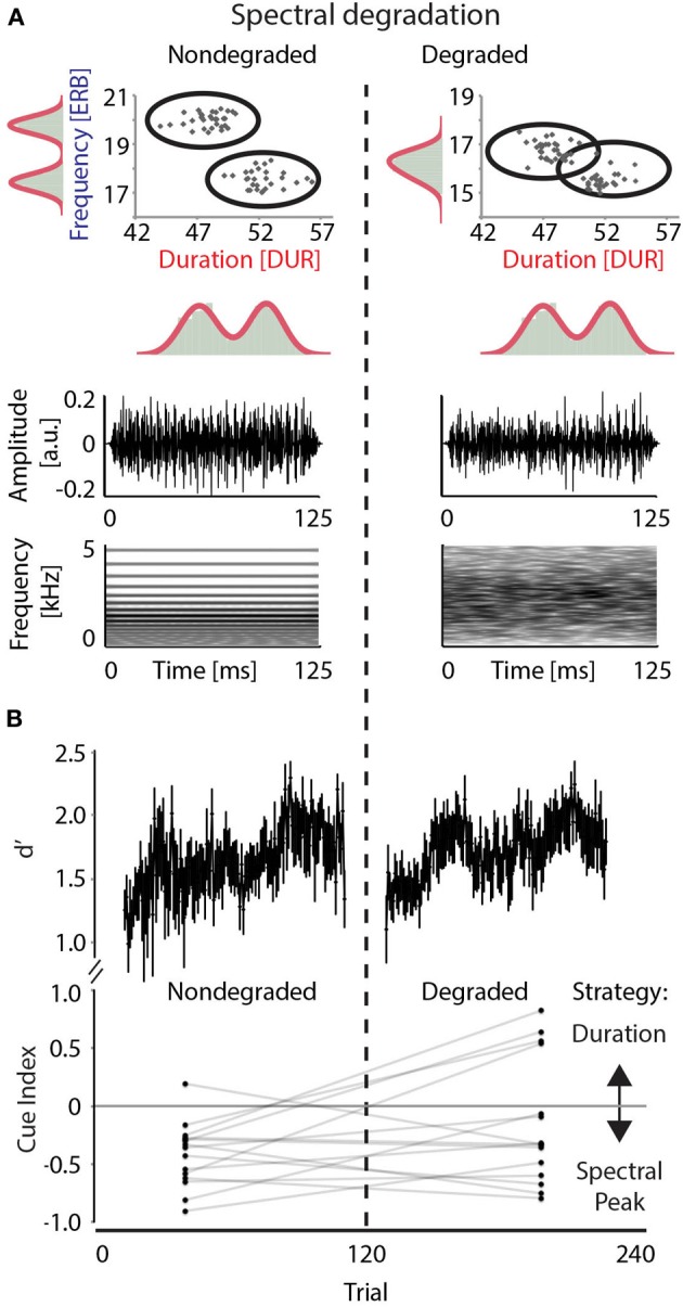
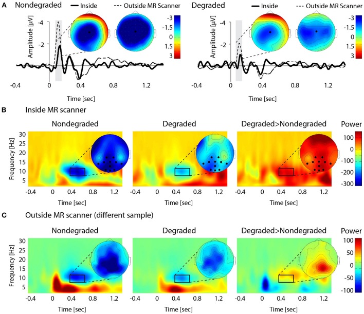
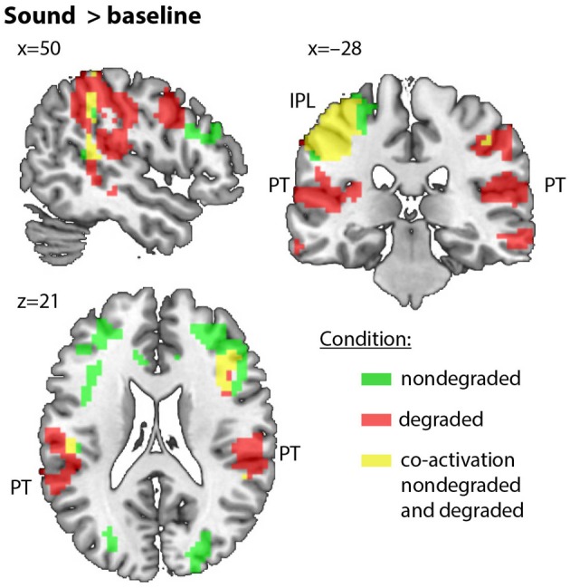
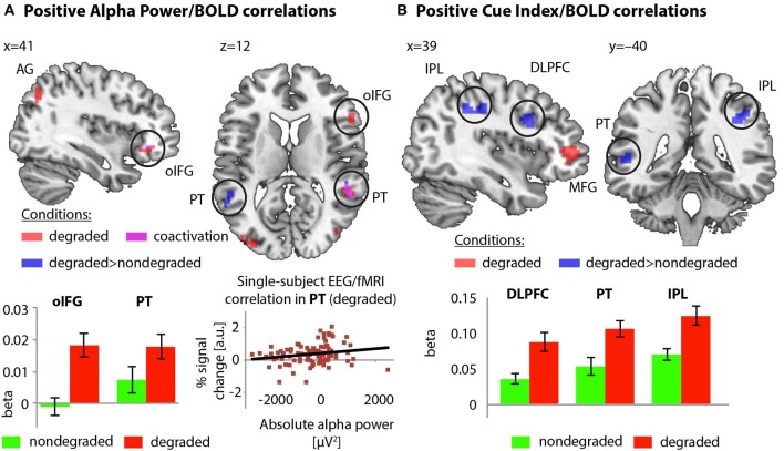
Optimal utilization of acoustic cues during auditory categorization is a vital skill, particularly when informative cues become occluded or degraded. Consequently, the acoustic environment requires flexible choosing and switching amongst available cues. The present study targets the brain functions underlying such changes in cue utilization. Participants performed a categorization task with immediate feedback on acoustic stimuli from two categories that varied in duration and spectral properties, while we simultaneously recorded Blood Oxygenation Level Dependent (BOLD) responses in fMRI and electroencephalograms (EEGs). In the first half of the experiment, categories could be best discriminated by spectral properties. Halfway through the experiment, spectral degradation rendered the stimulus duration the more informative cue. Behaviorally, degradation decreased the likelihood of utilizing spectral cues. Spectrally degrading the acoustic signal led to increased alpha power compared to nondegraded stimuli. The EEG-informed fMRI analyses revealed that alpha power correlated with BOLD changes in inferior parietal cortex and right posterior superior temporal gyrus (including planum temporale). In both areas, spectral degradation led to a weaker coupling of BOLD response to behavioral utilization of the spectral cue. These data provide converging evidence from behavioral modeling, electrophysiology, and hemodynamics that (a) increased alpha power mediates the inhibition of uninformative (here spectral) stimulus features, and that (b) the parietal attention network supports optimal cue utilization in auditory categorization. The results highlight the complex cortical processing of auditory categorization under realistic listening challenges.

Keywords: alpha suppression; attention; audition; categorization; cue weighting; spectro-temporal information.


Figures

Figure 1
Figure 1
Stimulus characteristics and behavioral results. (A) Top: Complex sounds differing in spectral peak (expressed in ERB; y-axis) and duration (expressed in DUR; x-axis). Distributions are indicated by ellipses, with black dots illustrating distributions for a representative participant. Bottom: Stimulus wave form and spectrogram illustrate the complex structure of sounds in the nondegraded condition (left) and the spectral smearing as a result of vocoding in the degraded condition (right). Duration and amplitude envelope were unaffected by degradation. (B) Results of behavioral discrimination. Top: Perceptual sensitivity (d′) over time, obtained from sliding windows over nondegraded and degraded trials per participant (window size = 20 trials, step size = 1 trial). Bottom: Comparison between by-subject cue indices in the nondegraded and degraded conditions. Mean cue index values for the nondegraded and the degraded conditions are connected for each participant.
Figure 2
Figure 2
EEG results. (A) Grand-average of evoked responses in the nondegraded (left) and degraded (right) condition. ERP-differences between conditions were seen for the N1, with a central/midline distribution (100–150 ms, indicated by gray bars). (B) Averaged time-frequency representations for the nondegraded (left) and degraded (middle) condition, and difference between averages (degraded > nondegraded; right). The strongest effect of alpha suppression (compared to baseline) occurred at central-posterior electrodes (selection marked with black dots; 400–700 ms, 7–11 Hz), where it also significantly differed between conditions. (C) Averaged time-frequency representations from the control experiment outside the MR scanner (nondegraded: left, degraded: middle, difference: right). Differences and topographies are comparable to within-scanner recordings. Note that overall magnitude differences should not be compared between the experiments inside and outside the MR scanner, due to different recording equipment.
Figure 3
Figure 3
Regions of the sounds>baseline contrast in nondegraded (green) and degraded (red) condition (co-activation in nondegraded and degraded condition: yellow). Slices focus on the temporo-parietal network. Note that overlap/co-activation is shown as illustrative means and is not based on statistical measures.
Figure 4
Figure 4
(A) Positive correlation of alpha power with BOLD activity in the degraded condition (red), and correlation differences between conditions (blue; co-activation: magenta). Betas extracted from orbital inferior frontal gyrus and PT ROI visualize correlation differences between conditions. Data taken from a representative participant illustrate the positive single-subject alpha power/BOLD correlation in the degraded condition. (B) Positive correlations of cue index and BOLD in the degraded condition (red) and correlation differences between conditions (blue). Betas extracted from IFG, dorso-lateral prefrontal cortex and IPL visualize the correlation differences between conditions.

Similar articles

Cited by

References

    1. Adrian E. D., Matthews B. H. C. (1934). The interpretation of potential waves in the cortex. J. Physiol. 81, 440–471 - PMC - PubMed
    1. Alho K., Rinne T., Herron T. J., Woods D. L. (2014). Stimulus-dependent activations and attention-related modulations in the auditory cortex: a meta-analysis of fMRI studies. Hear. Res. 307, 29–41 10.1016/j.heares.2013.08.001 - DOI - PubMed
    1. Ashburner J., Friston K. J. (2004). “Computational neuroanatomy,” in Human Brain Function, eds Frackowiak R. S., Friston K. J., Frith C. D., Dolan R. J., Price C., Zeki S. (Amsterdam: Academic Press; ), 655–672
    1. Ashburner J., Good C. D. (2003). “Spatial registration of images,” in Qualitative MRI of the Brain: Measuring Changes Caused by Disease, ed Tofts P. (Chichester: John Wiley and Sons; ), 503–531 10.1002/0470869526.ch15 - DOI
    1. Becker R., Pefkou M., Michel C. M., Hervais-Adelman A. G. (2013). Left temporal alpha-band activity reflects single word intelligibility. Front. Syst. Neurosci. 7:121 10.3389/fnsys.2013.00121 - DOI - PMC - PubMed

LinkOut - more resources