. 2014 Jun 5:8:43.
doi: 10.3389/fnana.2014.00043. eCollection 2014.

Ascending midbrain dopaminergic axons require descending GAD65 axon fascicles for normal pathfinding

Affiliations

Ascending midbrain dopaminergic axons require descending GAD65 axon fascicles for normal pathfinding

Claudia M García-Peña et al. Front Neuroanat. .

Abstract

The Nigrostriatal pathway (NSP) is formed by dopaminergic axons that project from the ventral midbrain to the dorsolateral striatum as part of the medial forebrain bundle. Previous studies have implicated chemotropic proteins in the formation of the NSP during development but little is known of the role of substrate-anchored signals in this process. We observed in mouse and rat embryos that midbrain dopaminergic axons ascend in close apposition to descending GAD65-positive axon bundles throughout their trajectory to the striatum. To test whether such interaction is important for dopaminergic axon pathfinding, we analyzed transgenic mouse embryos in which the GAD65 axon bundle was reduced by the conditional expression of the diphtheria toxin. In these embryos we observed dopaminergic misprojection into the hypothalamic region and abnormal projection in the striatum. In addition, analysis of Robo1/2 and Slit1/2 knockout embryos revealed that the previously described dopaminergic misprojection in these embryos is accompanied by severe alterations in the GAD65 axon scaffold. Additional studies with cultured dopaminergic neurons and whole embryos suggest that NCAM and Robo proteins are involved in the interaction of GAD65 and dopaminergic axons. These results indicate that the fasciculation between descending GAD65 axon bundles and ascending dopaminergic axons is required for the stereotypical NSP formation during brain development and that known guidance cues may determine this projection indirectly by instructing the pathfinding of the axons that are part of the GAD65 axon scaffold.

Keywords: NCAM; Robo; axon interaction; dopaminergic; fasciculation; nigrostriatal pathway.


Figures
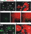
Figure 1
Figure 1
Early projecting midbrain TH axons grow into territories containing pre-existing non-dopaminergic axon bundles. Whole brains from E12 (A–E) and E13 (F,G) rat embryos were immunostained, cut along dorsal and ventral midlines and the resulting hemibrains were mounted flat. Confocal images were collected and are shown here as projections of Z-stacks. (A,C,E,F) Tyrosine Hydroxylase (TH, green) immunostaining. Approximate location of dopaminergic cell groups is indicated from A9 to A15. (B,D,E,G) β III-Tubulin-stained fiber tracts (red). (C–E) Show a magnified view of a field in the ventral MB region and part of the diencephalon (Di) indicated in (B). Arrow in (B) indicates the point of entry of TH axons into the striatal anlage. Abbreviations: Tel, Telencephalon; Di, Diencephalon; I, Isthmus; HB, hindbrain; MB, midbrain; Lc, locus ceruleus; TPOC, tract of the post-optic commissure; HT, habenular tract; MTG, mammillotegmental tract; TmesV, mesencephalic nucleus of the trigeminal nerve. Scale bar: 100 μm.
Figure 2
Figure 2
Retrograde labeling in embryonic rat brain reveals that ascending TH axons interact with descending GAD65 axons. Dextran-Rhodamine was applied at the locations indicated by asterisks in E12.5 (A) and E13 (D,F) rat embryos (red) and whole brains were immunostained for TH (B,C, green) or GAD65 (H,I, green). Flat-mounted hemibrains are shown. (A,D–F) are panoramic views; (B,C) show a magnified view of the frame indicated in (A) revealing apposition of descending Dextran-labeled axons with ascending TH axons (example axon indicated by three arrows). (F) shows dextran labeling in the diencephalon at the DA axon entry point into the telencephalon and (E) shows the rostral region of the same preparation revealing neurons with descending axons. (G–I) are magnified views of the frame indicated in (D) and show colocalization of GAD65 signal (green) with all Dextran-labeled axons (red, examples indicated by arrows) consistent with expression of GAD65 in descending axons. Abbreviations as in Figure 1. Scale bars: 100 μm.
Figure 3
Figure 3
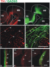
Dopaminergic axons grow in close apposition to GAD65 axon bundles during mesostriatal projection (E12, E13). Whole brains (A–D) or frozen sections (E–G) from E13 (A–E) and E14 (F,G) embryonic rat brains were immunostained for TH (red) and GAD65 (green). (A,B) Show panoramic views of a flat-mounted hemibrain and the location of magnified field shown in (C,D) is indicated in (B). The apposition observed in (C,D) was confirmed in single focal planes and in histological sections through the diencephalon in E13 (E) and E14 (F,G) embryos. Arrows in (C–G) indicate TH axons adjacent to GAD65 axons. Scale bar: 100 μm.
Figure 4
Figure 4
Conditional depletion of GAD65 axon fibers alters dopaminergic meso-striatal projection. Brains or sections of E13.5 control mouse embryos (ROSA26DTA/+, A–G) or of embryos carrying both alleles (ROSA26DTA/+:GAD65CRE/+, H–N) were immunostained for GAD65 (red) and TH (green). (A,H) are views of flat-mounted hemibrains showing the more abundant ventral projection in embryos carrying both alleles (arrows), (B–E,I–L) are of parasaggital sections. (C–E,J–L) Are magnified views of frames indicated in (B,I), respectively. (L) Shows a dramatic reduction in the GAD65 staining compared to (E). (F,M) Are parasaggital sections though the telencephalon showing the location of the magnified views in (G,N) respectively. Note that the rostro-caudal level in (F,M) is different as the location of the TH axons bundle varies between control and double transgenic brains. Scale bar: 100 μm.
Figure 5
Figure 5
NCAM is expressed in midbrain dopaminergic and descending GAD65 neurons and non-dopaminergic longitudinal axon bundle expresses Robo proteins. Whole rat brains or frozen sections were immunostained for NCAM (red) and either TH or GAD65 (both in green). (A,B) Show sagittal sections of the ventral midbrain (MB-v) of an E12 embryo. (C–E) Are images of a frame in the diencephalon of an E13 embryo. (F–H) Are images of a sagittal section in the ventral midbrain region of an E13 brain. (H1′,H2′) Are orthogonal views of a confocal Z-stack along the lines (H1, red; H2, green). Arrows in (H1′,H2′) indicate colocalization of GAD65 and NCAM signal. (I–K) Coronal sections of rat E12, E13 and E15, were immunostained for TH (green) and either Robo1 or Robo2 (red). (I,J) Midbrain (arrows indicate location of longitudinal axon bundle dorsal to the dopaminergic neurons), (K) diencephalon. Scale bar: (A–J), 100 μm; (K), 50 μm.
Figure 6
Figure 6
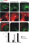
Dopaminergic misprojection in mouse embryos lacking either Robo1/Robo2 or Slit1/Slit2 is accompanied by dramatic alterations in the GAD65 axon bundles. Brains from control (A,D,G), Robo1/Robo2 double knock out (dko) (B,E,H), or Slit1/Slit2 dko (C,F,I) E12 mouse embryos were double immunostained for TH (green) and GAD65 (red) and hemibrains were mounted flat for observation. Normal projection at this early stage of TH+ axon pathfinding can be observed in (A) (control). Dramatic reduction and misprojection of TH axons is observed in (B) (Robo1/Robo2 dko), and (C) (Slit1/Slit2 dko). (D–F) Show GAD65 immunostaining of the same areas; in (D), normal GAD65 axon scaffolds are observed while notable alterations in the scaffold are observed in double knockouts (dko) (E,F) being more dramatic in Robo1/Robo2 dko (E). (G–I) Show single focal planes of the same regions with ortohogonal projections at the red and green lines shown. Apposition of TH axons on the orthogonal projections shown in (G–I) was quantified and is shown in (J). Significance (*) in (J) is p < 0.05. Scale bar: 100 μm.
Figure 7
Figure 7
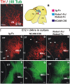
NCAM and Robo1/2 ectodomains impair projection of midbrain dopaminergic axons in whole embryo culture. Whole E12 rat embryos were cultured for 24 h in control medium (C,F) or in medium with either NCAM or Robo1/Robo2 soluble ectodomains (D,G,E,H, respectively) followed by TH (red) or β III-Tubulin (green) immunostaining. Flat-mounted hemibrains are shown. (A) Shows TH signal in a normal E12 embryo at the beginning of the culture. (C–F) Show control growth and (D,G,E,H) show halted growth or misprojection in cultures with NCAM or Robo1/2 ectodomains, respectively. The diagram in (B) shows the growth observed in each condition. Arrows in (C) indicate normal growth of TH axons into the diencephalon and in (D,E) indicate aberrant dorsally projecting axons. Scale bar: 100 μm.
Figure 8
Figure 8
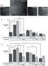
NCAM on the substrate enhances dopaminergic axon growth in culture and soluble Robo1/Robo2 ectodomains block such effect. The ventral region of the midbrain containing dopaminergic neurons was extracted from E12 rat brains, dissociated and seeded onto coverslips coated with control substrates [fibronectin (FNIII) or laminin] or the ectodomains indicated. (A–D) Show examples of fluorescent TH immunostaining signal (red) overlaid on phase contrast images (b/w) of the same fields of some of the different culture conditions tested. The effect of soluble ectodomains was assessed by adding them to the culture medium as indicated. After culture, TH immunostaining was performed and the length of axons of individual neurons that were not in contact with other axons was quantified. (E) Results obtained with fibronectin as control substrate. (F) Results obtained with laminin as control substrate. The number of axons quantified for each conditions were as follows. (E): FNIII/control: 261; NCAM/Control: 39; NCAM/Ig-Fc: 38; NCAM/NCAM; 47; NCAM/Robo1&2: 33; Robo1&2/Control: 54; Robo1&2/Ig-Fc: 54; Robo1&2/NCAM: 53; Robo1&2/Robo1&2:41; FNIII/NCAM: 70; FNIII/Robo1&2; 69. (F): Lam/Control: 56; NCAM/Control: 50; NCAM/Ig-Fc: 11; NCAM/NCAM: 58; NCAM/Robo1&2: 58; Robo1&2/Control: 36; Robo1&2/Ig-Fc: 35; Robo1&2/NCAM: 81; Robo1&2/Robo1&2: 58; Lam/NCAM: 36; Lam/Robo1&2: 22. Scale bars: 50 μm.

Similar articles

Cited by

References

    1. Albanese A., Altavista M. C., Rossi P. (1986). Organization of central nervous system dopaminergic pathways. J. Neural Transm. Suppl. 22, 3–17 - PubMed
    1. Anderson R. B., Key B. (1999). Role of acetylcholinesterase in the development of axon tracts within the embryonic vertebrate brain. Int. J. Dev. Neurosci. 17, 787–793 10.1016/S0736-5748(99)00064-7 - DOI - PubMed
    1. Auclair F., Marchand R., Glover J. C. (1999). Regional patterning of reticulospinal and vestibulospinal neurons in the hindbrain of mouse and rat embryos. J. Comp. Neurol. 411, 288–300 - PubMed
    1. Bagri A., Marin O., Plump A. S., Mak J., Pleasure S. J., Rubenstein J. L., et al. (2002). Slit proteins prevent midline crossing and determine the dorsoventral position of major axonal pathways in the mammalian forebrain. Neuron 33, 233–248 10.1016/S0896-6273(02)00561-5 - DOI - PubMed
    1. Bennett K. L., Bradshaw J., Youngman T., Rodgers J., Greenfield B., Aruffo A., et al. (1997). Deleted in colorectal carcinoma (DCC) binds heparin via its fifth fibronectin type III domain. J. Biol. Chem. 272, 26940–26946 10.1074/jbc.272.43.26940 - DOI - PubMed

LinkOut - more resources