Review
. 2014 Apr;2(1):7-21.
doi: 10.14791/btrt.2014.2.1.7. Epub 2014 Apr 29.

Altered histone modifications in gliomas

Affiliations
Review

Altered histone modifications in gliomas

Young Zoon Kim. Brain Tumor Res Treat. 2014 Apr.

Abstract

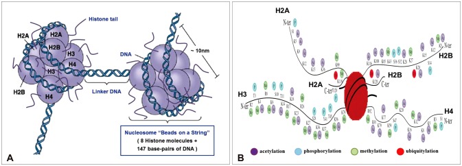
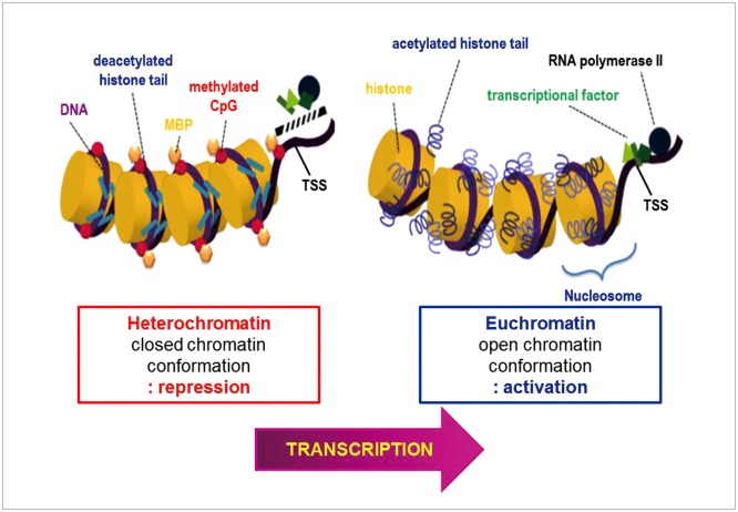
Gliomas are the most frequently occurring primary brain tumors in adults. Although they exist in different malignant stages, including histologically benign forms and highly aggressive states, most gliomas are clinically challenging for neuro-oncologists because of their infiltrative growth patterns and inherent relapse tendency with increased malignancy. Once this disease reaches the glioblastoma multiforme stage, the prognosis of patients is dismal: median survival time is 15 months. Extensive genetic analyses of glial tumors have revealed a variety of deregulated genetic pathways involved in DNA repair, apoptosis, cell migration/adhesion, and cell cycle. Recently, it has become evident that epigenetic alterations may also be an important factor for glioma genesis. Of epigenetic marks, histone modification is a key mark that regulates gene expression and thus modulates a wide range of cellular processes. In this review, I discuss the neuro-oncological significance of altered histone modifications and modifiers in glioma patients while briefly overviewing the biological roles of histone modifications.

Keywords: Acetylation; Epigenetics; Glioblastoma; Glioma; Histone; Methylation.


Conflict of interest statement

The author has no financial conflicts of interest.

Figures

Fig. 1
Fig. 1
Three major mechanisms of inheritable epigenetics. Mammalian gene expression is tightly controlled by genetic as well as epigenetic mechanisms. Epigenetics modifies the phenotype without altering the genotype of a cell. Shown here are some well-defined epigenetic mechanisms that include histone modifications, DNA methylation, and the noncoding RNA-mediated modulation of gene expression. Some of these mechanisms are inheritable through successive cell divisions and contribute to the maintenance of cellular phenotype. Recent studies show that the association of components of transcriptional regulatory machinery with target genes on mitotic chromosomes is a novel epigenetic mechanism that poises genes involved in key cellular processes, such as growth, proliferation, and lineage commitment, for expression in progeny cells (adapted by Zaidi et al. Mol Cell Biol 2010;30:4758-66 [87], and modified by author).
Fig. 2
Fig. 2
Schematic representation of the nucleosome and mammalian core histone modifications. A: Histones provide the basis for the nucleosome, the basic unit of chromatin structure, as seen as "beads-on-a-string" structures on electron micrographs. The nucleosome core is comprised of a histone octomer [(H2A-H2B)×2, (H3-H4)×2]. The DNA double helix is wrapped around (~1.7 times) the histone octomer. With nuclease digestion, 146 bps of DNA are tightly associated with the nucleosome but ~200 bps of DNA in total are associated with the nucleosome (modified image which was obtained at the website of http://www.mun.ca/biology/desmid/brian/BIOL2060/BIOL2060-18/18_21.jpg [88]). B: N- and C-terminal histone tails extend from the globular domains of histones H2A, H2B, H3, and H4. DNA is wrapped around the nucleosome octamer made up of two H2A-H2B dimers and an H3-H4 tetramer. Post-translational covalent modifications include acetylation, methylation, phosphorylation, and ubiquitylation. Human histone tail amino acid sequences are shown. Lysine positions 56 and 79 on histone H3 are located within the globular domain of the histone (adapted by Mercurio et al. Epigenetics in human disease 2012 [89], and modified by author).
Fig. 3
Fig. 3
Schematic diagram illustrating euchromatin and heterochromatin. Heterochromatin on the left is characterized by DNA methylation and deacetylated histones, is condensed and inaccesible to transcription factors (closed chromatin conformation), which is repressive regulation of transcription. On the contrary, euchromatin on the right is in a loose form and transcriptionally active; DNA is unmethylated and histone tails acetylated (open chromatin conformation), which is active regulation of transcription (adapted by Hatzimichael et al. J Drug Deliv 2013;2013:529312 [90], and modified by author).
Fig. 4
Fig. 4
Illustration of histone code according to active and repressive markers. DNA is wrapped around histone octamer of the four core histones H2A, H2B, H3, and H4. Histone H1, the linker protein, is bound to DNA between nucleosomes. Different amino acids constituting histone tails are represented along with the different covalent modification specific of each residue. Active marks are represented in the upper part of the figure and repressive marks are represented in the lower part of the figure. Lysine (K), arginine (R), serine (S), and threonine (T) (adapted by Sawan et al.A dv Genet 2010;70:57-85 [91], and modified by author).
Fig. 5
Fig. 5
Summaries of cellular role of histone modification. Functional implications in transcription regulation (A.C), DNA damage response (D) and DNA replication (E) are illustrated. The labels "Ub", "Ac", "Me", and "P" refer to mono-ubiquitination, acetylation, di- and trimethylation, and phosphorylation respectively (adapted by Vissers et al. Cell Div 2008;3:8 [92], and modified by author).
Fig. 6
Fig. 6
Recursive partitioning analysis (RPA) results individualizing 10 different prognostic groups among the 230 samples from glioma patients who underwent resection included. Each node, where the branches of the RPA tree bifurcate, divides patients according to whether the value of a specific feature (predictor) is above or below a selected cutoff value. The first node is represented by the tumor grade. In low-grade glioma patients, histologic subtype provides the second node, and histone modifications (e.g., the percentage of cells stained positively for H3K9Ac) provide the third node. High-grade glioma patients were further divided into World Health Organization (WHO) grade 3 and 4 ones. Histologic subtype and pathogenesis provide the third node, respectively, and histone modifications of H3K4me2, H3K18Ac, or H4K20me3 provide the fourth node (adapted by Liu et al. Cancer Epidemiol Biomarkers Prev 2010;19:2888-96 [75]). GBM: glioblastoma multiforme.

Similar articles

Cited by

References

    1. Clark SJ, Harrison J, Frommer M. CpNpG methylation in mammalian cells. Nat Genet. 1995;10:20–27. - PubMed
    1. Okano M, Xie S, Li E. Cloning and characterization of a family of novel mammalian DNA (cytosine-5) methyltransferases. Nat Genet. 1998;19:219–220. - PubMed
    1. Hansen RS, Gartler SM. 5-Azacytidine-induced reactivation of the human X chromosome-linked PGK1 gene is associated with a large region of cytosine demethylation in the 5' CpG island. Proc Natl Acad Sci U S A. 1990;87:4174–4178. - PMC - PubMed
    1. Li E, Beard C, Jaenisch R. Role for DNA methylation in genomic imprinting. Nature. 1993;366:362–365. - PubMed
    1. Eden A, Gaudet F, Waghmare A, Jaenisch R. Chromosomal instability and tumors promoted by DNA hypomethylation. Science. 2003;300:455. - PubMed