. 2014 Aug:57:227-43.
doi: 10.1016/j.cortex.2014.04.016. Epub 2014 May 9.

The DCDC2/intron 2 deletion and white matter disorganization: focus on developmental dyslexia

Affiliations

The DCDC2/intron 2 deletion and white matter disorganization: focus on developmental dyslexia

Cecilia Marino et al. Cortex. 2014 Aug.

Abstract

Introduction: The DCDC2 gene is involved in neuronal migration. Heterotopias have been found within the white matter of DCDC2-knockdown rats. A deletion in DCDC2/intron 2 (DCDC2d), which encompasses a regulatory region named 'regulatory element associated with dyslexia 1' (READ1), increases the risk for dyslexia. We hypothesized that DCDC2d can be associated to alterations of the white matter structure in general and in dyslexic brains.

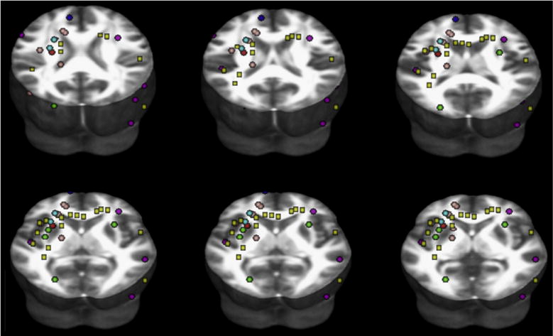
Methods: Based on a full-factorial analysis of covariance (ANCOVA) model, we investigated voxel-based diffusion tensor imaging (VB-DTI) data of four groups of subjects: dyslexia with/without DCDC2d, and normal readers with/without DCDC2d. We also tested DCDC2d effects upon correlation patterns between fractional anisotropy (FA) and reading scores.

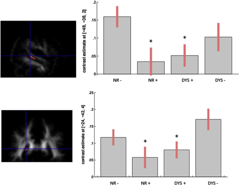
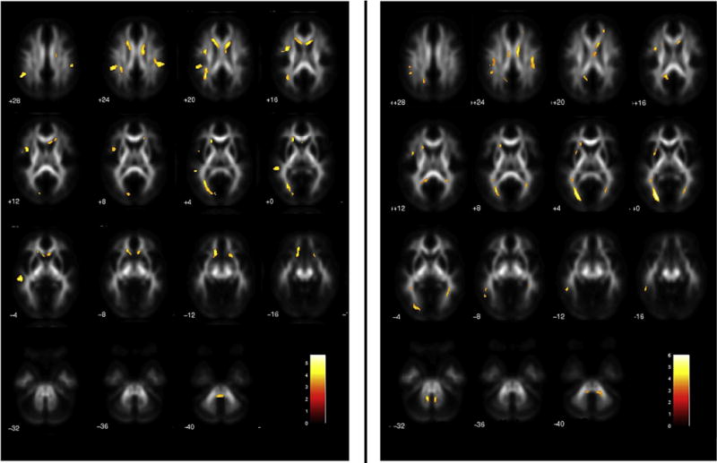
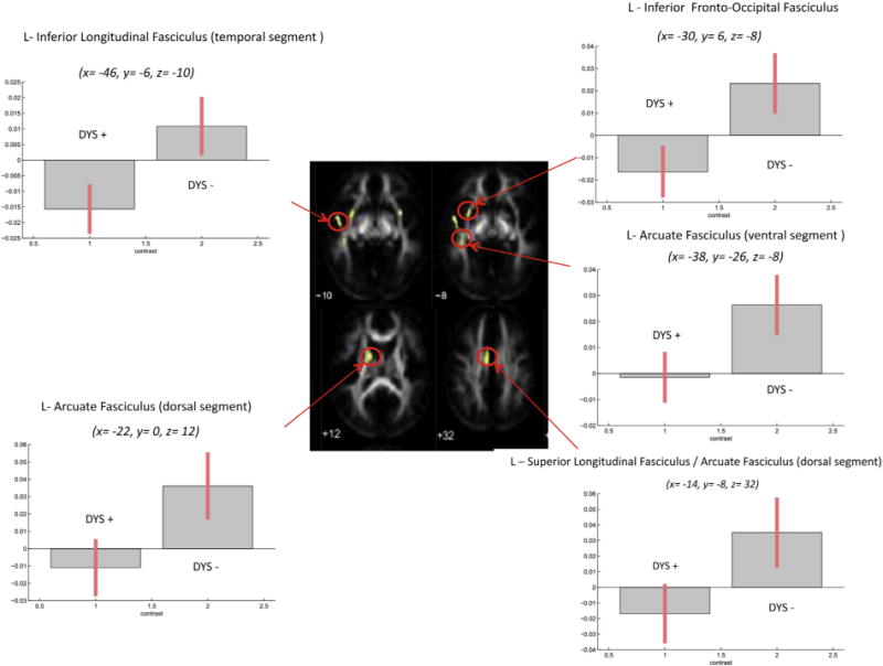
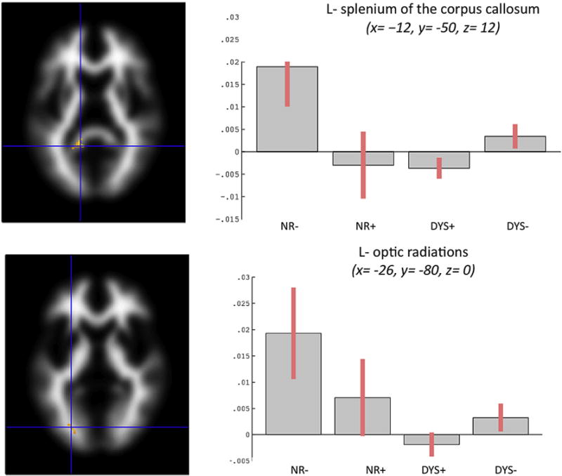
Results: We found that FA was reduced in the left arcuate fasciculus and splenium of the corpus callosum in subjects with versus without DCDC2d, irrespective of dyslexia. Subjects with dyslexia and DCDC2d showed reduced FA, mainly in the left hemisphere and in the corpus callosum; their counterpart without DCDC2d showed similar FA alterations. Noteworthy, a conjunction analysis in impaired readers revealed common regions with lower FA mainly in the left hemisphere. When we compared subjects with dyslexia with versus without DCDC2d, we found lower FA in the inferior longitudinal fasciculus and genu of the corpus callosum, bilaterally. Normal readers with versus without DCDC2d had FA increases and decreases in both the right and left hemisphere.

Discussion: The major contribution of our study was to provide evidence relating genes, brain and behaviour. Overall, our findings support the hypothesis that DCDC2d is associated with altered FA. In normal readers, DCDC2-related anatomical patterns may mark some developmental cognitive vulnerability to learning disabilities. In subjects with dyslexia, DCDC2d accounted for both common - mainly located in the left hemisphere - and unique - a more severe and extended pattern - alterations of white matter fibre tracts.

Keywords: DCDC2; Developmental dyslexia; Diffusion tensor imaging; Neuronal migration; READ1.


Figures

Fig. 1
Fig. 1
Map of coordinates in Montreal Neurological Institute space of significant findings of FA reductions in subjects with dyslexia compared to normal readers, as reviewed across DTI studies. We performed an online search in the database PUBMED from January 1996 to February 2011, using the keywords “diffusion tensor” and “developmental dyslexia”. We then checked the references lists for DTI additional studies. We included studies that reported (1) a minimum of six directions (2) anisotropy-based statistical comparisons between subjects with dyslexia and normal readers (3) stereotactic coordinates of FA whole-brain results (4) thresholds for significance, i.e., corrected for multiple comparisons or uncorrected with spatial extent thresholds. We also included one study that employed Tract-Based Spatial Statistics (Richards et al., 2008), and one that applied a priori defined anatomical regions of interest (Niogi & McCandliss, 2006). Six studies were included (Table 1). From each cluster of significant differences, we selected the millimetre-space coordinates of the voxel where the difference between subjects with dyslexia and normal readers was maximum. Coordinates reported in Talairach space were converted to Montreal Neurological Institute space. Spherical regions of interest (2 mm radius; see colours in Table 1) were then created and centred at the peak coordinates reported for each study. Yellow box regions of interest were instead created and centred at the coordinates reported in Table 4A (FA differences between DYS+ and NR− in our study). Finally, all regions of interest were re-referenced to the FMRIB58_FA 3D template in Montreal Neurological Institute space provided with the FSL package (FMRIB’s Software Library, www.fmrib.ox.ac.uk/fsl). The placement of the regions of interest projected to different axial planes overlaid to the FMRIB58_FA 3D template is shown (www.fmrib.ox.ac.uk/fsl).
Fig. 2
Fig. 2
Main effect of DCDC2d for FA values in the arcuate fasciculus (upper part: x = −48, y = −38, z = 2) and in the splenium of the corpus callosum (lower part: x = −24, y = −42, z = 4) in the four groups. Plots of parameter estimates of FA values are represented for each tract for the four groups.
Fig. 3
Fig. 3
Statistical parametric maps of decreased FA in DYS + versus NR− (left panel), and in DYS − versus NR− (right panel). Threshold: p < .05 FDR corrected at the voxel level (k > 100 voxels).
Fig. 4
Fig. 4
Fibre tracts exhibiting significant positive correlations of FA and average reading in DYS − versus DYS +. Plots of parameter estimates are represented for each tract for the two groups. Bars indicate correlation coefficients, which are measures of the strength of the relationship of FA and average reading.
Fig. 5
Fig. 5
Fibre tracts exhibiting significant positive correlations of FA and average reading in NR− (identified by inclusive masking procedure). Bars indicate correlation coefficients, which are measures of the strength of the relationship of FA and average reading. Significant differences in correlation patterns were found in DYS − versus DYS + in both the splenium of the corpus callosum and optic radiations. Furthermore, significant differences emerged in NR− versus NR+ in the splenium of the corpus callosum but not in the optic radiations, and in NR− versus both DYS + and DYS − in both the splenium of the corpus callosum and optic radiations.

References

    1. Beaulieu C. The basis of anisotropic water diffusion in the nervous system – a technical review. NMR in Biomedicine. 2002;15(7–8):435–455. - PubMed
    1. Becker J, Czamara D, Scerri TS, Ramus F, Csépe V, Talcott JB, et al. Genetic analysis of dyslexia candidate genes in the European cross-linguistic NeuroDys cohort. European Journal of Human Genetics. 2014;22(5):675–680. - PMC - PubMed
    1. Brambati SM, Termine C, Ruffino M, Danna M, Lanzi G, Stella G, et al. Neuropsychological deficits and neural dysfunction in familial dyslexia. Brain Research. 2006;1113(1):174–185. - PubMed
    1. Brambati SM, Termine C, Ruffino M, Stella G, Fazio F, Cappa SF, et al. Regional reductions of gray matter volume in familial dyslexia. Neurology. 2004;63(4):742–745. - PubMed
    1. Briggs GG, Nebes RD. Patterns of hand preference in a student population. Cortex. 1975;11(3):230–238. - PubMed

Substances

LinkOut - more resources