

On the crucial cerebellar wound healing-related pathways and their cross-talks after traumatic brain injury in Danio rerio
- PMID: 24926785
- PMCID: PMC4057083
- DOI: 10.1371/journal.pone.0097902
On the crucial cerebellar wound healing-related pathways and their cross-talks after traumatic brain injury in Danio rerio
Abstract
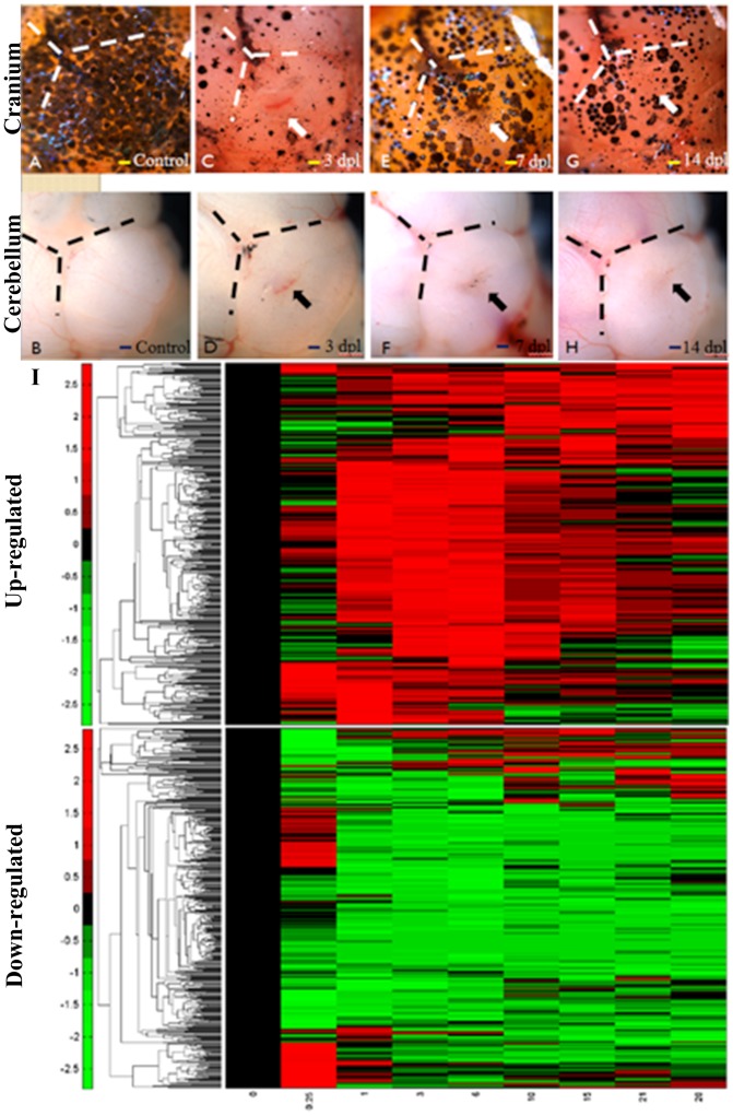
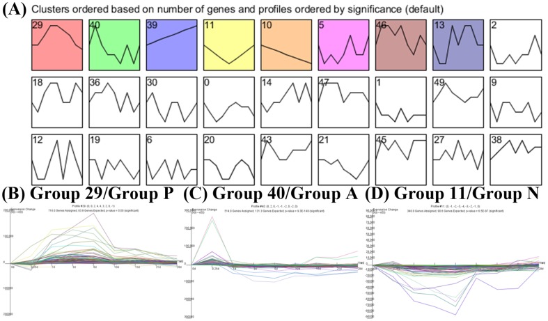
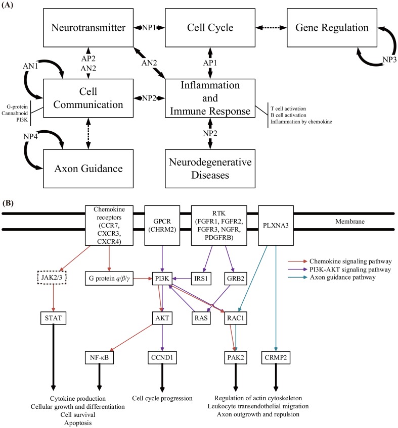
Upon injury, the direct damage and the subsequent secondary injury in the brain often result in chronic neurological disorders. Due to multifactorial nature of secondary injury and subsequent complex cellular responses, much of the underlying mechanisms are unclear. This study used an adult zebrafish cerebellum injury model to investigate the phenotypes and the secondary injury responses for recovery mechanisms of injured brain. Using the time course microarray analysis, a candidate protein-protein interaction (PPI) network was refined as cerebellar wound healing PPI network by dynamic modeling and big data mining. Pathway enrichment and ontological analysis were incorporated into the refined network to highlight the main molecular scheme of cerebellar wound healing. Several significant pathways, including chemokine, Phosphatidylinositide 3-kinases, and axon guidance signaling pathway and their cross-talks through PI3K, PAK2, and PLXNA3 were identified to coordinate for neurogenesis and angiogenesis, which are essential for the restoration of the injured brain. Our finding provides an insight into the molecular restoration mechanisms after traumatic brain injury, and open up new opportunity to devise the treatment for traumatic brain injury in human.
Conflict of interest statement
Figures


Similar articles
-
Induction of oxidative stress and apoptosis in the injured brain: potential relevance to brain regeneration in zebrafish.Mol Biol Rep. 2021 Jun;48(6):5099-5108. doi: 10.1007/s11033-021-06506-7. Epub 2021 Jun 24. Mol Biol Rep. 2021. PMID: 34165768
-
Regenerative neurogenesis from neural progenitor cells requires injury-induced expression of Gata3.Dev Cell. 2012 Dec 11;23(6):1230-7. doi: 10.1016/j.devcel.2012.10.014. Epub 2012 Nov 15. Dev Cell. 2012. PMID: 23168169
-
Microglia are essential for tissue contraction in wound closure after brain injury in zebrafish larvae.Life Sci Alliance. 2024 Oct 17;8(1):e202403052. doi: 10.26508/lsa.202403052. Print 2025 Jan. Life Sci Alliance. 2024. PMID: 39419547 Free PMC article.
-
Models of traumatic cerebellar injury.Cerebellum. 2009 Sep;8(3):211-21. doi: 10.1007/s12311-009-0114-8. Epub 2009 Jun 5. Cerebellum. 2009. PMID: 19495901 Free PMC article. Review.
-
Modeling traumatic brain and neural injuries: insights from zebrafish.Front Mol Neurosci. 2025 Mar 27;18:1552885. doi: 10.3389/fnmol.2025.1552885. eCollection 2025. Front Mol Neurosci. 2025. PMID: 40213159 Free PMC article. Review.
Cited by
-
Delivering Traumatic Brain Injury to Larval Zebrafish.Methods Mol Biol. 2024;2707:3-22. doi: 10.1007/978-1-0716-3401-1_1. Methods Mol Biol. 2024. PMID: 37668902
-
The role of TGF-β signaling and apoptosis in innate and adaptive immunity in zebrafish: a systems biology approach.BMC Syst Biol. 2014 Oct 24;8:116. doi: 10.1186/s12918-014-0116-0. BMC Syst Biol. 2014. PMID: 25341656 Free PMC article.
-
Identification of network-based biomarkers of cardioembolic stroke using a systems biology approach with time series data.BMC Syst Biol. 2015;9 Suppl 6(Suppl 6):S4. doi: 10.1186/1752-0509-9-S6-S4. Epub 2015 Dec 9. BMC Syst Biol. 2015. PMID: 26679092 Free PMC article.
-
Key Immune Events of the Pathomechanisms of Early Cardioembolic Stroke: Multi-Database Mining and Systems Biology Approach.Int J Mol Sci. 2016 Feb 27;17(3):305. doi: 10.3390/ijms17030305. Int J Mol Sci. 2016. PMID: 26927091 Free PMC article.
-
The utilization of small non-mammals in traumatic brain injury research: A systematic review.CNS Neurosci Ther. 2021 Apr;27(4):381-402. doi: 10.1111/cns.13590. Epub 2021 Feb 4. CNS Neurosci Ther. 2021. PMID: 33539662 Free PMC article.
References
-
- Faul M, Xu L, Wald MM, Coronado V, Dellinger AM (2010) Traumatic Brain Injury in the United States: National Estimates of Prevalence and Incidence, 2002-2006. Injury Prevention 16: A268–A268.
-
- Kizil C, Kaslin J, Kroehne V, Brand M (2012) Adult neurogenesis and brain regeneration in zebrafish. Developmental Neurobiology 72: 429–461. - PubMed
-
- Tanaka EM, Ferretti P (2009) Considering the evolution of regeneration in the central nervous system. Nature Reviews Neuroscience 10: 713–723. - PubMed
-
- Horner PJ, Gage FH (2000) Regenerating the damaged central nervous system. Nature 407: 963–970. - PubMed
-
- Antos CL, Tanaka EM (2010) Vertebrates That Regenerate as Models for Guiding Stem Cells. Cell Biology of Stem Cells 695: 184–214. - PubMed
Publication types
MeSH terms
Substances
LinkOut - more resources
Full Text Sources
Other Literature Sources
Molecular Biology Databases
Miscellaneous
