. 2014 Jun 13;9(6):e98685.
doi: 10.1371/journal.pone.0098685. eCollection 2014.

Protective immunity and safety of a genetically modified influenza virus vaccine

Affiliations

Protective immunity and safety of a genetically modified influenza virus vaccine

Rafael Polidoro Alves Barbosa et al. PLoS One. .

Abstract

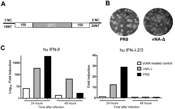
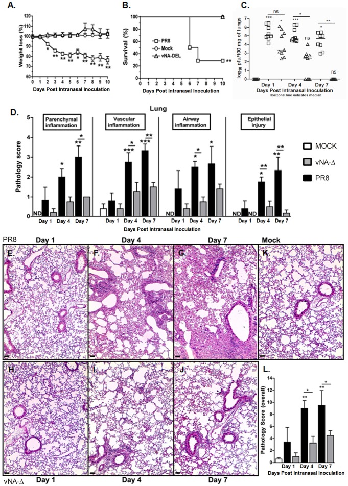
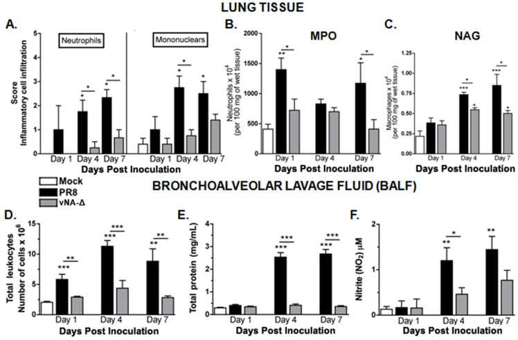
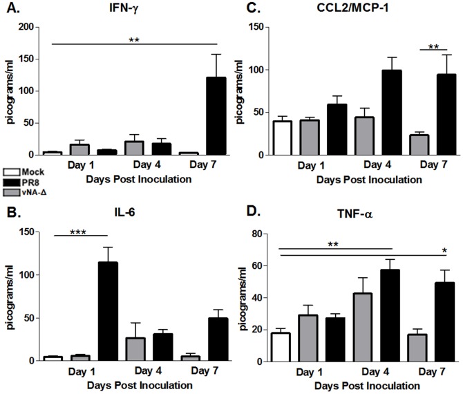
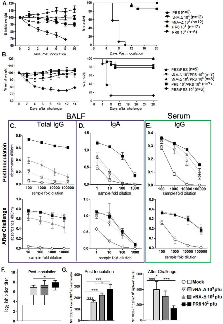
Recombinant influenza viruses are promising viral platforms to be used as antigen delivery vectors. To this aim, one of the most promising approaches consists of generating recombinant viruses harboring partially truncated neuraminidase (NA) segments. To date, all studies have pointed to safety and usefulness of this viral platform. However, some aspects of the inflammatory and immune responses triggered by those recombinant viruses and their safety to immunocompromised hosts remained to be elucidated. In the present study, we generated a recombinant influenza virus harboring a truncated NA segment (vNA-Δ) and evaluated the innate and inflammatory responses and the safety of this recombinant virus in wild type or knock-out (KO) mice with impaired innate (Myd88 -/-) or acquired (RAG -/-) immune responses. Infection using truncated neuraminidase influenza virus was harmless regarding lung and systemic inflammatory response in wild type mice and was highly attenuated in KO mice. We also demonstrated that vNA-Δ infection does not induce unbalanced cytokine production that strongly contributes to lung damage in infected mice. In addition, the recombinant influenza virus was able to trigger both local and systemic virus-specific humoral and CD8+ T cellular immune responses which protected immunized mice against the challenge with a lethal dose of homologous A/PR8/34 influenza virus. Taken together, our findings suggest and reinforce the safety of using NA deleted influenza viruses as antigen delivery vectors against human or veterinary pathogens.


Conflict of interest statement

Competing Interests: The authors have declared that no competing interests exist.

Figures

Figure 1
Figure 1. Generation and characterization of recombinant viruses harboring truncated neuraminidase.
Deleted neuraminidase segments displaying deletions at 3′ and 5′ extremities were generated as described in Material and Methods. The remaining neuraminidase coding regions are shown in white squares. A spacer sequence was inserted between the 3′ and 5′ moieties (A). Wild type PR8 virus and recombinant vNA-Δ influenza viruses were generated by reverse genetics as described in Material and Methods. The plaque phenotypes of these viruses were assessed by standard agarose plaque assay in MDCK infected cells after 3 days of incubation (B). Confluent monolayers of A549 cells were cultured with DMEM media containing BSA, Trypsin and neuraminidase (VcNA treated control) or infected with wild type PR8 virus or recombinant vNA-Δ at M.O.I of two. The induction of hu-IFN-β (type I, A) and hu-IFN λ2/3 (type III, B) was assessed at different time points by quantitative PCR using lightcycler Real Time PCR Machine (Applied Biosystems; C). Analysis was performed using SDS 2 software. All data are expressed as a ratio relative to VcNA treated control.
Figure 2
Figure 2. Characterization of virulence and lung inflammation in mice inoculated vNA-Δ.
C57BL/6 mice were infected intranasally with 105 PFU Influenza PR8, vNA-Δ or PBS (mock) inoculated (n  =  4–6 in each group). Weight loss (A) and lethality (B) were evaluated over 10 days. Mice were euthanized 1, 4 and 7 dpi and virus titers were quantified in lung (C). The figure shows one representative experiment. Lung pathologic score after infection with influenza PR8 virus or vNA-Δ was assessed in lung slices stained with H&E by a pathologist showing parenchyma, vascular and airway inflammation, and epithelial injury (D). Representative slides of PR8 virus (E, F and G), vNA-Δ (H, I and J) and mock (K) inoculated mice at 1, 4 and 7 dpi. The pathology overall score was determined (L). n  =  5 for all groups. Data are presented as mean ± SEM. * and ** for p<0.05 and p<0.01, respectively, when compared to mock or indicated groups (one-way ANOVA, Newman-Keuls).
Figure 3
Figure 3. Leukocyte recruitment to the lungs and BAL following vNA-Δ infection.
C57BL/6 mice were inoculated with PBS (mock) or infected intranasally with influenza 105 PFU of PR8 virus or vNA-Δ (n = 5). Mice were euthanized and lungs removed 1, 4 and 7 dpi. The recruitment of neutrophils and macrophages/mononuclear (A) cells to the lungs was assessed in lung H&E stained slides. Frozen lungs sections were assessed for Myeloperoxidase (B) and N-acetylglucosaminidase (C) contents, indirect measurements for neutrophils and macrophages, respectively. Mice were euthanized (n  =  6–8 in each group) 1, 4 and 7 dpi and bronchoalveolar lavage was performed. Absolute numbers of airways leukocytes after infection with 105 PFU (D). Total proteins (E) and nitrite (NO2; F) were also determined in BALF. Data are presented as mean ± SEM. *, ** and *** for p<0.05, p<0.01 and p<0.001, respectively, when compared to mock or indicated groups (one-way ANOVA, Newman-Keuls).
Figure 4
Figure 4. Measurement of cytokines in the lung.
C57BL/6 mice were inoculated with PBS (mock) or infected intranasally with 105 PFU influenza PR8 virus or vNA-Δ (n = 5) and euthanized 1, 4 and 7 dpi. The induction of murine IFN-β and IFN-λ2 (A) was measured in lungs by qRT-PCR as described in Material and Methods. The levels of cytokines IFN-γ (B), TNF-α (C), IL-1β (D), IL-6 (E), IL-4 (F) and IL-10 (G) were measured in lung tissue by ELISA. n  =  5 for all groups at days 1 and 4, n  =  5, 4, 6 for mock, PR8 and vNA-Δ viruses at day 7. Data are presented as mean ± SEM. * ** and *** for p<0.05, p<0.01 and p<0.001, respectively, when compared to mock or indicated groups (one-way ANOVA, Newman-Keuls or unpaired t test (qRT-PCRs).
Figure 5
Figure 5. Measurement of cytokines in sera of inoculated mice.
C57BL/6 mice were inoculated with PBS (mock; n = 9) or infected intranasally with 105 PFU of influenza PR8 (n = 10-12) or vNA-Δ (n = 9–11) viruses. By CBA, the levels of IFN-γ (A), CCL2/MCP-1 (B), IL-6 (C) and TNF-α (D) were measured in the sera collected from mice at days 1, 4 and 7 after inoculation. Data represents two independent experiments and are presented as mean ± SEM. * ** and *** for p<0.05, p<0.01 and p<0.001, respectively, when compared to mock or indicated groups (one-way ANOVA, Newman-Keuls).
Figure 6
Figure 6. Evaluation of protection and acquired immune response elicited by inoculation with vNA-Δ.
C57BL/6 mice were inoculated with PBS (mock) or infected intranasally with 105 PFU Influenza A/PR8 virus, 103 or 105 PFU of vNA-Δ virus. Twenty-one days after the prime-inoculation the animals were challenged with a lethal dose of 105 PFU of PR8. The weight loss and survival were determined after the prime-inoculation (A) and the challenge (B) infection (data represents two independent experiments). Blood and BALF were collected fourteen days after prime-inoculation and challenge infections. Tenfold dilutions of BALF samples were used to determine total IgG (C) and IgA (D) in BALF and total IgG in serum by ELISA (E)(Data depict one representative experiment). Two fold serial dilution of serum was used for the hemagglutinin-inhibition assay (F). n  =  4 for BALF IgA and IgG measures after inoculation. n  =  4, 7, 7, 6 for mock, vNA-Δ 103, vNA-Δ 105, PR8 103 for serum IgG. n  =  4, 5, 6, 5 for mock, vNA-Δ 103, vNA-Δ 105, PR8 103 for hemagglutinin-inhibition assay (Data represents two independent experiments). Spleens of mice (n  =  4) were obtained two weeks after the inoculation or challenge infection. Specific NP CD8+ T cells were assessed by ELISPOT using nucleoprotein (NP) of PR8 ASNENMETM peptide (NP; aa 366–374) as stimulus (G) Data represents two (inoculum) or three (challenge) independent experiments. Data were evaluated by Mann-Whitney test *, ** and *** for p<0.05, p<0.01 and p<0.001 respectively.
Figure 7
Figure 7. Characterization of recombinant vNA-Δ virus in immunodeficient mice.
C57BL/6 mice, MyD88 -/- and RAG -/- mice were anesthetized and inoculated with PBS (mock) or 5×10³ PFU of the PR8 or 5×10⁴ PFU of the recombinant vNA-Δ virus. The weight loss (A and B) and mortality (C) were followed (n  =  9–12 in each group; data represents two independent experiments). Results depicted in figure A and B were obtained from the same experiment.

References

    1. Lamb RA, Krug RM (2007) Orthomyxoviridae: the viruses and their replication. In: B.N F, editor. Virology. 5 ed. Philadelphia: Lippincott-Raven. pp. 1647–1690.
    1. Rollier CS, Reyes-Sandoval A, Cottingham MG, Ewer K, Hill AV (2011) Viral vectors as vaccine platforms: deployment in sight. Curr Opin Immunol 23: 377–382. - PubMed
    1. Li J, Arevalo MT, Zeng M (2013) Engineering influenza viral vectors. Bioengineered 4: 9–14. - PMC - PubMed
    1. Robert-Guroff M (2007) Replicating and non-replicating viral vectors for vaccine development. Curr Opin Biotechnol 18: 546–556. - PMC - PubMed
    1. Rocha CD, Caetano BC, Machado AV, Bruna-Romero O (2004) Recombinant viruses as tools to induce protective cellular immunity against infectious diseases. Int Microbiol 7: 83–94. - PubMed

Publication types

MeSH terms