. 2014 Jul-Aug;6(4):879-93.
doi: 10.4161/mabs.28762.

A new tool for monoclonal antibody analysis: application of IdeS proteolysis in IgG domain-specific characterization

Affiliations

A new tool for monoclonal antibody analysis: application of IdeS proteolysis in IgG domain-specific characterization

Yan An et al. MAbs. 2014 Jul-Aug.

Abstract

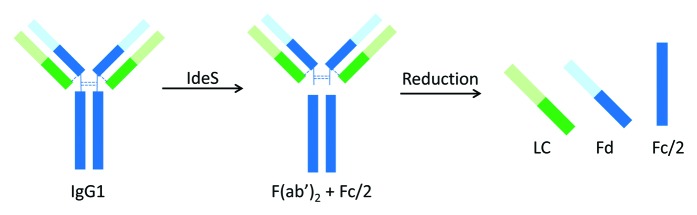
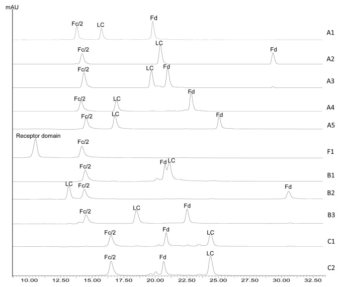
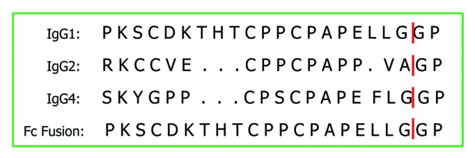
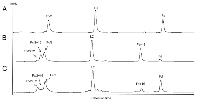
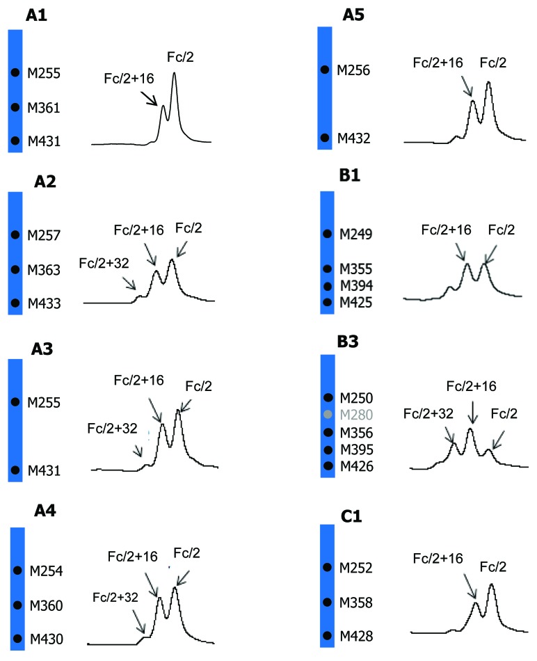
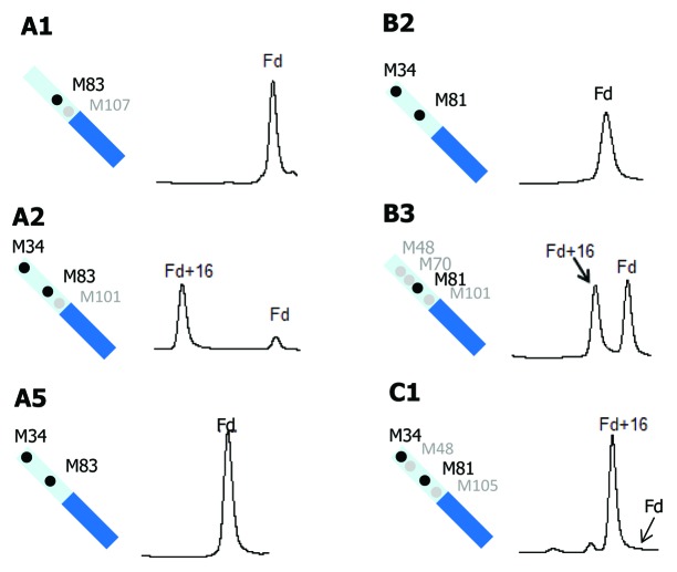
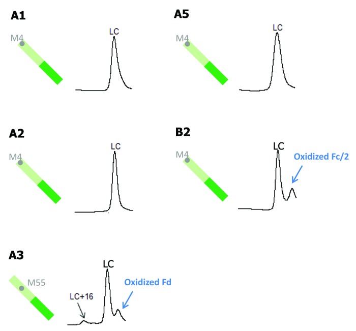
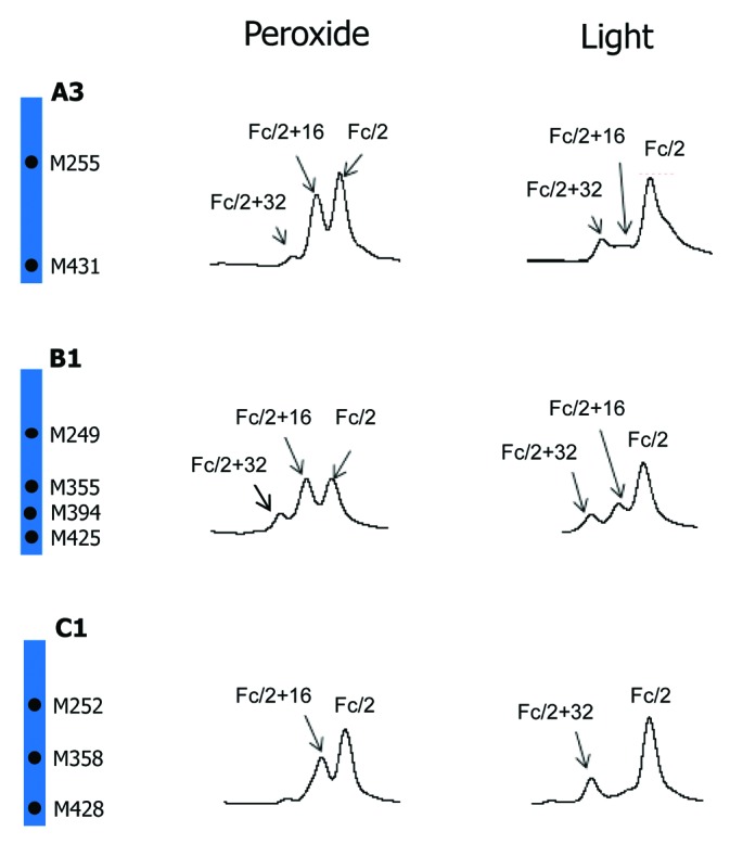
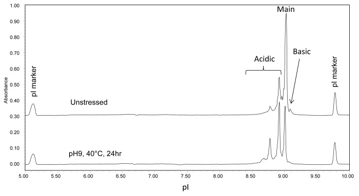
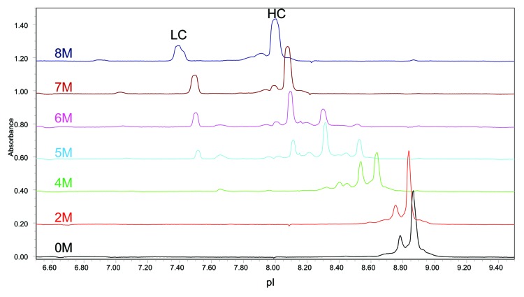
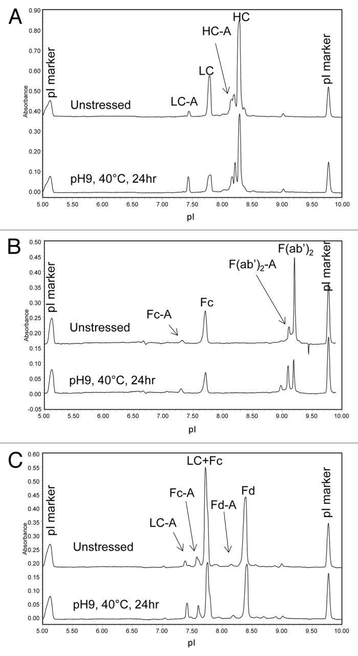
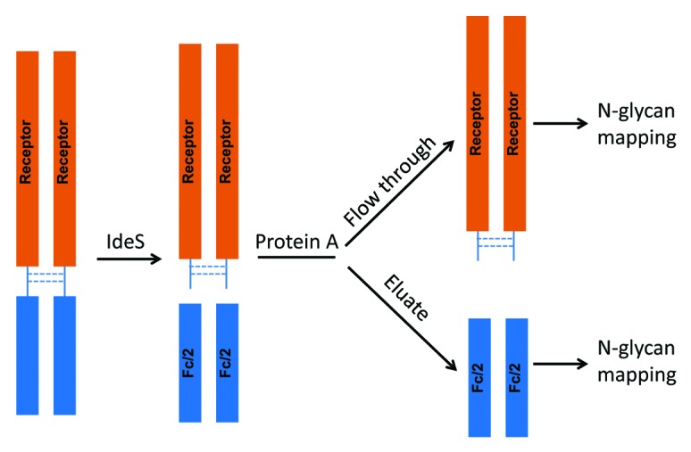
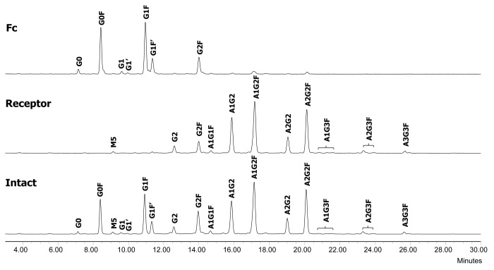
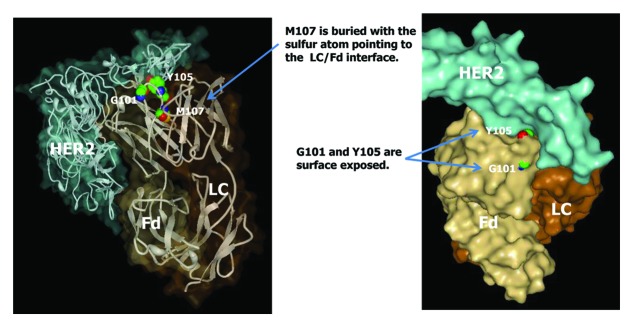
Monoclonal antibody (mAb) products are extraordinarily heterogeneous due to the presence of a variety of enzymatic and chemical modifications, such as deamidation, isomerization, oxidation, glycosylation, glycation, and terminal cyclization. The modifications in different domains of the antibody molecule can result in different biological consequences. Therefore, characterization and routine monitoring of domain-specific modifications are essential to ensure the quality of the therapeutic antibody products. For this purpose, a rapid and informative methodology was developed to examine the heterogeneity of individual domains in mAb products. A recently discovered endopeptidase, IdeS, cleaves heavy chains below the hinge region, producing F(ab') 2 and Fc fragments. Following reduction of disulfide bonds, three antibody domains (LC, Fd, and Fc/2) can be released for further characterization. Subsequent analyses by liquid chromatography/mass spectrometry, capillary isoelectric focusing, and glycan mapping enable domain-specific profiling of oxidation, charge heterogeneity, and glycoform distribution. When coupled with reversed phase chromatography, the unique chromatographic profile of each molecule offers a simple strategy for an identity test, which is an important formal test for biopharmaceutical quality control purposes. This methodology is demonstrated for a number of IgGs of different subclasses (IgG1, IgG2, IgG4), as well as an Fc fusion protein. The presented technique provides a convenient platform approach for scientific and formal therapeutic mAb product characterization. It can also be applied in regulated drug substance batch release and stability testing of antibody and Fc fusion protein products, in particular for identity and routine monitoring of domain-specific modifications.

Keywords: Fc fusion protein; IdeS; IgG; batch release; charge variant; domain-specific modification; formal identity; glycosylation; methionine oxidation; monoclonal antibody; stability testing.


Figures

None
Figure 1. Limited proteolysis of IgG1 by IdeS.
None
Figure 2. RP HPLC of IdeS digested IgG1 (A1-A5), IgG2 (B1-B3), IgG4 (C1-C2), and Fc fusion protein (F1) molecules.
None
Figure 3. IgG hinge region sequence comparison. IdeS cleavage sites identified by LC-MS are indicated by vertical lines.
None
Figure 4. IdeS domain mapping of mAb A2 by RP LC-MS. A, untreated; B, tBHP treated; C, exposed to 0.5X ICH light exposure level.
None
Figure 5. Comparison of tBHP induced oxidation on Fc. RP chromatographic regions of Fc/2 related peaks are presented. Highly conserved Fc methionine residues are denoted by black dots and the non-conserved methionine residue is denoted by gray dot.
None
Figure 6. Comparison of tBHP induced oxidation on Fd. RP chromatographic regions of Fd related peaks are presented. Highly conserved Fd methionine residues are denoted by black dots and non-conserved methionine residues are denoted by gray dots.
None
Figure 7. Comparison of tBHP induced oxidation on LC. RP chromatographic regions of LC related peaks are presented. Grey dots denote non-conserved methionine residues on LC.
None
Figure 8. Comparison of tBHP (left) and light (right, 1X ICH light exposure level) induced oxidation on Fc. RP chromatographic regions of Fc/2 related peaks are presented. Highly conserved Fc methionine residues are denoted by black dots.
None
Figure 9. cIEF electropherogram overlay of unstressed (top) and high pH stressed (bottom) mAb A1.
None
Figure 10. Effect of urea concentration on cIEF analysis of partially reduced mAb A1.
None
Figure 11. cIEF electropherogram overlays of unstressed (top) and high pH stressed (bottom) mAb A1 following (A) partial reduction, (B) IdeS digestion, and (C) IdeS digestion and partial reduction. Acidic species are denoted by “-A.”
None
Figure 12. Illustration of the experimental procedure for Receptor and Fc domain-specific N-glycan mapping of the Fc fusion protein F1.
None
Figure 13. N-glycan profiles of the Fc fusion protein F1 (bottom) and its receptor (middle) and Fc (top) domains. G, non-reducing end galactose; F, core fucose; A, sialic acid; M, high mannose.
None
Figure 14. Crystal structure of trastuzumab (Herceptin®) Fab in complex with the extracellular domain of human HER2 (pdb code: 1N8Z). Sphere presentations are shown for Fd amino acid residues G101, Y105, and M107.

References

    1. Reichert JM. Marketed therapeutic antibodies compendium. MAbs. 2012;4:413–5. doi: 10.4161/mabs.19931. - DOI - PMC - PubMed
    1. Salfeld JG. Isotype selection in antibody engineering. Nat Biotechnol. 2007;25:1369–72. doi: 10.1038/nbt1207-1369. - DOI - PubMed
    1. Jiang X-R, Song A, Bergelson S, Arroll T, Parekh B, May K, Chung S, Strouse R, Mire-Sluis A, Schenerman M. Advances in the assessment and control of the effector functions of therapeutic antibodies. Nat Rev Drug Discov. 2011;10:101–11. doi: 10.1038/nrd3365. - DOI - PubMed
    1. Liu H, Gaza-Bulseco G, Faldu D, Chumsae C, Sun J. Heterogeneity of monoclonal antibodies. J Pharm Sci. 2008;97:2426–47. doi: 10.1002/jps.21180. - DOI - PubMed
    1. Beck A, Wagner-Rousset E, Ayoub D, Van Dorsselaer A, Sanglier-Cianférani S. Characterization of therapeutic antibodies and related products. Anal Chem. 2013;85:715–36. doi: 10.1021/ac3032355. - DOI - PubMed