

Postoperative recovery of visual function after macula-off rhegmatogenous retinal detachment
- PMID: 24927502
- PMCID: PMC4057275
- DOI: 10.1371/journal.pone.0099787
Postoperative recovery of visual function after macula-off rhegmatogenous retinal detachment
Abstract
Purpose: To determine which factors affect the recovery of visual function in macula off rhegmatogenous retinal detachment (RRD).
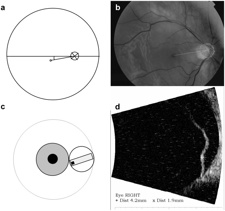
Methods: In a prospective study of forty-five patients with a primary macula-off RRD of 24 hours to 6 weeks duration, the height of the macular detachment was determined by ultrasonography. At 12 months postoperatively, best corrected visual acuity (BCVA), contrast acuity, and color confusion indexes (CCI) were obtained.
Results: Macular detachment was present for 2-32 (median 7) days before repair. A shorter duration of macular detachment was correlated with a better CCI saturé (p = 0.0026) and lower LogMAR BCVA (better Snellen visual acuity) (p = 0.012). Also, a smaller height of macular detachment was correlated with a lower LogMAR BCVA (p = 0.0034). A younger age and lower pre-operative LogMAR BCVA at presentation were both correlated with better postoperative contrast acuity in the total group (age: p = 1.7×10(-4) and pre-operative LogMAR BCVA: p = 0.0034).
Conclusion: Functional recovery after macula-off RRD is affected by the duration and the height of the macular detachment. Recovery of contrast acuity is also affected by age and BCVA at presentation.
Meeting presentation: ARVO annual meeting 2013, May 7, Seattle, Washington, United States of America.
Trial registration: trialregister.nl NTR839.
Conflict of interest statement
Figures
References
-
- van de Put MAJ, Hooymans JM, Los LI (2013) The Dutch Rhegmatogenous Retinal Detachment Study Group (2013) The incidence of rhegmatogenous retinal detachment in the Netherlands. Ophthalmology 120: 616–622. - PubMed
-
- Tani PT, Robertson DM, Langworthy A (1981) Prognosis for central vision and anatomic reattachment in rhegmatogenous retinal detachment with macula detached. Am J Ophthalmol 92: 611–620. - PubMed
-
- Pastor JC, Fernández I, Rodríguez de la Rúa E, Coco R, Sanabria-Ruiz Colmenares MR, et al. (2008) Surgical outcomes for primary rhegmatogenous retinal detachments in phakic and pseudophakic patients: the Retina 1 Project – report 2. Br J Ophthalmol 92: 378–382. - PubMed
-
- D'Amico DJ (2008) Primary retinal detachment. N Engl J Med 359: 2346–2354. - PubMed
Publication types
MeSH terms
Supplementary concepts
Associated data
LinkOut - more resources
Full Text Sources
Other Literature Sources
Medical
