. 2014 Jun 24;111(25):E2524-9.
doi: 10.1073/pnas.1403182111. Epub 2014 Jun 9.

Cofactor-dependent conformational heterogeneity of GAD65 and its role in autoimmunity and neurotransmitter homeostasis

Affiliations

Cofactor-dependent conformational heterogeneity of GAD65 and its role in autoimmunity and neurotransmitter homeostasis

Itamar Kass et al. Proc Natl Acad Sci U S A. .

Abstract

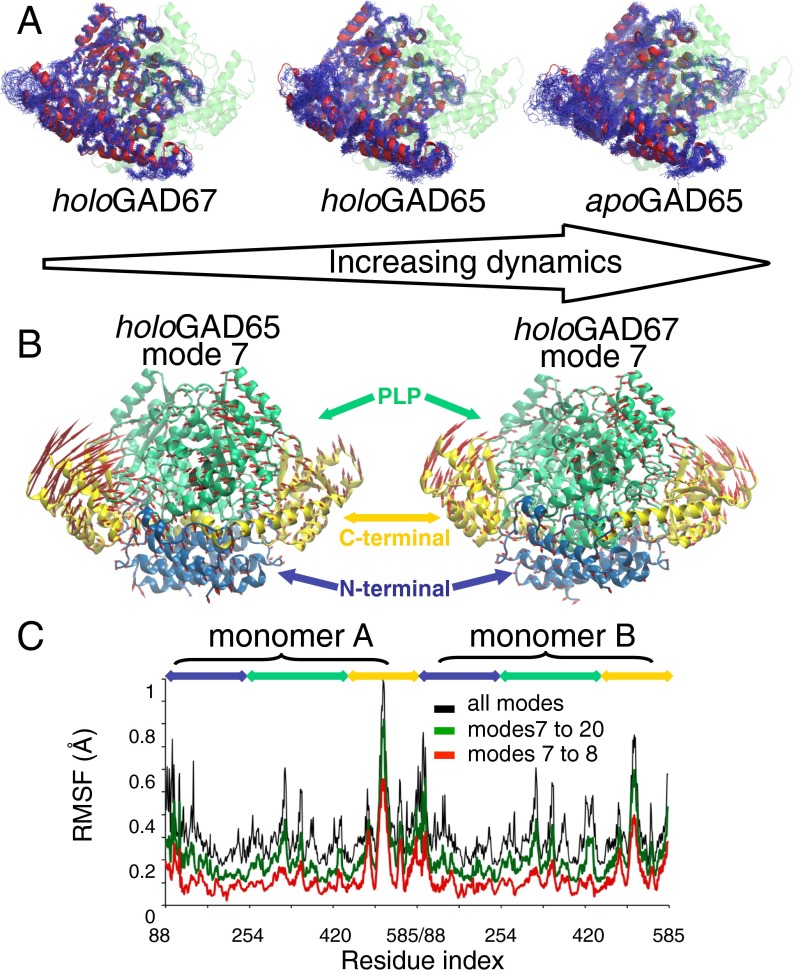
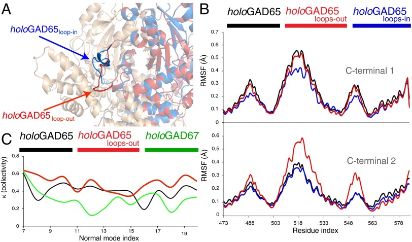
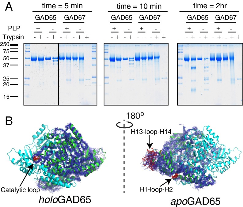
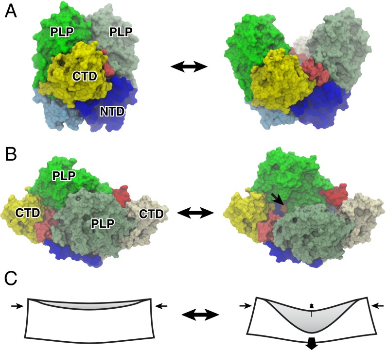
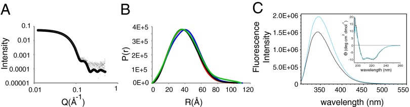
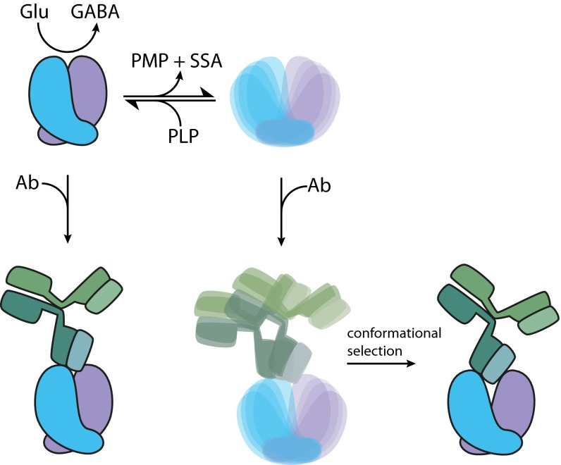
The human neuroendocrine enzyme glutamate decarboxylase (GAD) catalyses the synthesis of the inhibitory neurotransmitter gamma-aminobutyric acid (GABA) using pyridoxal 5'-phosphate as a cofactor. GAD exists as two isoforms named according to their respective molecular weights: GAD65 and GAD67. Although cytosolic GAD67 is typically saturated with the cofactor (holoGAD67) and constitutively active to produce basal levels of GABA, the membrane-associated GAD65 exists mainly as the inactive apo form. GAD65, but not GAD67, is a prevalent autoantigen, with autoantibodies to GAD65 being detected at high frequency in patients with autoimmune (type 1) diabetes and certain other autoimmune disorders. The significance of GAD65 autoinactivation into the apo form for regulation of neurotransmitter levels and autoantibody reactivity is not understood. We have used computational and experimental approaches to decipher the nature of the holo → apo conversion in GAD65 and thus, its mechanism of autoinactivation. Molecular dynamics simulations of GAD65 reveal coupling between the C-terminal domain, catalytic loop, and pyridoxal 5'-phosphate-binding domain that drives structural rearrangement, dimer opening, and autoinactivation, consistent with limited proteolysis fragmentation patterns. Together with small-angle X-ray scattering and fluorescence spectroscopy data, our findings are consistent with apoGAD65 existing as an ensemble of conformations. Antibody-binding kinetics suggest a mechanism of mutually induced conformational changes, implicating the flexibility of apoGAD65 in its autoantigenicity. Although conformational diversity may provide a mechanism for cofactor-controlled regulation of neurotransmitter biosynthesis, it may also come at a cost of insufficient development of immune self-tolerance that favors the production of GAD65 autoantibodies.

Keywords: GABA biosynthesis; autoepitopes; conformational dynamics; immunogenicity; normal mode analysis.


Conflict of interest statement

The authors declare no conflict of interest.

Figures

Fig. 1.
Fig. 1.
MD and NM analyses of GAD proteins. (A) Atomistic MD of GAD65 vs. GAD67 indicating the higher flexibility of apoGAD65 compared with holoGAD65 and holoGAD67. Backbone atoms are shown after a superposition of 25 structures sampled every 10 ns from a 250-ns simulation of (Right) apoGAD65 and (Center) holoGAD65 and (Left) 20 structures sampled every 10 ns from a 200-ns simulation of holoGAD67. (B) Motions described by the lowest frequency NM (mode 7) of holoGAD65 and holoGAD67. The directions and amplitudes of the motions are represented by arrows. (C) Flexibility profile [root mean square fluctuation (RMSF)] provided by distinct sets of holoGAD65 NMs. Blue, N terminus; green, PLP; yellow, C terminus.
Fig. 2.
Fig. 2.
(A) Modeling and dynamics of the distinct CL conformations and effect on the collectivity of the CTD dynamics of GAD65. The in conformation (similar to that in holoGAD67) is represented in blue, and the out conformation is represented in red. (B) RMSFs of the CTDs provided by modes 7–20: holoGAD65 (black), holoGAD65loops-in (blue), and holoGAD65loops-out (red). Residues are numbered as in Fig. 1. (C) Collectivity of motions described by low frequency NMs. The collectivity index κ represents the fraction of atoms participating significantly in a given displacement.
Fig. 3.
Fig. 3.
Limited proteolysis of GAD65/GAD67. (A) GAD proteins purified in the presence/absence of PLP were exposed to trypsin or buffer only. At the indicated times, an aliquot of the reaction mixture was boiled in SDS sample buffer, and the reactions were resolved by SDS/PAGE. Trypsin (∼23 kDa) was run alone for comparison as indicated. (B) Models (MD snapshots) of GAD65 mobility are shown with the cleavage sites (red) and labeled.
Fig. 4.
Fig. 4.
Open and closed dimers of GAD65 and the mechanism of dimer opening in holoapo conversion. (A) Molecular surface representations of (Left) the closed form of holoGAD65 and (Right) model of apoGAD65 in an open conformation. The CL (red/pink) connects the CTD and PLP-binding domain, mediating the dimer opening as supported by our MD and proteolysis data and ref. . (B) Orthogonal view facing the dimer interface. The active site lysine (396) that is buried in the closed form but solvent-accessible in the open form is shown in orange and indicated by an arrow (only one of the two sites is visible). (C) Origami analogy illustrating the mechanism of dimer opening in same view as in B. Coupled orthogonal motions of the PLP-binding domains and CTDs that accompany dimer opening are indicated by arrows. Domains are colored and labeled: NTD (blue, monomer A; cyan, monomer B), PLP (green, monomer A; pea green, monomer B), and CTD (yellow). NTD, N-terminal domain.
Fig. 5.
Fig. 5.
(A and B) SAXS analysis of apo- and holoGAD65. (A) HoloGAD65 scattering data fitted to the crystal structure (χ2 = 3.3). (B) P(r) functions from measured data (black, holoGAD65; green, apoGAD65) and scattering intensities calculated from models (blue, apoGAD65; red, holoGAD65). (C) Fluorescence emission spectra on excitation at 295 nm and CD spectra (Inset) of holoGAD65 (black) and apoGAD65 (cyan).
Fig. 6.
Fig. 6.
Implications of GAD65 autoinactivation for neurotransmitter biosynthesis and autoantigenicity. HoloGAD65 readily loses its PLP cofactor and autoinactivates through a secondary reaction, yielding a diverse ensemble. Supply of PLP shifts the equilibrium in favor of the primary reaction that catalyzes the conversion of Glu to GABA, thus regulating GABA production. This PLP-dependent autoinactivation may play a role in GAD65 autoantigenicity. The rigid closed holoGAD65 dimer binds an anti-GAD65 mAb with high affinity. The antibody also engages with the diverse open ensemble of apoGAD65, with conformational selection and/or induced fit permitting a mode of binding not available with the relatively rigid closed holo dimer. The PLP-dependent dynamic properties of GAD65 may be implicated in the insufficient development of immune self-tolerance that favors the production of GAD65 autoantibodies. PMP, pyridoxamine 5′-phosphate; SSA, succinic semialdehyde.

References

    1. Erlander MG, Tillakaratne NJ, Feldblum S, Patel N, Tobin AJ. Two genes encode distinct glutamate decarboxylases. Neuron. 1991;7(1):91–100. - PubMed
    1. Bu DF, et al. Two human glutamate decarboxylases, 65-kDa GAD and 67-kDa GAD, are each encoded by a single gene. Proc Natl Acad Sci USA. 1992;89(6):2115–2119. - PMC - PubMed
    1. Sandmeier E, Hale TI, Christen P. Multiple evolutionary origin of pyridoxal-5'-phosphate-dependent amino acid decarboxylases. Eur J Biochem. 1994;221(3):997–1002. - PubMed
    1. Giardina G, et al. Open conformation of human DOPA decarboxylase reveals the mechanism of PLP addition to Group II decarboxylases. Proc Natl Acad Sci USA. 2011;108(51):20514–20519. - PMC - PubMed
    1. Martin DL, Rimvall K. Regulation of gamma-aminobutyric acid synthesis in the brain. J Neurochem. 1993;60(2):395–407. - PubMed

Publication types

MeSH terms

LinkOut - more resources