. 2014 Jun 24;111(25):9157-62.
doi: 10.1073/pnas.1317630111. Epub 2014 Jun 9.

Targeting c-MYC by antagonizing PP2A inhibitors in breast cancer

Affiliations

Targeting c-MYC by antagonizing PP2A inhibitors in breast cancer

Mahnaz Janghorban et al. Proc Natl Acad Sci U S A. .

Abstract

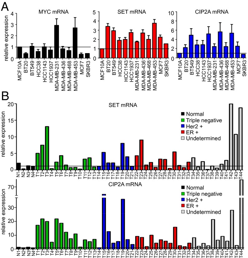
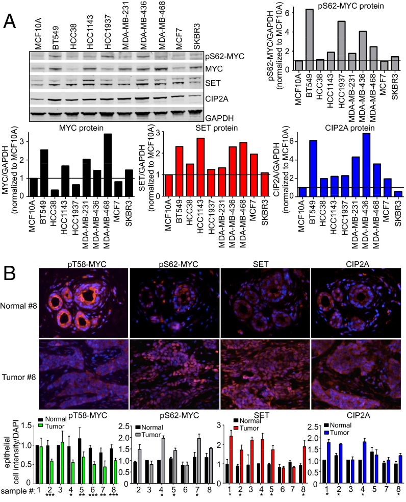
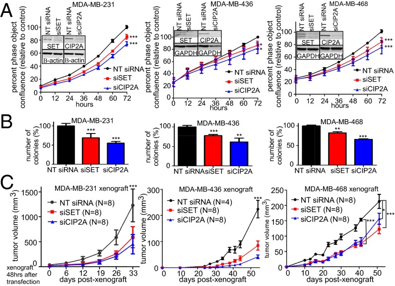
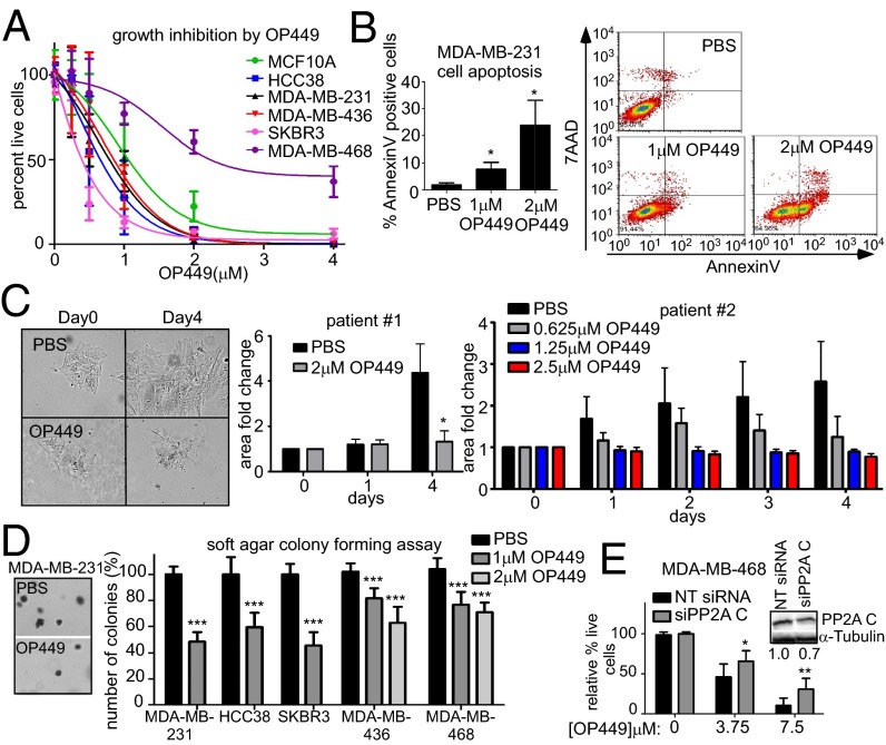
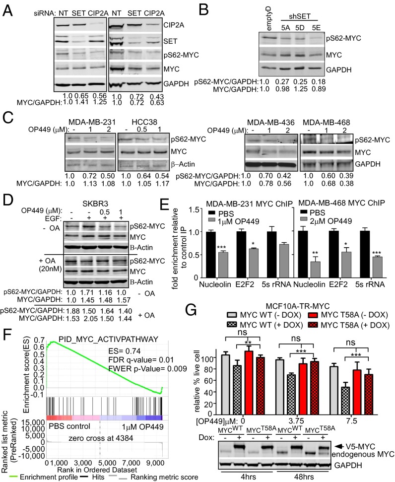
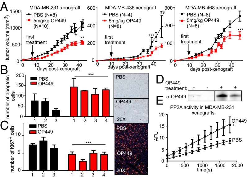
The transcription factor c-MYC is stabilized and activated by phosphorylation at serine 62 (S62) in breast cancer. Protein phosphatase 2A (PP2A) is a critical negative regulator of c-MYC through its ability to dephosphorylate S62. By inactivating c-MYC and other key signaling pathways, PP2A plays an important tumor suppressor function. Two endogenous inhibitors of PP2A, I2PP2A, Inhibitor-2 of PP2A (SET oncoprotein) and cancerous inhibitor of PP2A (CIP2A), inactivate PP2A and are overexpressed in several tumor types. Here we show that SET is overexpressed in about 50-60% and CIP2A in about 90% of breast cancers. Knockdown of SET or CIP2A reduces the tumorigenic potential of breast cancer cell lines both in vitro and in vivo. Treatment of breast cancer cells in vitro or in vivo with OP449, a novel SET antagonist, also decreases the tumorigenic potential of breast cancer cells and induces apoptosis. We show that this is, at least in part, due to decreased S62 phosphorylation of c-MYC and reduced c-MYC activity and target gene expression. Because of the ubiquitous expression and tumor suppressor activity of PP2A in cells, as well as the critical role of c-MYC in human cancer, we propose that activation of PP2A (here accomplished through antagonizing endogenous inhibitors) could be a novel antitumor strategy to posttranslationally target c-MYC in breast cancer.

Keywords: breast cancer therapy; phosphatase activator.


Conflict of interest statement

Conflict of interest statement: D.J.C. is an employee and shareholder of Oncotide Pharmaceuticals, Inc. J.O. is an employee of Oncotide Pharmaceuticals, Inc.

Figures

Fig. 1.
Fig. 1.
SET and CIP2A are frequently overexpressed in human breast cancer. (A) qRT-PCR analysis of MYC, SET, and CIP2A mRNA expression in 12 breast cell lines grown in 0.1% serum. Relative expression is calculated by ∆CT normalized to MCF10A. (B) qPCR analysis of SET and CIP2A expression in 44 breast tumors and four normal samples grouped by histologic subtypes, obtained from TissueScan Breast Cancer and Normal Tissue cDNA array (array 4).
Fig. 2.
Fig. 2.
Increased SET, CIP2A, and pS62-MYC protein levels occur in human breast cancer. (A) Representative Western blots of SET, CIP2A, pS62-MYC, and MYC protein expression in 10 breast cell lines grown in 0.1% serum. Quantification of SET, CIP2A, pS62-MYC, and MYC protein expression over GAPDH was done using a LICOR scanner and software. Quantification is graphed relative to expression in MCF10A. (B) Immunofluorescence of serial formalin-fixed, paraffin-embedded sections of breast tumors stained for pT58-MYC, pS62-MYC, SET, and CIP2A compared with their adjacent normal breast tissue. The graphs represent quantification of the mean staining intensity per epithelial cell for each protein over DAPI across multiple regions of interest and then normalized to the adjacent normal. Error bars represent SD. *P < 0.05, **P < 0.01, and ***P < 0.001.
Fig. 3.
Fig. 3.
SET and CIP2A knockdown decreases tumorigenic potential of breast cancer cell lines. (A) Population expansion analysis of the indicated cell lines over 72 h after transfection with SET or CIP2A siRNA compared with the control NT siRNA from three independent experiments using live cell imaging and IncuCyte analysis software. Representative Western blots show knockdown. (B and C) Soft agar colony assay and xenograft of these cells into the fourth mammary glands of NSG mice. Experimental details and statistics are described in Materials and Methods.
Fig. 4.
Fig. 4.
The SET antagonist OP449 decreases the growth of breast cancer cells and induces apoptosis. (A) Cytotoxicity of multiple breast cancer cell lines after 24 h of OP449 treatment. Cell viability was assessed by Trypan blue exclusion. (B) Apoptosis assay for MDA-MB-231 cells using AnnexinV-7-aminoactinomycin D after 6 h treatment with OP449 or PBS control. (C) Cell colony expansion of two primary invasive breast carcinoma samples treated with OP449 or PBS. Fold colony area ± SD is shown. The images are representative cell colonies (patient 1) taken at day 0 and 4 d with and without OP449 treatment. (D) Soft agar colony assay of breast cancer cell lines after treatment with OP449 or PBS. Representative images of colonies are shown. (E) The 3-(4,5-dimethylthiazol-2-yl)-5-(3-carboxymethoxyphenyl)-2-(4-sulfophenyl)-2H-tetrazolium, inner salt (MTS) assay for MDA-MB-468 cells transfected with control NT siRNA or siRNA against PP2A C subunit for 48 h. Cells were then treated with OP449 for another 48 h. A representative Western blot of knockdown is shown. Error bars represent SD from three independent experiments except in C, as indicated, and E, which is from two experiments performed in triplicate. *P < 0.05, **P < 0.01, and ***P < 0.001.
Fig. 5.
Fig. 5.
OP449 decreases S62-phosphorylated MYC and MYC transcriptional activity contributing to cytotoxicity in breast cancer cells. (A) Western blot analysis of pS62-MYC and MYC proteins in MDA-MB-231 and MDA-MB-436 cells after siRNA knockdown of SET or CIP2A. The levels of pS62-MYC and total MYC were quantified on a LICOR scanner and calculated over GAPDH. (B) Western blot analysis of pS62-MYC and MYC protein in MDA-MB-231 stable clones with control or SET knockdown (shown in Fig. S3B). (C) Western blot analysis of pS62-MYC and MYC after treatment of the indicated cells with OP449 for 4 h. (D) Western blot analysis of pS62-MYC and MYC from SKBR3 cells starved for 24 h and then treated with OP449 without or with 20 nM okadaic acid (OA) for 2 h, followed by 10 min EGF (100 ng/mL) treatment. (E) qChIP for MYC at the Nucleolin, E2F2, and 5s rRNA promoters after 24 h of treatment with OP449. The fold enrichment of bound DNA was graphed as the fold enrichment in MYC IP relative to the fold enrichment in IgG control IP. (F) Gene Set Enrichment Analysis of a MYC gene signature (30) in RNA-seq data from MDA-MB-231 cells treated with 1 µM OP449 or PBS for 12 h. The positive enrichment score (ES) and statistical values are listed. (G) MTS assay for MCF10A-TR-MYC cells treated with 1 µg/mL doxycycline (Dox) for 4 h to induce ectopic expression of wild-type (MYCWT) or mutant MYC (MYCT58A). Cells were then treated with different concentrations of OP449 as indicated for 48 h. Representative Western blot of ectopic MYC in these cells with 4 or 48 h of Dox treatment. Error bars represent SD. *P < 0.05, **P < 0.01, and ***P < 0.001.
Fig. 6.
Fig. 6.
OP449 suppresses breast tumor growth in vivo associated with increased PP2A activity. (A) Tumor growth curve for MDA-MB-231, MDA-MB-436, and MDA-MB-468 xenografts in the fourth mammary gland of NSG mice following treatment with OP449 or PBS. (B) TUNEL assay and (C) IF for Ki67 for MDA-MB-231 harvested xenografts from A. The mean and SD of total apoptotic cells in 75 random fields or Ki67 positive cells in 25 fields for three control mice (six tumors) and four OP449-treated mice (eight tumors) are graphed. (D) Western blot analysis of OP449 with peptide-specific antibody in lysates from MDA-MB-231 xenografts (Fig. S5A). (E) Tumor lysates from D were used to measure PP2A activity as described previously (22).

References

    1. Chrzan P, Skokowski J, Karmolinski A, Pawelczyk T. Amplification of c-myc gene and overexpression of c-Myc protein in breast cancer and adjacent non-neoplastic tissue. Clin Biochem. 2001;34(7):557–562. - PubMed
    1. Wolfer A, et al. MYC regulation of a “poor-prognosis” metastatic cancer cell state. Proc Natl Acad Sci USA. 2010;107(8):3698–3703. - PMC - PubMed
    1. Malempati S, et al. Aberrant stabilization of c-Myc protein in some lymphoblastic leukemias. Leukemia. 2006;20(9):1572–1581. - PMC - PubMed
    1. Zhang X, et al. Mechanistic insight into Myc stabilization in breast cancer involving aberrant Axin1 expression. Proc Natl Acad Sci USA. 2012;109(8):2790–2795. - PMC - PubMed
    1. Sears R, et al. Multiple Ras-dependent phosphorylation pathways regulate Myc protein stability. Genes Dev. 2000;14(19):2501–2514. - PMC - PubMed

Publication types

MeSH terms

Associated data