. 2014 Jun 24;111(25):9109-14.
doi: 10.1073/pnas.1404148111. Epub 2014 Jun 9.

Integration of bacterial lytic polysaccharide monooxygenases into designer cellulosomes promotes enhanced cellulose degradation

Affiliations

Integration of bacterial lytic polysaccharide monooxygenases into designer cellulosomes promotes enhanced cellulose degradation

Yonathan Arfi et al. Proc Natl Acad Sci U S A. .

Abstract

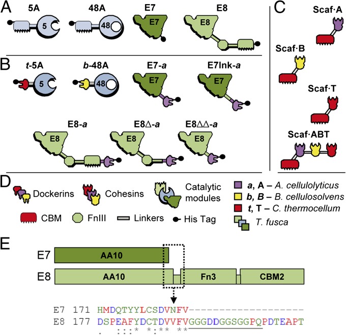
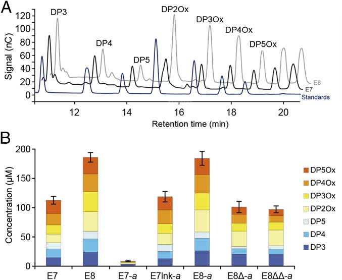
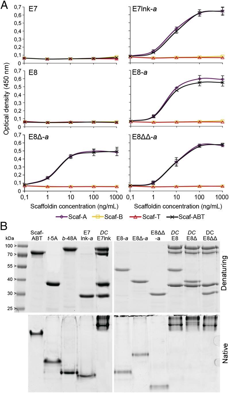
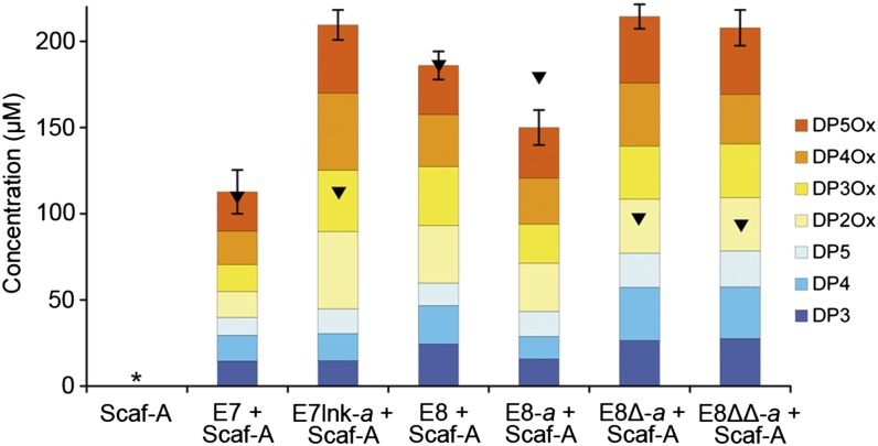
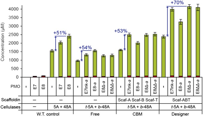
Efficient conversion of cellulose into soluble sugars is a key technological bottleneck limiting efficient production of plant-derived biofuels and chemicals. In nature, the process is achieved by the action of a wide range of cellulases and associated enzymes. In aerobic microrganisms, cellulases are secreted as free enzymes. Alternatively, in certain anaerobic microbes, cellulases are assembled into large multienzymes complexes, termed "cellulosomes," which allow for efficient hydrolysis of cellulose. Recently, it has been shown that enzymes classified as lytic polysaccharide monooxygenases (LPMOs) were able to strongly enhance the activity of cellulases. However, LPMOs are exclusively found in aerobic organisms and, thus, cannot benefit from the advantages offered by the cellulosomal system. In this study, we designed several dockerin-fused LPMOs based on enzymes from the bacterium Thermobifida fusca. The resulting chimeras exhibited activity levels on microcrystalline cellulose similar to that of the wild-type enzymes. The dockerin moieties of the chimeras were demonstrated to be functional and to specifically bind to their corresponding cohesin partner. The chimeric LPMOs were able to self-assemble in designer cellulosomes alongside an endo- and an exo-cellulase also converted to the cellulosomal mode. The resulting complexes showed a 1.7-fold increase in the release of soluble sugars from cellulose, compared with the free enzymes, and a 2.6-fold enhancement compared with free cellulases without LPMO enhancement. These results highlight the feasibility of the conversion of LPMOs to the cellulosomal mode, and that these enzymes can benefit from the proximity effects generated by the cellulosome architecture.

Keywords: biomass conversion; enzyme synergy.


Conflict of interest statement

The authors declare no conflict of interest.

Figures

Fig. 1.
Fig. 1.
Recombinant proteins used in this study. Schematic diagram of the wild-type enzymes (A), chimeric enzymes (B), engineered scaffoldins (C), and key to the diagram (D). Each protein is color coded according to the source of the different modules, as follows: light green, dark green, and light blue, Thermobifida fusca; purple, Acetovibrio cellulolyticus; red, Clostridium thermocellum; yellow, Bacteroides cellulosovens. The numbers 5 and 48 refer to the corresponding CAZY family classification of the catalytic modules (GH5 and GH48). (E, Upper) Diagram of the modular architecture of E7 (Upper, dark green) and E8 (Lower, light green), to scale. (E, Lower) An excerpt of the amino acid sequence alignment (ClustalW) between E7 and E8 is provided, corresponding to the boxed section (dashed line) of the diagram. The linker segment is underlined. The color coding of the residues and the consensus symbols follow the standard ClustalW schemes.
Fig. 2.
Fig. 2.
Oxidative cleavage of cellulose by E7, E8, and their dockerin variants. (A) HPAEC analysis of the soluble sugars released by the action of E7 (black) and E8 (gray) on Avicel, compared with reduced and oxidized standards (dark blue). Peak annotation was performed according to the standards’ retention times (DP2Ox, cellobionic acid; DP3, cellotriose; DP3Ox, cellotrionic acid; DP4, cellotetraose; DP4Ox, cellotetraonic acid; DP5, cellopentaose; DP5Ox, cellopentaonic acid). Two unlabeled peaks were visible at retention times greater than 19 min for which no standards were available (presumably cellohexaonic and celloheptaonic acids). (B) Quantification by HPAEC analysis of the soluble sugar released from the cleavage of microcrystalline cellulose (Avicel, 10 mg/mL) by 1 µM LPMO, at 50 °C for 72 h. The concentration (in micromolars) of each sugar was determined by integration of the peak area and comparison with a standard curve. Unlabeled peaks were not quantified. Values are the mean of three biological replicates (n = 3). Error bars correspond to one cumulated SD (error bar = ±SDtot; with SDtot = √(SD12 + SD22 + …). For clarity, data are only shown for retention times between 10.5 and 20.5 min. No peak was observed for longer retention times. Detailed concentrations and associated SDs are available in Fig. S1.
Fig. 3.
Fig. 3.
Cohesin–dockerin interactions. (A) Affinity-based ELISA analysis of the interaction between the LPMO variants and the engineered scaffoldins. The LPMOs are individually coated in a plate well. The amount of bound scaffoldin is detected by using HRP-labeled antibodies directed specifically against their CBM modules. Higher amounts of scaffoldin bound are indicated by higher optical densities. Each data point is the mean value of three replicates (n = 3). Error bars correspond to one SD. (B) Electrophoretic mobility of individual components and assembled complexes in denaturing and native PAGE. Equimolar amounts of each protein were analyzed either individually or in combination (DCE7lnk, DCE8, DCE8∆, and DCE8∆∆).
Fig. 4.
Fig. 4.
Restoration of the substrate targeting by scaffoldin-borne CBM. Quantification by HPAEC analysis of the soluble sugar released from the cleavage of microcrystalline cellulose (Avicel, 10 mg/mL) by 1 µM of LPMO + 1 µM Scaf-A, at 50 °C for 72 h. Black triangles indicate the total amount of sugars released under the same conditions without the scaffoldin (Fig. 2). See Fig. 2B for additional information. Detailed concentrations and associated SDs are available in Fig. S2.
Fig. 5.
Fig. 5.
Degradation of cellulose by LPMO-containing designer cellulosomes. Quantification by HPAEC analysis of the soluble sugars released from the cleavage of microcrystalline cellulose (Avicel, 10 mg/mL), at 50 °C for 72 h. Different architectures were assayed: free enzymes (W.T. control and Free), enzymes bound to monovalent scaffoldins (CBM), and enzymes bound to trivalent scaffoldin (Designer). Each protein was added at a final concentration of 0.5 µM. Concentrations of reducing sugars (green) and oxidized sugars (red) are shown. Values for reducing sugars were obtained by adding the concentrations of DP1–DP5 (where DP1 = glucose, DP2 = cellobiose, DP3 = cellotriose, DP4 = cellotetraose, and DP5 = cellopentaose). Values for oxidized sugars were obtained by adding the concentrations of DP1Ox-DP5Ox. The detailed concentrations are available in SI Materials and Methods. Blue arrows and the associated values indicate the percent increase (“boost effect”) in reducing sugars, related to the addition of a LPMO, compared to that of the control. Detailed concentrations and associated SDs are available in Fig. S3A. Values for oxidized sugars were obtained by adding the concentrations of DP1Ox-DP5Ox. The detailed concentrations of oxidized sugars and the associated SDs are available in Fig. S3B.

References

    1. Lynd LR, et al. How biotech can transform biofuels. Nat Biotechnol. 2008;26(2):169–172. - PubMed
    1. Ragauskas AJ, et al. The path forward for biofuels and biomaterials. Science. 2006;311(5760):484–489. - PubMed
    1. Himmel ME, Bayer EA. Lignocellulose conversion to biofuels: Current challenges, global perspectives. Curr Opin Biotechnol. 2009;20(3):316–317. - PubMed
    1. Sims REH, Mabee W, Saddler JN, Taylor M. An overview of second generation biofuel technologies. Bioresour Technol. 2010;101(6):1570–1580. - PubMed
    1. Mba Medie F, Davies GJ, Drancourt M, Henrissat B. Genome analyses highlight the different biological roles of cellulases. Nat Rev Microbiol. 2012;10(3):227–234. - PubMed

Publication types

MeSH terms