. 2014 Aug;35(26):7346-54.
doi: 10.1016/j.biomaterials.2014.05.014. Epub 2014 Jun 10.

Tough and flexible CNT-polymeric hybrid scaffolds for engineering cardiac constructs

Affiliations

Tough and flexible CNT-polymeric hybrid scaffolds for engineering cardiac constructs

Mahshid Kharaziha et al. Biomaterials. 2014 Aug.

Abstract

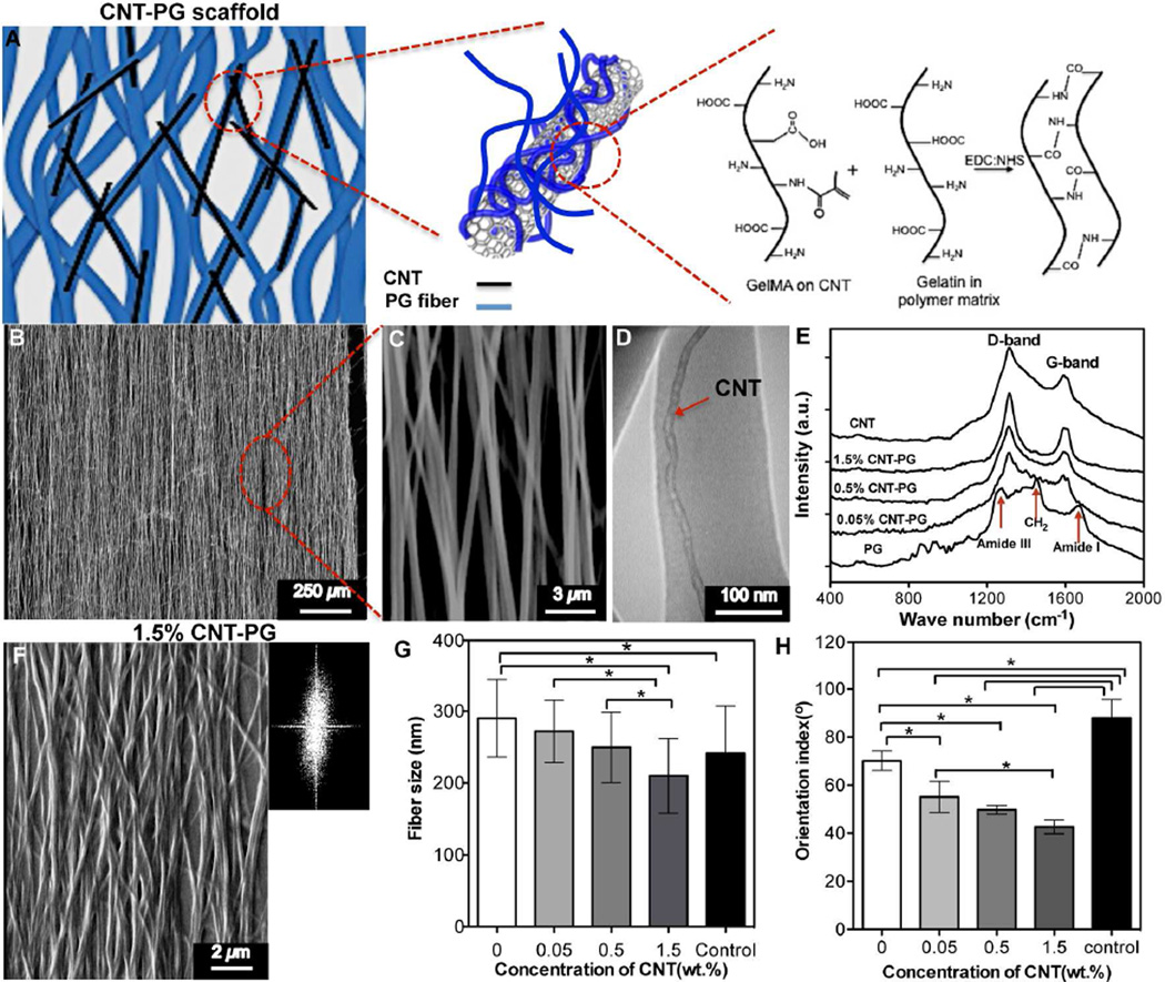
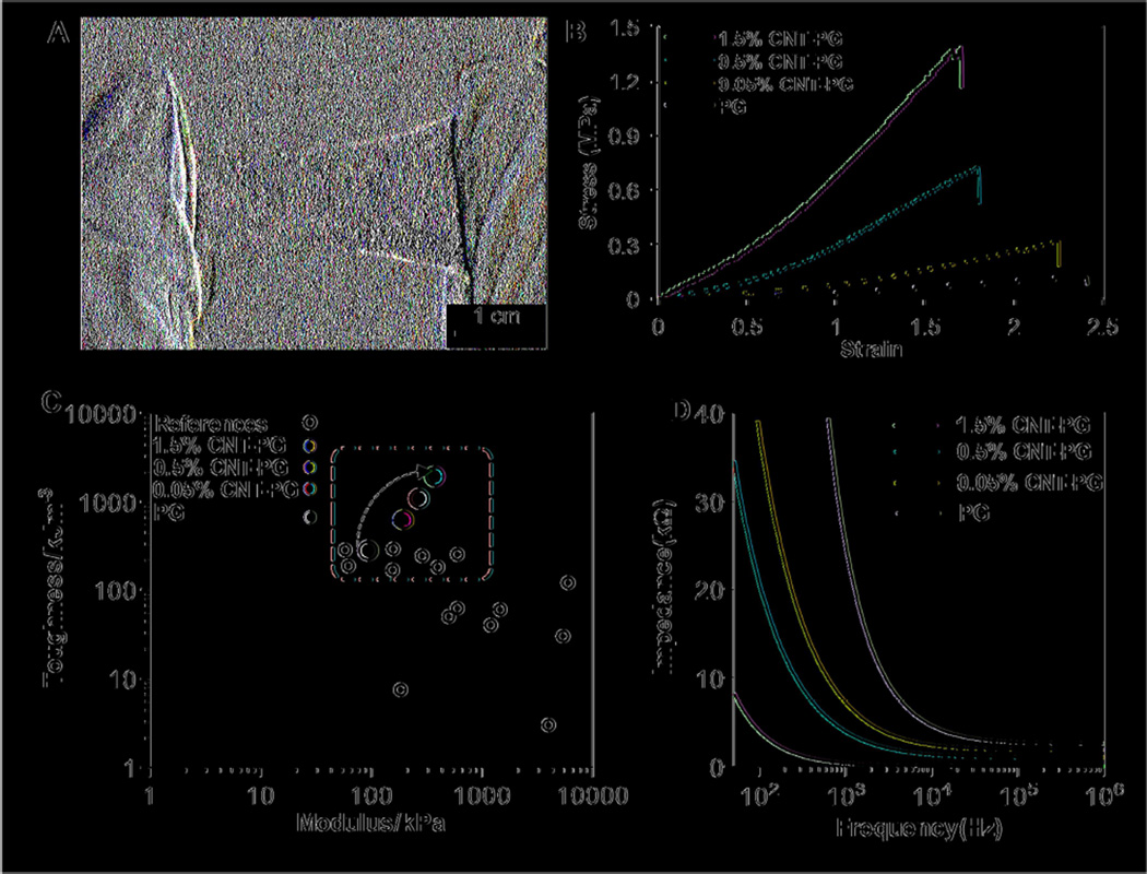
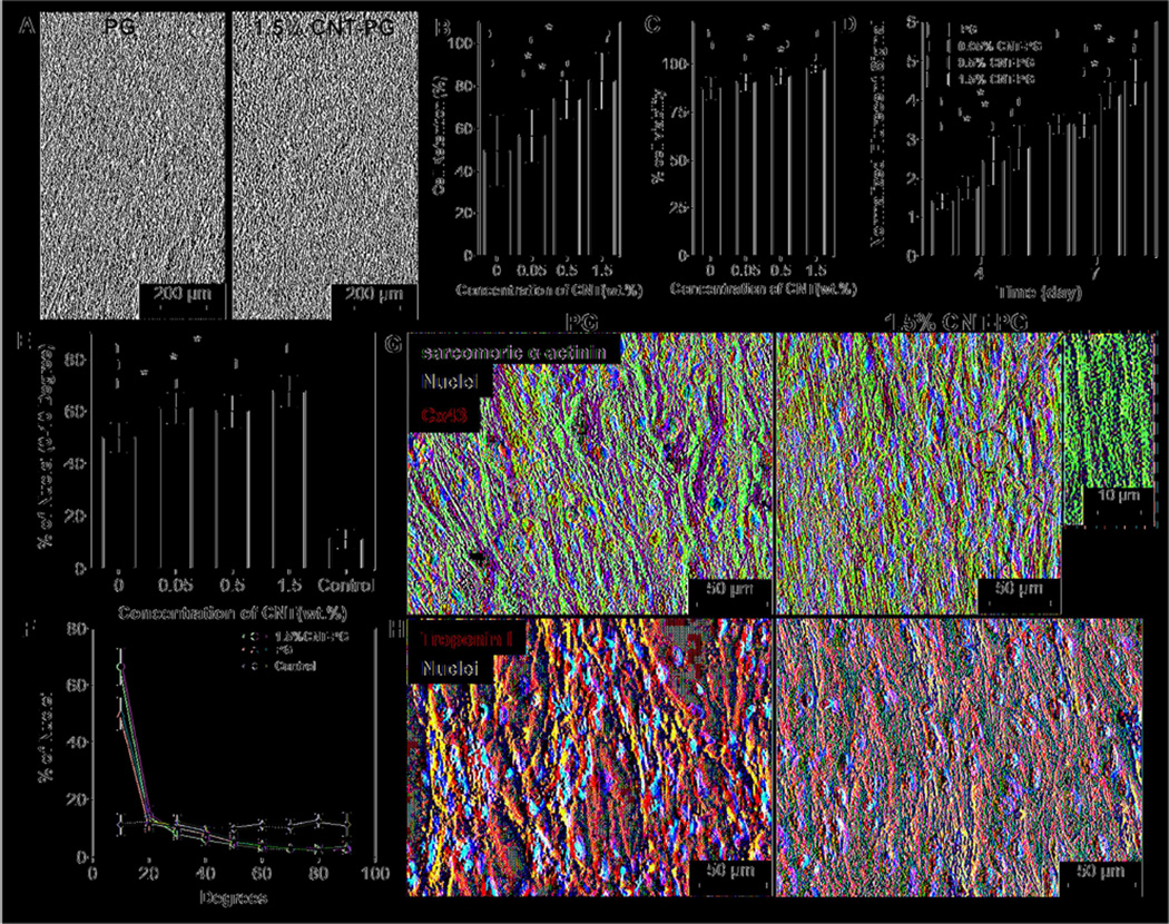
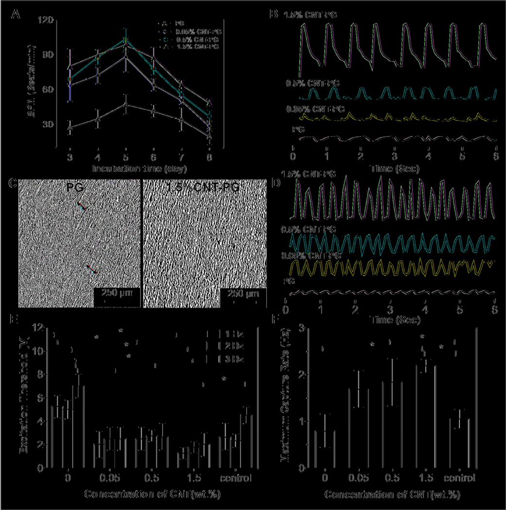
In the past few years, a considerable amount of effort has been devoted toward the development of biomimetic scaffolds for cardiac tissue engineering. However, most of the previous scaffolds have been electrically insulating or lacked the structural and mechanical robustness to engineer cardiac tissue constructs with suitable electrophysiological functions. Here, we developed tough and flexible hybrid scaffolds with enhanced electrical properties composed of carbon nanotubes (CNTs) embedded aligned poly(glycerol sebacate):gelatin (PG) electrospun nanofibers. Incorporation of varying concentrations of CNTs from 0 to 1.5% within the PG nanofibrous scaffolds (CNT-PG scaffolds) notably enhanced fiber alignment and improved the electrical conductivity and toughness of the scaffolds while maintaining the viability, retention, alignment, and contractile activities of cardiomyocytes (CMs) seeded on the scaffolds. The resulting CNT-PG scaffolds resulted in stronger spontaneous and synchronous beating behavior (3.5-fold lower excitation threshold and 2.8-fold higher maximum capture rate) compared to those cultured on PG scaffold. Overall, our findings demonstrated that aligned CNT-PG scaffold exhibited superior mechanical properties with enhanced CM beating properties. It is envisioned that the proposed hybrid scaffolds can be useful for generating cardiac tissue constructs with improved organization and maturation.

Keywords: Carbon Nanotubes (CNTs); Cardiac tissue engineering; Cardiomyocyte; Poly(glycerol sebacate):gelatin; Scaffold.


Figures

Figure 1
Figure 1
Structural characteristics of CNT-PG scaffolds. A) Schematic drawing showing the interactions of CNTs with PG scaffold upon crosslinking with EDC/NHS. B and C) SEM images showing uncrosslinked 1.5% CNT-PG scaffold. D) A representative TEM image of 1.5% CNT-PG confirming the well dispersion of CNTs (arrow) aligned along the nanofibers axis. E) Raman spectra of the CNTs and CNT-PG scaffolds. F) SEM and FFT (Inset) images of crosslinked 1.5% CNT-PG. G and H) The average fiber size and orientation index of nanofibrous scaffolds, demonstrating that increasing CNTs content resulted in reduced fiber size and enhanced fiber orientation (random 1.5% CNT-PG scaffold was considered as control) (*: P<0.05).
Figure 2
Figure 2
Physical and electrical characteristics of CNT-PG scaffolds. A) A representative optical image of tough 1.5% CNT-PG scaffold demonstrating easy manipulation of the scaffold. B) Representative stress-strain curves of crosslinked scaffolds after 3 days of soaking in DPBS demonstrating significant improvement in mechanical properties of the scaffolds by increasing the CNT concentration. C) Comparison of the elastic moduli and toughness for previously reported compliant fibrous scaffolds (open circle) [, , –46] and the proposed CNT-PG scaffolds consisting of 0% (purple circle), 0.05% (blue circle), 0.5% (red circle) and 1.5% (light green circle for aligned and dark green circle for random nanofibrous scaffolds) CNTs in hydrated state; The dashed window demonstrates the region including the previously developed and the presented CNT-PG tough fibrous scaffolds. As indicated by the arrow, the toughness of the PG scaffolds significantly enhanced through increasing the CNTs concentration. D) The overall impedance of 60µm thick scaffolds as a function of CNTs concentration showing a drastic decrease in impedance through CNTs content.
Figure 3
Figure 3
CM adhesion, viability, metabolic activity, and maturation. A) Phase contrast images of CMs after one day of culture indicating enhanced CMs retention on 1.5% CNT-PG compared to PG scaffold. Quantified results of B) cell retention (day 1), C) live/dead assay (day 1) and D) normalized metabolic activity, confirming significantly improved cell survival and proliferation as a function of CNT content (*: P< 0.05). E) Quantified average preferred nuclei alignment between 0–10 degree and F) nuclei alignment distribution on the developed scaffolds after day 7 of culture, indicated significantly enhancement in cellular alignment as a function of CNT content (*: P< 0.05) (Random 1.5% CNT-PG scaffold was considered as control). Representative images of CMs proteins expression stained for G) Sarcomeric α-actinin (green), Cx43 (red) and DAPI (blue), and H) Troponin I (red) and DAPI (blue) after 7 days of culture, revealing organized sarcomeres with higher Cx43 expression on 1.5% CNT-PG than PG scaffold. A high magnification image (inset in Fig. 3G) shows interconnected sarcomeric structures perpendicular to the direction of the nanofibers.
Figure 4
Figure 4
Electrophysiological functions of engineered cardiac constructs. A) Beating frequency (beats/min, BPM) of constructs as a function of CNT concentration and incubation time. B) Representative spontaneous contraction patterns of CMs cultured on PG scaffolds and CNT incorporated scaffolds recorded after 7 days of cultivation. C) Phase contrast images indicating organized tissue construct and non-continuous aligned tissue (red arrows) on the CNT-PG and PG scaffolds, respectively after 7 days of culture. D) Representative contraction patterns of electrically stimulated CMs on PG scaffold compared to CNT-PG scaffold after 7 days of culture (Frequency = 1). E) Excitation threshold and F) maximum capture rate of CMs seeded on scaffolds, indicating that increasing the CNT concentration and aligned structures significantly reduced excitation threshold and enhanced maximum capture rate (CMs cultured on random 1.5% CNT-PG scaffold was considered as control). (*:P<0.05).

Similar articles

Cited by

References

    1. Chien KR, Domian IJ, Parker KK. Cardiogenesis and the complex Biology of regenerative cardiovascular Medicine. Science. 2008;322:1494–1497. - PubMed
    1. Tandon V, Zhang B, Radisic M, Murthy SK. Generation of tissue constructs for cardiovascular regenerative medicine: From cell procurement to scaffold design. Biotechnol Adv. 2013;31:722–735. - PMC - PubMed
    1. Rockwood DN, Akins RE, Jr., Parrag IC, Woodhouse KA, Rabolt JF. Culture on electrospun polyurethane scaffolds decreases atrial natriuretic peptide expression by cardiomyocytes in vitro. Biomaterials. 2008;29:4783–4791. - PMC - PubMed
    1. Annabi N, Tsang K, Mithieux SM, Nikkhah M, Ameri A, Khademhosseini A, et al. Highly elastic micropatterned hydrogel for engineering functional cardiac tissue. Adv Funct Mater. 2013;23:4950–4959. - PMC - PubMed
    1. Dvir T, Timko BP, Brigham MD, Naik SR, Karajanagi SS, Levy O, et al. Nanowired three-dimensional cardiac patches. Nat Nanotechnol. 2011;6:720–725. - PMC - PubMed

Publication types