. 2014 Aug;55(8):1702-10.
doi: 10.1194/jlr.M048918. Epub 2014 Jun 13.

Hsp90 modulates PPARγ activity in a mouse model of nonalcoholic fatty liver disease

Affiliations

Hsp90 modulates PPARγ activity in a mouse model of nonalcoholic fatty liver disease

Matthew C Wheeler et al. J Lipid Res. 2014 Aug.

Abstract

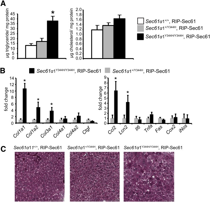
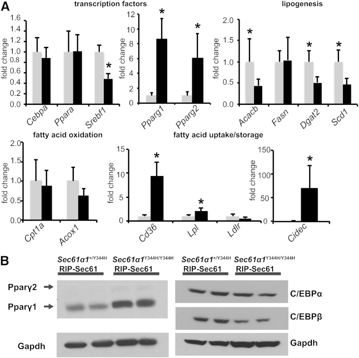
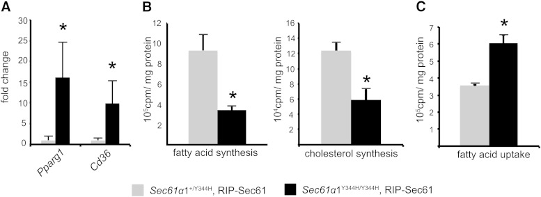
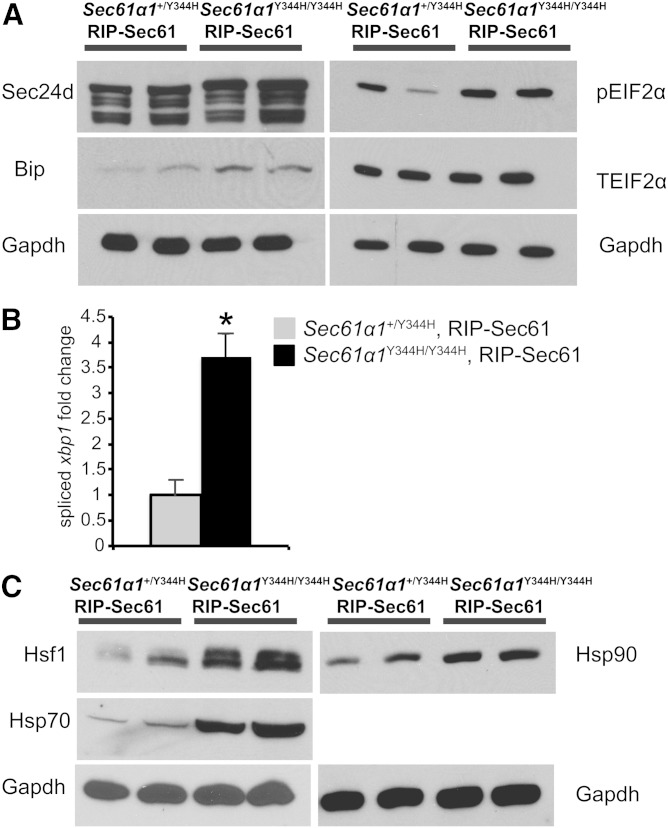
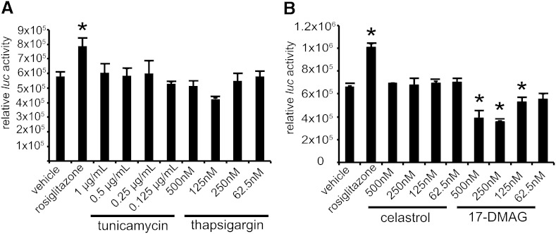
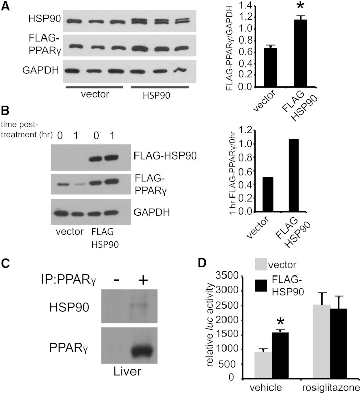
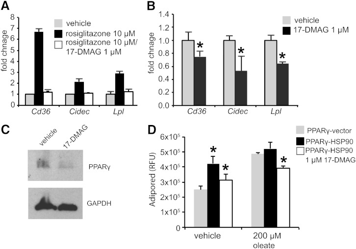
Nonalcoholic fatty liver disease (NAFLD) is a highly prevalent complication of obesity, yet cellular mechanisms that lead to its development are not well defined. Previously, we have documented hepatic steatosis in mice carrying a mutation in the Sec61a1 gene. Here we examined the mechanism behind NAFLD in Sec61a1 mutant mice. Livers of mutant mice exhibited upregulation of Pparg and its target genes Cd36, Cidec, and Lpl, correlating with increased uptake of fatty acid. Interestingly, these mice also displayed activation of the heat shock response (HSR), with elevated levels of heat shock protein (Hsp) 70, Hsp90, and heat shock factor 1. In cell lines, inhibition of Hsp90 function reduced Pparγ signaling and protein levels. Conversely, overexpression of Hsp90 increased Pparγ signaling and protein levels by reducing degradation. This may occur via a physical interaction as Hsp90 and Pparγ coimmunoprecipitated in vivo. Furthermore, inhibition of Hsp90 in Sec61a1 mutant hepatocytes also reduced Pparγ protein levels and signaling. Finally, overexpression of Hsp90 in liver cell lines increased neutral lipid accumulation, and this accumulation was blocked by Hsp90 inhibition. Our results show that the HSR and Hsp90 play an important role in the development of NAFLD, opening new avenues for the prevention and treatment of this highly prevalent disease.

Keywords: heat shock protein 90; heat shock response; peroxisome proliferator-activated receptor γ; steatosis.


Figures

Fig. 1.
Fig. 1.
Sec61a1Y344H/Y344H, RIP-Sec61 mice are steatotic. A: Biochemical assay for triglyceride and cholesterol from lipids extracted from livers of Sec61a1+/+, RIP-Sec61 (n = 5); Sec61a1+/Y344H, RIP-Sec61 (n = 5); and Sec61a1Y344H/Y344H, RIP-Sec61 (n = 6) mice. B: Abundance of the indicated mRNA as measured by quantitative PCR (qPCR) from livers of Sec61a1+/Y344H, RIP-Sec61 (n = 4) and Sec61a1Y344H/Y344H, RIP-Sec61 (n = 4) mice. C: Masson trichrome stained liver sections from mice of the indicated genotype. Bar = 100 μM. Error bars represent SEM, and * indicates P ≤ 0.05.
Fig. 2.
Fig. 2.
Upregulation of Pparg and its targets in Sec61a1Y344H/Y344H, RIP-Sec61 mice. A: qPCR analysis of metabolically relevant genes from RNA isolated from livers of mice of the indicated genotype (n = 4 mice per genotype). B: Western blot analysis of Pparγ, C/EBPα, and C/EBPβ proteins from liver tissue homogenates. Error bars represent SEM, and * indicates P ≤ 0.05.
Fig. 3.
Fig. 3.
Sec61a1Y344H/Y344H, RIP-Sec61 mice display increased uptake of free fatty acid. A: qPCR analysis on RNA isolated from hepatocytes from Sec61a1+/Y344H, RIP-Sec61 or Sec61a1Y344H/Y344H, RIP-Sec61 mice (n = 4 per genotype). Error bars represent SEM, and * indicates P ≤ 0.05. B: Primary hepatocytes were isolated from age- and sex-matched, fasted mice. Rates of sterol and triglyceride synthesis were determined by labeling with 14C-acetate for 16 h. Triplicate assays were performed from each of four (Sec61a1+/Y344H, RIP-Sec61) or three (Sec61a1Y344H/Y344H, RIP-Sec61) mice. C: Primary hepatocytes were isolated as in B. Rate of fatty acid uptake was determined in triplicate after cells were incubated with 14C-labeled oleate for 5 min, washed, and lysed. Cell-associated radioactivity was read by a scintillation counter (n = 2 per genotype).
Fig. 4.
Fig. 4.
Increased ER stress and HSR in Sec61a1Y344H/Y344H, RIP-Sec61 mice. A: Western blot analysis for markers of ER stress activation of liver extracts from mice of the indicated genotype. B: qPCR analysis of spliced Xbp1 message on RNA isolated from livers of the indicated genotype (n = 4 per genotype). Error bars represent SEM, and * indicates P ≤ 0.05 by Student’s t-test. C: Western blot analysis of Hsf1, Hsp70, and Hsp90 from liver homogenates prepared from mice of the indicated genotype.
Fig. 5.
Fig. 5.
Hsp90 inhibition reduces Pparγ signaling. A: F442A cells stably transfected with luciferase under the control of the AP2 promoter (F442A-Ap2Luc cells) were treated with rosiglitazone, tunicamycin, or thapsigargin at the indicated concentrations for 16 h, followed by lysis and homogeneous assay for luciferase activity. B: Cells were treated and assayed as in A, except that the HSR inducers celastrol and 17-DMAG were used. Error bars represent SD, and * indicates P ≤ 0.05 by Student’s t-test.
Fig. 6.
Fig. 6.
Hsp90 regulates Pparγ activity. A: Western blot analysis of HEK293 cells cotransfected with CMV:FLAG-Pparγ and either FLAG-Hsp90 or empty vector. Densitometry is shown to the right. B: Cycloheximide treatment (50 μg/ml) of HEK293 cells transfected with CMV:FLAG-Pparγ and either CMV:FLAG-Hsp90 or vector. C: Immunoprecipitation of Pparγ from liver of chow-fed wild-type mice; (-) indicates isotype control. D: HEK293 cells transfected with both a CMV:FLAG-Pparγ and a PPRE luciferase construct and either vector or a FLAG:Hsp90 for 24 h followed by 16 h of treatment with vehicle or rosiglitazone (10 μM) before luciferase assay. Error bars represent SD, and * indicates P ≤ 0.05.
Fig. 7.
Fig. 7.
Inhibition of Hsp90 reduces Pparγ target gene induction in Sec61a1Y344H/Y344H, RIP-Sec61 mice. A: qPCR analysis of RNA extracted from primary hepatocytes from Sec61a1+/Y344H, RIP-Sec mice (n = 2) after treatment with 10 μM rosiglitazone for 20 h following 4 h of pretreatment with or without 1 μM 17-DMAG. B: qPCR analysis on RNA extracted from primary hepatocytes from Sec61a1Y344H/Y344H, RIP-Sec mice (n = 2) after treatment with 1 μM 17-DMAG for 24 h. C: Western blot analysis of Pparγ in primary hepatocytes from Sec61a1+/Y344H, RIP-Sec mice treated with 1 μM 17-DMAG for 24 h. D: Huh7 cells, transfected with CMV:FLAG-Pparγ, and either vector or CMV:FLAG:Hsp90, were treated with 200 μM oleate before staining with AdipoRed. The 17-DMAG treatment was at 1 μM for the duration of oleate or vehicle treatment. Error bars represent SEM in A and B, and SD in D, and * indicates P ≤ 0.05 compared with cells transfected with only Pparγ.

References

    1. Cohen J. C., Horton J. D., Hobbs H. H. 2011. Human fatty liver disease: old questions and new insights. Science. 332: 1519–1523. - PMC - PubMed
    1. Browning J. D., Szczepaniak L. S., Dobbins R., Nuremberg P., Horton J. D., Cohen J. C., Grundy S. M., Hobbs H. H. 2004. Prevalence of hepatic steatosis in an urban population in the United States: impact of ethnicity. Hepatology. 40: 1387–1395. - PubMed
    1. Targher G., Day C. P., Bonora E. 2010. Risk of cardiovascular disease in patients with nonalcoholic fatty liver disease. N. Engl. J. Med. 363: 1341–1350. - PubMed
    1. Knebel B., Haas J., Hartwig S., Jacob S., Kollmer C., Nitzgen U., Muller-Wieland D., Kotzka J. 2012. Liver-specific expression of transcriptionally active SREBP-1c is associated with fatty liver and increased visceral fat mass. PLoS ONE. 7: e31812. - PMC - PubMed
    1. Koonen D. P., Jacobs R. L., Febbraio M., Young M. E., Soltys C. L., Ong H., Vance D. E., Dyck J. R. 2007. Increased hepatic CD36 expression contributes to dyslipidemia associated with diet-induced obesity. Diabetes. 56: 2863–2871. - PubMed

MeSH terms

LinkOut - more resources