. 2014 Jun 14:9:85.
doi: 10.1186/1750-1172-9-85.

Pulmonary alveolar proteinosis in children on La Réunion Island: a new inherited disorder?

Affiliations

Pulmonary alveolar proteinosis in children on La Réunion Island: a new inherited disorder?

Laurent Enaud et al. Orphanet J Rare Dis. .

Abstract

Background: Pulmonary alveolar proteinosis (PAP) is very rare in children. Only a few small series have been published, with little information about long-term progression. The objective of our study was to describe the clinical, radiological and pathological features, and the long-term course of PAP in a cohort of 34 children from La Réunion Island.

Methods: Data were retrospectively collected from medical files. Radiological and pathological elements were reviewed by two pediatric radiologists and three pathologists, respectively.

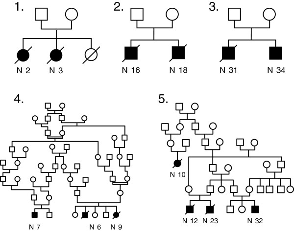
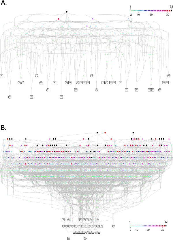
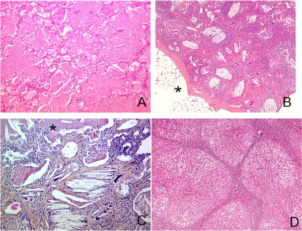
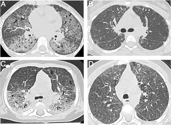
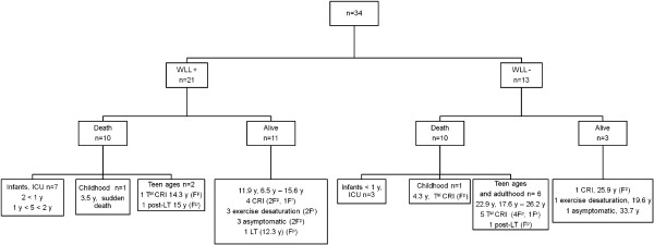
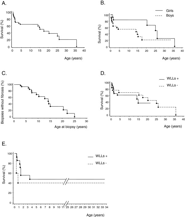
Results: Thirteen cases were familial and 32/34 (94%) cases were family connected. Disease onset occurred in the first six months of life in 82% of the patients. Thoracic computed tomography scans showed the typical "crazy-paving" pattern in 94% of cases. Respiratory disease was associated with a liver disorder, with the detection of liver enlargement at diagnosis in 56% of cases. The course of the disease was characterized by frequent progression to chronic respiratory insufficiency, accompanied by the appearance of cholesterol granulomas and pulmonary fibrosis. Overall prognosis was poor, with a mortality of 59% and an overall five-year survival rate from birth of 64%. Whole-lung lavages were performed in 21 patients, with no significant effect on survival. Liver disease progressed to cirrhosis in 18% of children, with no severe complication.

Conclusions: PAP in children from la Réunion Island is characterized by an early onset, associated liver involvement, poor prognosis and frequent progression to lung fibrosis, despite whole-lung lavages treatment. The geographic clustering of patients and the detection of many familial links between most of the cases strongly suggest a genetic etiology, with an autosomal recessive mode of inheritance.


Figures

Figure 1
Figure 1
Genealogical trees of familial cases of pulmonary alveolar proteinosis from La Réunion Island. Genealogical trees of the 5 families with several cases of PAP. Familial forms of PAP in La Réunion Island in our study were characterized by the existence of at least one pair of affected siblings per family in addition of one or two cousins for pedigrees 4 and 5. The coexistence of affected and unaffected children among siblings, the fact that both sexes can be affected, the ascertained consanguinity in family 4 and the transversal distribution of the disease are highly suggestive of an autosomic recessive mode of inheritance.
Figure 2
Figure 2
Genealogical analysis of cases of pulmonary alveolar proteinosis from La Réunion Island. A. Genealogical network connecting the 32 patients for which a parental consent was obtained to their most recent couple of common ancestors. Each patient (squares for boys and circles for girls) is denoted by his/her identifier. Each couple of ancestors is represented by an octagon of varying size and color according to the number of patients for which they are ancestors (from 1 to 32 as illustrated by the inner scale). The most recent couple of common ancestors (black octagon) married in 1731. Therefore, we can assert that all studied cases for which a consent was obtained are family connected. B. Deepest analysis connecting the 32 patients for which a parental consent was obtained to all their common ancestors. We found 32 couples of ancestors common to all patients (black octagons), each of them being a potential carrier of a genetic factor reponsible of the disease.
Figure 3
Figure 3
Pathological features. A: Lung histological specimen from a 9 months old boy, post-mortem examination. Diffuse intra alveolar accumulation of granular eosinophilic proteinaceous material with rare cholesterol clefts and normal interalveolar septa typical of PAP. There was no inflammation and no fibrosis. HES (×100). B: Lung histological specimen from a 9 years old girl, OLB. Numerous intra-alveolar cholesterol clefts with giant cells associated with inflammatory cells and fibrosis, typical of PICG. Lesions predominantly observed in the subpleural part of the lung parenchyma. Fatty involution of the visceral pleura (*). Tiny foci of PAP were also observed. HES (×100). C: Lung histological specimen from a 25 years old woman, post-mortem examination. Lung sample displayed both PAP on the upper part of the figure (*) and PICG with cholesterol clefts, giant cells granuloma, interstitial fibrosis and inflammation in the lower part of the figure. HES (×200). D: Liver histological specimen from a 2 years and 9 months old boy. Micro nodular cirrhosis, steatosis, ductular proliferation without active inflammation. HES (×100).
Figure 4
Figure 4
Radiological features. A and B: CT scan from a girl at the ages of 9 (A) and 13 (B) years old. A: Typical aspect of crazy paving pattern on thin axial sections: extensive areas of ground glass opacities (asterisks) superimposed with interlobular septa thickening (black arrows) and intralobular lines (black arrowheads). Irregular thickening of fissures are also noted (white arrows). B: Ground glass opacities, interlobular septa thickening and intralobular lines are less extensive than on the first CT scan while cystic lesions of a few millimetres (white arrowheads) are observed, with a subpleural distribution. Irregular traction bronchiectasis (white arrows) appeared that are initial signs of pulmonary fibrosis. C and D: CT scan of a girl at the ages of 8 months (C) and 5 years old (D). C: This CT scan image shows a symmetric combination of extensive ground-glass opacities (asterisks), intralobular lines (arrowheads) and interlobular septa thickening (black arrows), associated to consolidation (black curved arrows) in posterior areas of the lungs. Note the airspace hyperinflation in the anterior compartment of the lung (white asterisks) responsible of an increasing density gradient from anterior to posterior areas. D: Extensive ground-glass opacities, intralobular lines and interlobular septa thickening are still present. Subpleural cystic lesions (white arrowheads) and signs of fibrosis: honey-combing (white curved arrows) and traction bronchiectasis (white arrows) appeared.
Figure 5
Figure 5
Treatment and outcome. Definition of abbreviations: WLL = whole lung lavages; ICU = intensive care unit; CRI = chronic respiratory insufficiency, Tal CRI = terminal chronic respiratory insufficiency; LT = lung transplantation; Fp = pathologically documented lung fibrosis; Fr = lesions suggestive of lung fibrosis on thoracic CT scan images; y = years. This figure consists in a flowchart of the study population according to treatment group. For group of patients, ages are expressed as median.
Figure 6
Figure 6
Survival and occurrence of fibrosis. All curves were performed using Kaplan-Meier method. A. Overall survival from birth showing a 5-year survival rate of 65%. B. Overall survival from birth according to gender. Five-year survival from birth was significantly lower in boys than in girls (54% in boys versus 83% in girls, logrank test, p = 0.04). C. Occurrence of fibrosis according to the age at which lung biopsy was performed showing a probability of significant pulmonary fibrosis of almost 50% by the age of 14 years old. D. Overall survival from birth with or without WLL. WLL did not significantly change global survival rates (logrank test, p = 0.46). E. Overall survival from birth with or without WLL in patients diagnosed before the age of one year with a larger number of WLL carried out earlier (WLL+, n = 14 / WLL-, n = 5). In this group, this treatment did not significantly change global survival rates (logrank test, p = 0.39).

References

    1. Rosen SH, Castleman B, Liebow AA. Pulmonary alveolar proteinosis. N Engl J Med. 1958;258:1123–1142. doi: 10.1056/NEJM195806052582301. - DOI - PubMed
    1. Holbert JM, Costello P, Li W, Hoffman RM, Rogers RM. CT features of pulmonary alveolar proteinosis. AJR Am J Roentgenol. 2001;176:1287–1294. doi: 10.2214/ajr.176.5.1761287. - DOI - PubMed
    1. Trapnell BC, Whitsett JA, Nakata K. Pulmonary alveolar proteinosis. N Engl J Med. 2003;349:2527–2539. doi: 10.1056/NEJMra023226. - DOI - PubMed
    1. Inoue Y, Trapnell BC, Tazawa R, Arai T, Takada T, Hizawa N, Kasahara Y, Tatsumi K, Hojo M, Ichiwata T, Tanaka N, Yamaguchi E, Eda R, Oishi K, Tsuchihashi Y, Kaneko C, Nukiwa T, Sakatani M, Krischer JP, Nakata K. Characteristics of a large cohort of patients with autoimmune pulmonary alveolar proteinosis in Japan. Am J Respir Crit Care Med. 2008;177:752–762. doi: 10.1164/rccm.200708-1271OC. - DOI - PMC - PubMed
    1. Seymour JF, Presneill JJ. Pulmonary alveolar proteinosis: progress in the first 44 years. Am J Respir Crit Care Med. 2002;166:215–235. doi: 10.1164/rccm.2109105. - DOI - PubMed