. 2014 Jun 14:13:150.
doi: 10.1186/1476-4598-13-150.

FW-04-806 inhibits proliferation and induces apoptosis in human breast cancer cells by binding to N-terminus of Hsp90 and disrupting Hsp90-Cdc37 complex formation

Affiliations

FW-04-806 inhibits proliferation and induces apoptosis in human breast cancer cells by binding to N-terminus of Hsp90 and disrupting Hsp90-Cdc37 complex formation

Wei Huang et al. Mol Cancer. .

Abstract

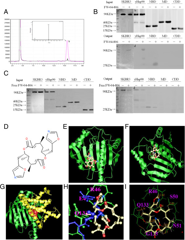
Background: Heat shock protein 90 (Hsp90) is a promising therapeutic target and inhibition of Hsp90 will presumably result in suppression of multiple signaling pathways. FW-04-806, a bis-oxazolyl macrolide compound extracted from China-native Streptomyces FIM-04-806, was reported to be identical in structure to the polyketide Conglobatin.

Methods: We adopted the methods of chemproteomics, computational docking, immunoprecipitation, siRNA gene knock down, Quantitative Real-time PCR and xenograft models on the research of FW-04-806 antitumor mechanism, through the HER2-overexpressing breast cancer SKBR3 and HER2-underexpressing breast cancer MCF-7 cell line.

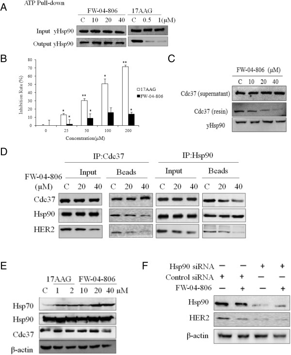
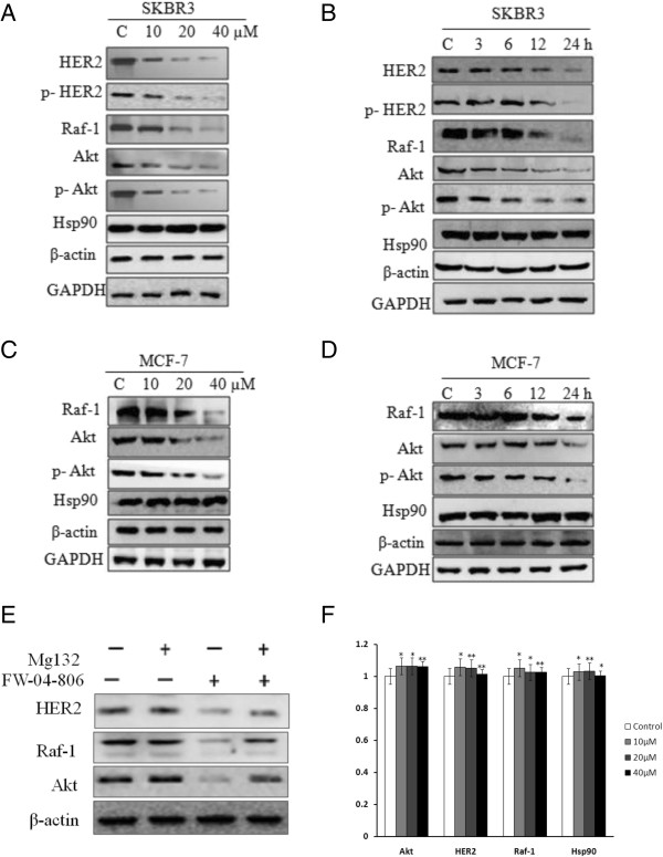
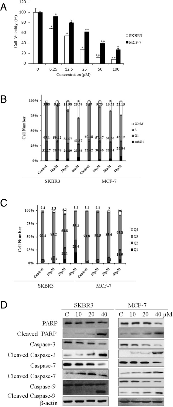
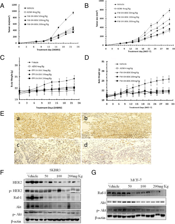
Results: We have verified the direct binding of FW-04-806 to the N-terminal domain of Hsp90 and found that FW-04-806 inhibits Hsp90/cell division cycle protein 37 (Cdc37) chaperone/co-chaperone interactions, but does not affect ATP-binding capability of Hsp90, thereby leading to the degradation of multiple Hsp90 client proteins via the proteasome pathway. In breast cancer cell lines, FW-04-806 inhibits cell proliferation, caused G2/M cell cycle arrest, induced apoptosis, and downregulated Hsp90 client proteins HER2, Akt, Raf-1 and their phosphorylated forms (p-HER2, p-Akt) in a dose and time-dependent manner. Importantly, FW-04-806 displays a better anti-tumor effect in HER2-overexpressed SKBR3 tumor xenograft model than in HER2-underexpressed MCF-7 model. The result is consistent with cell proliferation assay and in vitro apoptosis assay applied for SKBR-3 and MCF-7. Furthermore, FW-04-806 has a favorable toxicity profile.

Conclusions: As a novel Hsp90 inhibitor, FW-04-806 binds to the N-terminal of Hsp90 and inhibits Hsp90/Cdc37 interaction, resulting in the disassociation of Hsp90/Cdc37/client complexes and the degradation of Hsp90 client proteins. FW-04-806 displays promising antitumor activity against breast cancer cells both in vitro and in vivo, especially for HER2-overexpressed breast cancer cells.


Figures

Figure 1
Figure 1
FW-04-806 is identified as an Hsp90 binding medicine. (A) LC/MS spectrum detects the compound eluted from the Hsp90-loaded affinity column. Peak a and b represent elution samples from the control and FW-04-806 bound Hsp90-loaded column, respectively. MS spectrum displays M/z of Peak b. (B) Western blot confirms FW-04-806 binds to NBD of Hsp90. The “–” symbol represents no drug-loaded affinity column, the “+” symbol represents the drug-loaded affinity column. The test proteins are SKBR3 cell lysate, His-tagged yeast Hsp90, and His-tagged NBD, MD, CDD of yeast Hsp90. Human Hsp90 Antibody and His-probe antibody were used respectively. (C) Soluble FW-04-806 was added into SKBR3 cell lysate, His-tagged full-length, NBD, MD and CDD of yeast Hsp90 up to 10 μM before incubation with drug-loaded affinity resin. Picture a and c show Human Hsp90 Antibody; picture b and d show His-probe antibody. (D) Molecular structure of FW-04-806. (E) Geldanamycin (GA) binds to N-terminal Hsp90 (Protein Data Bank [PDB] ID 1YTE) in the ATP binding pocket. Hsp90 is shown in the green ribbon view; GA is shown in stick view. (F) FW-04-806 docks to N-terminal Hsp90 (PDB ID 2K5B). N-Hsp90 is shown in green ribbon view; FW-04-806 is shown in stick view. (G) FW-04-806 binds to the N-Hsp90/Cdc37 complex (PDB ID 1US7). N-Hsp90 is shown in green ribbon view; Cdc37 is shown in yellow ribbon view; FW-04-806 is shown in orange ball view. (H) Stick view of FW-04-806 bound to N-Hsp90/Cdc37 complex. N-Hsp90 is shown in green stick view; FW-04-806 is shown in white stick view; Cdc37 is shown in blue stick view. (I) Stick views of FW-04-806 and N-Hsp90 in white and green, respectively.
Figure 2
Figure 2
FW-04-806 inhibits Hsp90/Cdc37 chaperone/co-chaperone interactions. (A) FW-04-806 (10, 20, 40 μM) was added into recombinant NBD Hsp90 protein did not affect ATP binding capacity of Hsp90, while 17AAG (0.5, 1 μM) decreased ATP binding. Western blot was analyzed with His-probe antibody. (B) FW-04-806 had little effect on Hsp90 ATPase activity as determined by the malachite green reagent. The assay used 0.5 μM Hsp90 protein, 1 mM ATP, and FW-04-806 or 17AAG at 25, 50, 100, or 200 μM, or vehicle (DMSO) at 620 nm. Results are presented as means ± SD of three independent experiments. *p < 0.05: significant difference from control by analysis of variance; **p < 0.01: very significant difference from control by analysis of variance. (C) FW-04-806 directly affect Hsp90-Cdc37 interaction in Pull-down assay. 400 ug of purified Hsp90 (His-tag)protein was bound with Ni-NTA resin and divided into four groups evenly with the addition of recombinant Cdc37 protein 50 μg each and FW-04-806 0, 10, 20, 40 μM, respectively. After incubation and wash, The resins were boiled with loading buffer and analyzed by western blotting, His-probe antibody and Cdc37 antibody was used respectively. (D) SKBR3 cells were treated with FW-04-806 or DMSO for 24 h. Cdc37 or Hsp90 were immunoprecipitated from whole-cell lysates (500 μg each) with an anti-Cdc37 or anti-Hsp90 antibody respectively, then analyzed by immunoblotting with antibody against Hsp90, Cdc37 and HER2 (E) SKBR3 cells were treated with FW-04-806 at 10, 20, 40 μM for 24 h; 17AAG was used as a positive control at 1 and 2 μM. Hsp70, Hsp90, and Cdc37 protein level were analyzed with western blotting using relevant antibodies. (F) SKBR3 cells were transiently transfected with control siRNA or Hsp90 siRNA for 48 h. Whole-cell lysates were analyzed with western blotting against HER2, Hsp90, and β-actin.
Figure 3
Figure 3
FW-04-806 induces proteasome-dependent degradation and decreases Hsp90 client protein level. (A) SKBR3 cells were treated by FW-04-806 at 10, 20, 40 μM for 24 h; HER2, phosphorylated HER2 (p-HER2), Raf-1, Akt, and phosphorylated Akt (p-Akt) protein levels were analyzed with western blotting. (B) SKBR3 cells were treated with 20 μM of FW-04-806 for 0, 3, 6, 12, or 24 h; HER2, p-HER2, Raf-1, Akt, and p-Akt protein levels were analyzed with western blot. (C) MCF-7 cells were treated by FW-04-806 at 10, 20, 40 μM for 24 h; western blot detected the protein expression of Raf-1, Akt, and p-Akt. (D) MCF-7 cells were treated with 20 μM FW-04-806 for 0, 3, 6, 12, or 24 h; western blot detected the protein expression of Raf-1, Akt, and p-Akt. (E) SKBR3 was pretreated with 1 μM of MG132 for 2 h in the presence or absence of 20 μM of FW-04-806 for an additional 12 h. Whole-cell lysates were subjected to western blot analysis using antibodies against HER2, Raf-1, Akt, and β-actin. (F) SKBR3 was treated with DMSO or FW-04-806 at 10, 20, 40 μM for 24 h. The total RNA was extracted for quantitative Real-time PCR of AKT, HER2, Raf-1 and Hsp90, using GAPDH as control. *P < 0.05: significant difference from control by analysis of variance; **P < 0.01: very significant difference from control by analysis of variance.
Figure 4
Figure 4
FW-04-806 inhibits growth, induces cell cycle arrest, induces apoptosis, and downregulates the expression of anti-apoptotic proteins. (A) SKBR3 and MCF-7 cells were grown for 48 h in the absence or presence of increasing concentrations of FW-04-806. Cell growth inhibition was measured with MTS and was expressed as a percentage of vehicle-treated control; results are presented as means ± SD of three independent experiments. *p < 0.05: significant difference from control by analysis of variance; **p < 0.01: very significant difference from control by analysis of variance. (B) SKBR3 and MCF-7 cells were treated with increasing doses of FW-04-806 or vehicle DMSO for 24 h. The cells were then fixed with 70% ethanol at −20°C overnight, incubated with RNase A at 37°C for 30 min, stained with propidium iodide for 10 min, and analyzed with flow cytometry. (C) SKBR3 and MCF-7 cells were treated with FW-04-806 for 24 h, and apoptotic cell death was detected by staining cells with an Annexin-V: FITC Apoptosis Detection Kit for analysis with flow cytometry. (D) SKBR3 and MCF-7 cells were treated with increasing doses of FW-04-806 for 24 h, and the apoptosis signal proteins were detected with western blot analysis using an Apoptosis Antibody Sampler Kit; β-actin was used as a loading control.
Figure 5
Figure 5
FW-04-806 inhibits the tumor growth of SKBR3 and MCF-7 tumor xenograft models. (A and B) SKBR3 and MCF-7 tumor xenograft nude mice were randomized into treatment groups (n = 6/group) and received FW-04-806 at doses of 50, 100, 200 mg/kg i.g., q3d (in a vehicle of 10% ethanol, 10% polyethylene glycol 400, and 10% Tween 80) or ADM at doses of 4 mg/kg i.p., q3d, until the day that the tumor sizes of the vehicle control groups reached approximately 1000 mm3. Tumors were weighed to evaluate the anticancer activity of FW-04-806. Data are presented as means ± SD (n = 6, P < 0.01). (C and D) Mouse body weight was measured twice a week. (E) HER2 expression in SKBR3 tumors. Picture a shows vehicle controls; picture b, c and d show 50, 100 and 200 mg/kg groups, respectively. (F) Tumor tissues excised from the SKBR3 xenograft mice were lysed; changes in the levels of in HER2, p-HER2, Raf-1, Akt, and p-Akt protein were tested. (G) Tumor tissues excised from the MCF-7 xenograft mice were lysed; western blot detected the changes in the levels of in Raf-1, Akt, and p-Akt protein expressions.

References

    1. Young JC, Agashe VR, Siegers K, Hartl FU. Pathways of chaperone-mediated protein folding in the cytosol. Nat Rev Mol Cell Biol. 2004;5:781–791. doi: 10.1038/nrm1492. - DOI - PubMed
    1. Welch WJ. Heat shock proteins functioning as molecular chaperones: their roles in normal and stressed cells. Phil Trans Roy Soc Lond B Biol Sci. 1993;339:327–333. doi: 10.1098/rstb.1993.0031. - DOI - PubMed
    1. Hartl FU. Molecular chaperones in cellular protein folding. Nature. 1996;381:571–579. doi: 10.1038/381571a0. - DOI - PubMed
    1. Patel HJ, Modi S, Chiosis G, Taldone T. Advances in the discovery and development of heat-shock protein 90 inhibitors for cancer treatment. Expet Opin Drug Discov. 2011;6:559–587. doi: 10.1517/17460441.2011.563296. - DOI - PMC - PubMed
    1. Kamal A, Thao L, Sensintaffar J, Zhang L, Boehm MF, Fritz LC, Burrows FJ. A high-affinity conformation of Hsp90 confers tumour selectivity on Hsp90 inhibitors. Nature. 2003;425:407–410. doi: 10.1038/nature01913. - DOI - PubMed

Publication types

MeSH terms