. 2014 Sep:44:80-90.
doi: 10.1016/j.neuro.2014.06.001. Epub 2014 Jun 10.

4R-cembranoid protects against diisopropylfluorophosphate-mediated neurodegeneration

Affiliations

4R-cembranoid protects against diisopropylfluorophosphate-mediated neurodegeneration

P A Ferchmin et al. Neurotoxicology. 2014 Sep.

Abstract

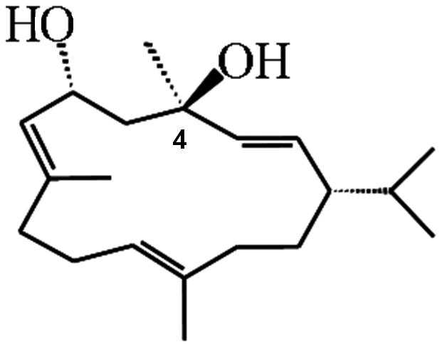
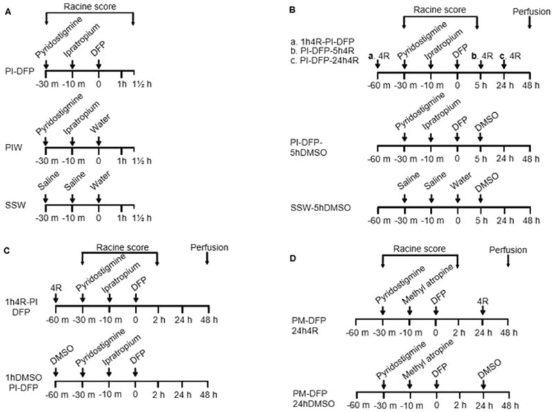
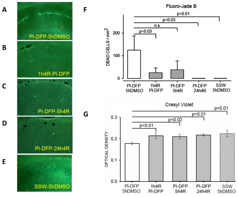
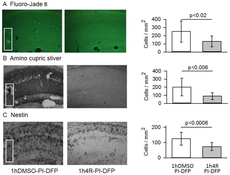
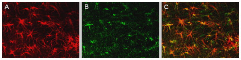
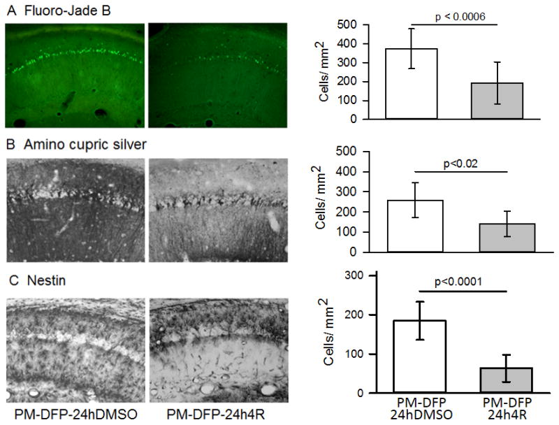
Many organophosphorous esters synthesized for applications in industry, agriculture, or warfare irreversibly inhibit acetylcholinesterase, and acute poisoning with these compounds causes life-threatening cholinergic overstimulation. Following classical emergency treatment with atropine, an oxime, and a benzodiazepine, surviving victims often suffer brain neurodegeneration. Currently, there is no pharmacological treatment to prevent this brain injury. Here we show that a cyclic diterpenoid, (1S,2E,4R,6R,7E,11E)-cembra-2,7,11-triene-4,6-diol (4R) ameliorates the damage caused by diisopropylfluorophosphate (DFP) in the hippocampal area CA1. DFP has been frequently used as a surrogate for the warfare nerve agent sarin. In rats, DFP is lethal at the dose used to cause brain damage. Therefore, to observe brain damage in survivors, the death rate was reduced by pre-administration of the peripherally acting antidotes pyridostigmine and methyl atropine or its analog ipratropium. Pyridostigmine bromide, methyl atropine nitrate, and ipratropium bromide were dissolved in saline and injected intramuscularly at 0.1mg/kg, 20mg/kg, and 23mg/kg, respectively. DFP (9mg/kg) dissolved in cold water was injected intraperitoneally. 4R (6mg/kg) dissolved in DMSO was injected subcutaneously, either 1h before or 5 or 24h after DFP. Neurodegeneration was assessed with Fluoro-Jade B and amino cupric silver staining; neuroinflammation was measured by the expression of nestin, a marker of activated astrocytes. Forty-eight hours after DFP administration, 4R decreased the number of dead neurons by half when injected before or after DFP. 4R also significantly decreased the number of activated astrocytes. These data suggest that 4R is a promising new drug that could change the therapeutic paradigm for acute poisoning with organophosphorous compounds by the implementation of a second-stage intervention after the classical countermeasure treatment.

Keywords: (1S,2E,4R,6R,7E,11E)-Cembra-2,7,11-triene-4,6-diol; Cembranoid; Diisopropylfluorophosphate; Neurodegeneration; Neuroprotection.


Figures

Figure 1
Figure 1. Structural formula of 4R-cembranoid
The chiral carbon 4, relevant to neuroprotection, is indicated.
Figure 2
Figure 2. Experimental design
The figure presents the treatment groups and the protocols used for experiments 1–4. The treatment names are abbreviated as the first letter of each compound, except in the case of DFP and DMSO, where the standard abbreviations are used; the compounds are named in the same sequence as they were injected. The protocols indicate the times of injection of the various drugs as well as the period during which the Racine scores were obtained. In experiments 2–4, the animals were perfused 48 hours after DFP. (A) Experiment 1. Pyridostigmine (P) and ipratropium (I) were injected before DFP in the experimental group PI-DFP; this group tested for the effects of DFP when preceded by pyridostigmine and ipratropium. The PIW group received pyridostigmine, ipratropium, and water (W). Water was the vehicle of DFP; this group tested for the effects of pyridostigmine and ipratropium in the absence of DFP. The SSW group was the all-vehicles control, which received saline (S), saline, and water. (B) Experiment 2 tested for the neuroprotective effect of 4R against DFP. 4R was administered within the PI-DFP paradigm 1 hour before DFP (a. 1h4R-PI-DFP), 5 hours after DFP (b. PI-DFP-5h4R), and 24 hours after DFP (c. PI-DFP-24h4R). The PI-DFP-5hDMSO group tested for the effect of PI-DFP in the presence of DMSO, the vehicle of 4R. The SSW-5hDMSO group was the all-vehicles control. (C) Experiment 3 replicated the study of 4R when injected 1 hour before DFP (1h4R-PI-DFP). The 1 hDMSO-PI-DFP group was the 4R vehicle control. (D) Experiment 4 replicated the study of 4R injected 24 hours after DFP using methyl atropine (M) instead of ipratropium. The experimental group was injected with pyridostigmine and methyl atropine before DFP, followed 24 hours later by 4R (PM-DFP-24h4R). The control group that did not receive 4R (PM-DFP-24hDMSO) was injected 24 hours after DFP with DMSO.
Figure 3
Figure 3. Experiment 1: Effect of DFP on brain AChE activity
AChE activity was measured in six brain areas of rats treated with the pyridostigmine-ipratropium-DFP protocol (PI-DFP); pyridostigmine-ipratropium-water (PIW), or saline-saline-water (SSW) (see Fig. 2A). Enzyme activity was the same in the PIW and SSW groups, which were pooled together in the “CONTROL” group. The bars represent the mean ± SEM of 3-6 rats per time point, except at 21 days when there were only two rats, and therefore SEM was not calculated.
Figure 4
Figure 4. Experiment 1: Effect of pyridostigmine, ipratropium, and DFP on the Racine scores
Racine scores were determined as described in Methods (experimental protocol in Fig. 2A). The symbols are the mean ± SEM of the scores for 10-min observation periods. The vertical lines labeled 1, 2, and 3 indicate the time of the three injections for each group. PI-DFP: (1) pyridostigmine, (2) ipratropium, and (3) DFP. PIW: (1) pyridostigmine, (2) ipratropium, and (3) water. SSW: (1) saline, (2) saline, and (3) water.
Figure 5
Figure 5. Experiment 2: Pre-application of 4R does not affect Racine scores
Racine scores were determined as described in Methods; for the experimental protocol, see Fig. 2B. The symbols are the mean ± SEM values of the scores for 10-min observation periods. PI-DFP stands for the combined PI-DFP-DMSO, PI-DFP-5h4R, and PI-DFP-24h4R groups and 1h4R-PI-DFP corresponds to the subjects injected with 4R 1 hour before DFP.
Figure 6
Figure 6. Experiment 2: 4R decreased neuronal death produced by the PI-DFP treatment
See Fig. 2B for experimental design. There were five experimental groups. The determinations were done in adjacent slices of the same brains. (A-E) Representative micrographs from the CA1 hippocampal area stained with Fluoro-Jade B. (F) The bar graph represents the mean ± SEM of dead cell density determined with Fluoro-Jade B. (G) The bars represent the mean ± SEM of the optical density of live cells stained with cresyl violet. The number of rats per group was PI-DFP-DMSO (10), 1h4R-PI-DFP (6), PI-DFP-5h4R (5), PI-DFP-24h4R (4), and SSW-5hDMSO (6). See statistical analysis in Methods.
Figure 7
Figure 7. 4R injected 1 hour before DFP decreased neuronal death and astrocyte activation
Micrographs (10X) are from a 4R vehicle control (1hDMSO-PI-DFP) and a rat preinjected with 4R (1h4R-PI-DFP). The magnification of the insets for Fluoro-Jade B was 30X and for amino cupric silver and nestin, 40X. (A) Fluoro-Jade B staining. The number of Fluoro-Jade B stained cells in the stratum pyramidale was significantly lower in the 4R group than in the 4R vehicle control (n =10 rats per group). (B) Amino cupric silver staining. The number of amino cupric silver-stained cells in the stratum pyramidale was significantly lower in the 4R group than in the 4R vehicle controls (n=10 per group). (C) Immunostaining for nestin. The number of activated astrocytes in the stratum radiatum was significantly lower in the 4R group than the 4R vehicle control which was injected with DMSO (n= 6 rats per group). The bars are means ± SEM. See statistics in Methods. See Fig. 2C for experimental design.
Figure 8
Figure 8. Nestin and GFAP immunostaining overlap in DFP-activated astrocytes
Confocal photomicrographs from coronal cortical sections from rats treated with DFP in the PI-DFP paradigm. (A) Immunostaining with antibody against GFAP. (B) Immunostaining with antibody against nestin. (C) Most GFAP-labeled cells overlap with nestin-positive cells. See Methods.
Figure 9
Figure 9. 4R was neuroprotective when injected 24 hours after DFP
Experiment 4 (see Fig. 2D for experimental design and abbreviations). Fluoro-Jade B staining (A), amino cupric silver staining (B), and nestin immunostaining (C) were applied to adjacent slices of the same brains. The images show representative micrographs from two rats; a rat injected 24 hours after DFP with DMSO, the vehicle of 4R (PM-DFP-24hDMSO), and the experimental rat injected at the same time with 4R (PM-DFP-24h4R). The bar graphs show dead neurons in (A) and (B) and activated astrocytes in (C). The number of rats used for Fluoro-Jade B staining included 9 4R vehicle controls and 10 rats injected with 4R; for ACS and nestin there were 8 rats per group. The bars are means ± SEM. See statistics in Methods.

References

    1. Banks CN, Lein PJ. A review of experimental evidence linking neurotoxic organophosphorus compounds and inflammation. Neurotoxicology. 2012;33:575–584. - PMC - PubMed
    1. Bradford MM. A rapid and sensitive method for the quantitation of microgram quantities of protein utilizing the principle of protein-dye binding. Analytical biochemistry. 1976;72:248–254. - PubMed
    1. Chapman S, Kadar T, Gilat E. Seizure duration following sarin exposure affects neuro-inflammatory markers in the rat brain. Neurotoxicology. 2006;27:277–283. - PubMed
    1. Chen Y. Organophosphate-induced brain damage: mechanisms, neuropsychiatric and neurological consequences, and potential therapeutic strategies. Neurotoxicology. 2012;33:391–400. - PubMed
    1. Clarke SR, Shetty AK, Bradley JL, Turner DA. Reactive astrocytes express the embryonic intermediate neurofilament nestin. Neuroreport. 1994;5:1885–1888. - PubMed

Publication types

MeSH terms

LinkOut - more resources