. 2014 Oct;37(2):377-86.
doi: 10.1007/s10827-014-0510-z. Epub 2014 Jun 14.

Effect of non-symmetric waveform on conduction block induced by high-frequency (kHz) biphasic stimulation in unmyelinated axon

Affiliations

Effect of non-symmetric waveform on conduction block induced by high-frequency (kHz) biphasic stimulation in unmyelinated axon

Shouguo Zhao et al. J Comput Neurosci. 2014 Oct.

Abstract

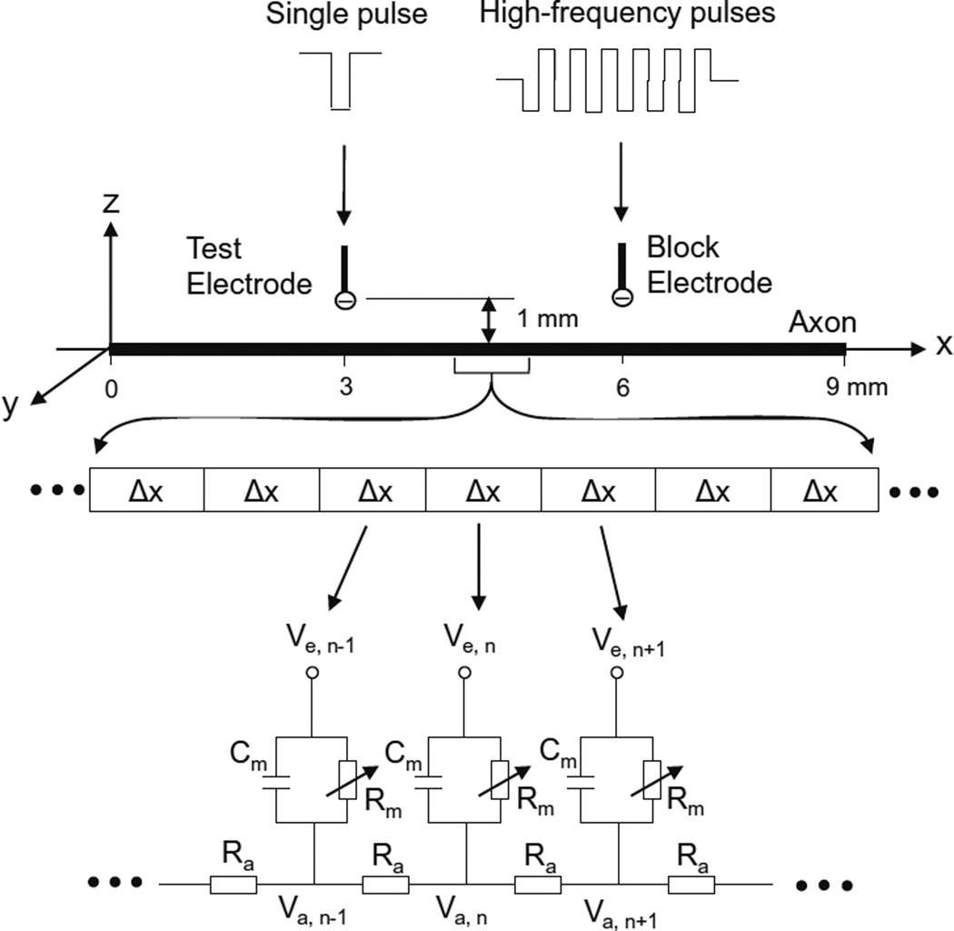
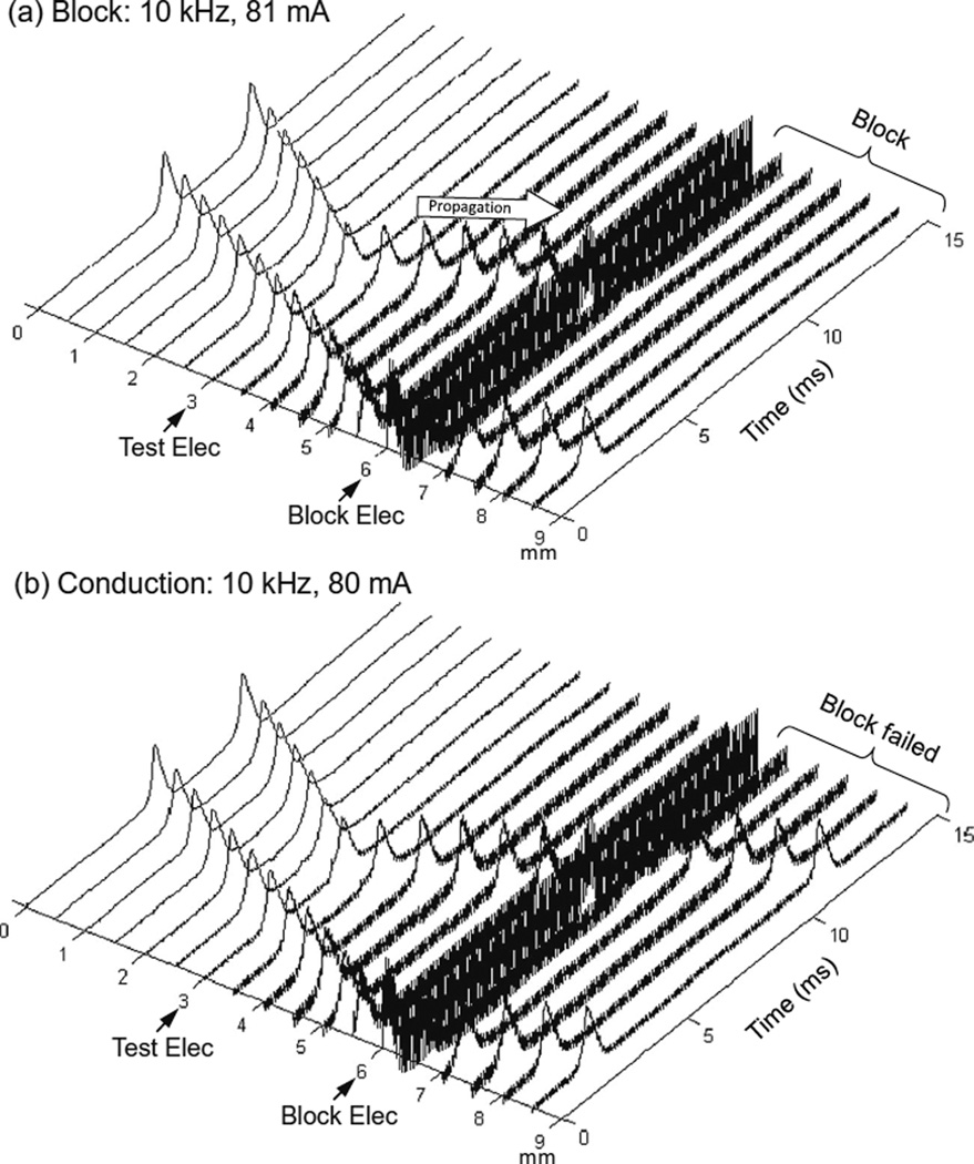
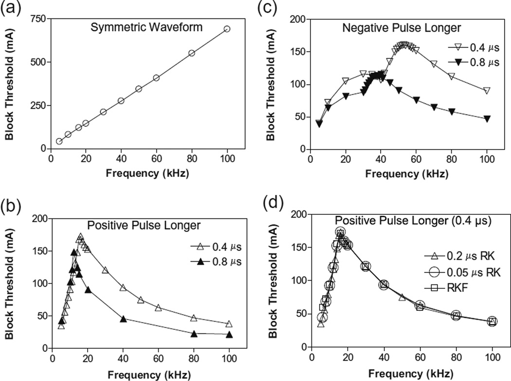
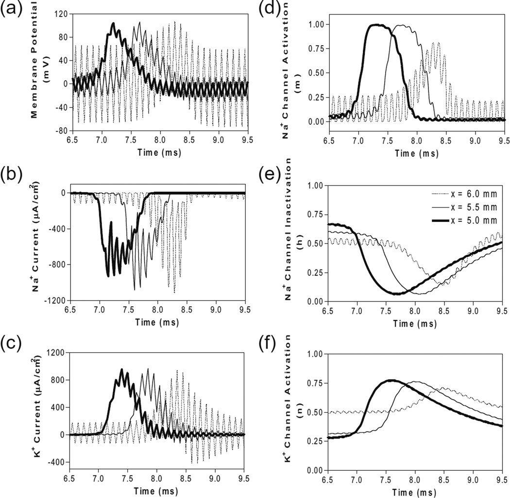
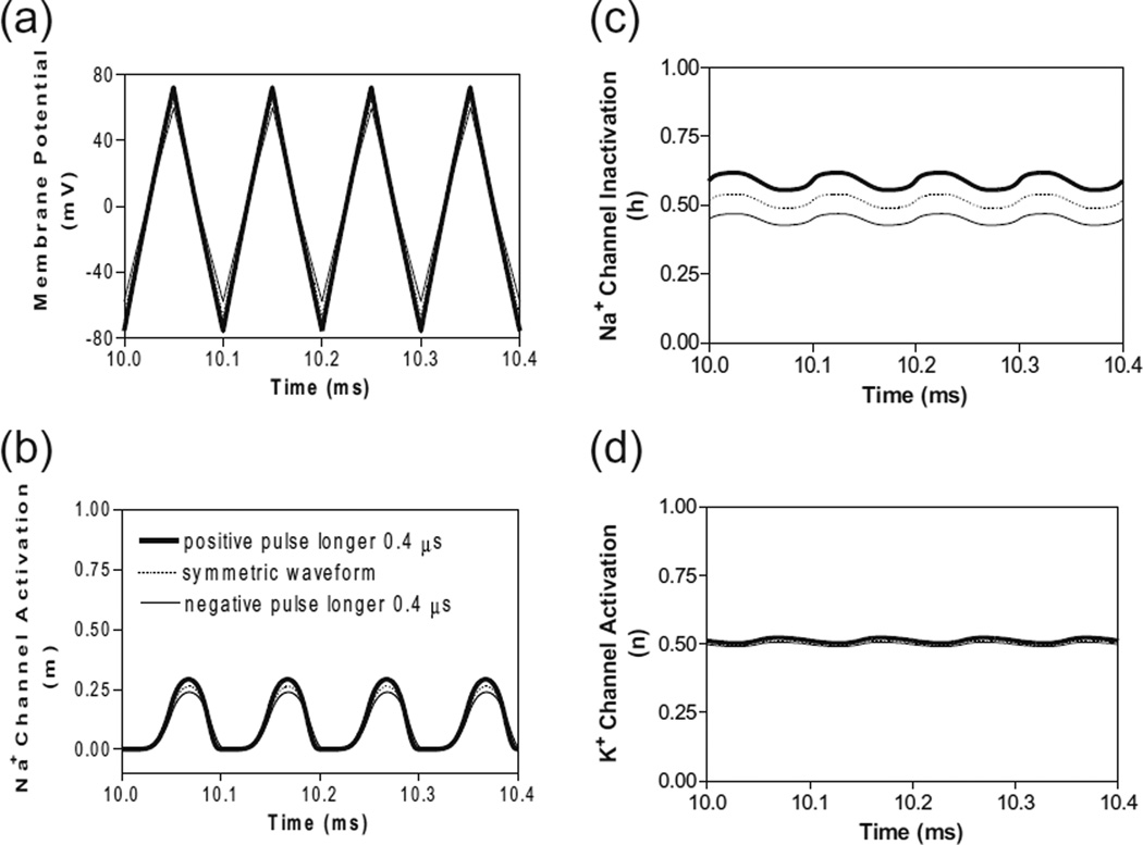
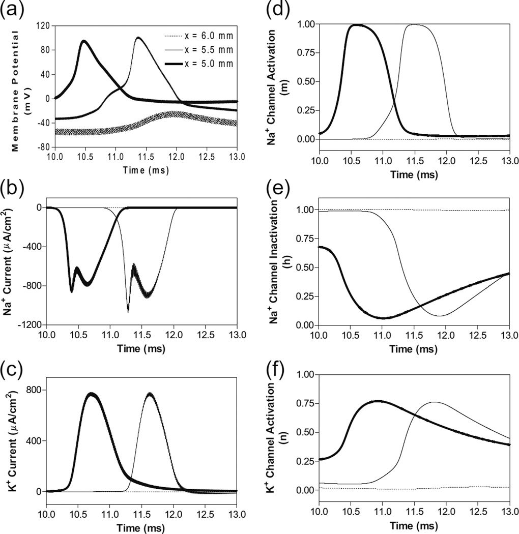
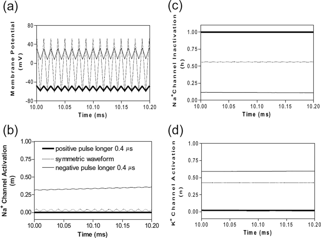
The effect of a non-symmetric waveform on nerve conduction block induced by high-frequency biphasic stimulation is investigated using a lumped circuit model of the unmyelinated axon based on Hodgkin-Huxley equations. The simulation results reveal that the block threshold monotonically increases with the stimulation frequency for the symmetric stimulation waveform. However, a non-monotonic relationship between block threshold and stimulation frequency is observed when the stimulation waveform is non-symmetric. Constant activation of potassium channels by the high-frequency stimulation results in the increase of block threshold with increasing frequency. The non-symmetric waveform with a positive pulse 0.4-0.8 μs longer than the negative pulse blocks axonal conduction by hyperpolarizing the membrane and causes a decrease in block threshold as the frequency increases above 12-16 kHz. On the other hand, the non-symmetric waveform with a negative pulse 0.4-0.8 μs longer than the positive pulse blocks axonal conduction by depolarizing the membrane and causes a decrease in block threshold as the frequency increases above 40-53 kHz. This simulation study is important for understanding the potential mechanisms underlying the nerve block observed in animal studies, and may also help to design new animal experiments to further improve the nerve block method for clinical applications.


Conflict of interest statement

Conflict of interest The authors declare that they have no conflict of interest.

Figures

Fig. 1
Fig. 1
Unmyelinated axon model to simulate conduction block induced by high-frequency biphasic electrical current. The unmyelinated axon is segmented into many small cylinders of length Δx, each of which is modeled by a resistance-capacitance circuit based on the Hodgkin–Huxley model. Ra: Axoplasm resistance. Rm: Membrane resistance. Cm: Membrane capacitance. Va: Intracellular potential. Ve: Extracellular Potential
Fig. 2
Fig. 2
Blocking the propagation of action potentials along an unmyelinated axon by high-frequency symmetric biphasic stimulation. High-frequency (10 kHz) stimulation is continuously delivered at the block electrode, which initiated an initial action potential. Another action potential is initiated via the test electrode at 5 ms after starting the high-frequency stimulation, and propagates towards both ends of the axon. The 10 kHz stimulation blocks nerve conduction at the intensity of 81 mA (a), but not at 80 mA (b). The short arrows mark the locations of test and block electrodes along the axon. The white arrow indicates propagation of the action potential to the location of the 10 kHz blocking stimulation. Axon diameter: 2 µm
Fig. 3
Fig. 3
The threshold intensity to block nerve conduction changes with the stimulation frequency. (a). For the symmetric waveform, the block threshold monotonically increases as the frequency increases. (b). If the positive pulse is longer (0.4 or 0.8 µs), the block threshold peaks at 12–16 kHz and then gradually decreases as the frequency increases. (c). If the negative pulse is longer (0.4 or 0.8 µs), the block threshold peaks at 40–53 kHz. (d). The same results were also obtained by Runge–Kutta (RK) numerical integration method with a smaller time step of 0.05 µs or by Runge–Kutta-Fehlberg (RKF) numerical integration method with an adaptive time step. Axon diameter: 2 µm
Fig. 4
Fig. 4
The changes in membrane potential, ionic currents and activation/inactivation of ion channels near the block electrode when conduction block occurs as shown in Fig. 2 (a) during stimulation with a symmetric waveform. The legends in (e) indicate the location of each axon segment. Node at 6.0 mm is under the block electrode. (a) Change in membrane potential, (b) Na+ current, (c) K+ current, (d) Na+ channel activation, (e) Na+ channel inactivation, (f) K+ channel activation. Symmetric stimulation waveform: 10 kHz, 81 mA. Axon diameter: 2 µm. Abscissa: time in ms after the start of blocking stimulation
Fig. 5
Fig. 5
The effects of non-symmetric waveforms on membrane potential and activation/inactivation of ion channels under the block electrode when stimulation frequency is 10 kHz. The legends in (b) indicate the types of waveform: symmetric and non-symmetric with a 0.4 µs difference in pulse width between the positive and negative pulses. (a) Change of membrane potential, (b) Na+ channel activation, (c) Na+ channel inactivation, (d) K+ channel activation. Stimulation waveforms: 10 kHz at block threshold intensities. Axon diameter: 2 µm. Abscissa: time in ms after the start of blocking stimulation
Fig. 6
Fig. 6
The changes in membrane potential, ionic currents and activation/inactivation of ion channels near the block electrode when conduction block is induced by a 80 kHz non-symmetric waveform with the positive pulse 0.4 µs longer than the negative pulse. The legends in (a) indicate the location of each axon segment. Node at 6.0 mm is under the block electrode. (a) Change in membrane potential, (b) Na+ current, (c) K+ current, (d) Na+ channel activation, (e) Na+ channel inactivation, (f) K+ channel activation. Non-symmetric stimulation waveform: 80 kHz, 48 mA. Axon diameter: 2 µm. Abscissa: time in ms after the start of blocking stimulation
Fig. 7
Fig. 7
The effects of non-symmetric waveforms on membrane potential and activation/inactivation of ion channels under the block electrode when stimulation frequency is 80 kHz. The legends in (b) indicate the types of waveform: symmetric and non-symmetric with a 0.4 µs difference in pulse width between the positive and negative pulses. (a) Change of membrane potentials, (b) Na+ channel activation, (c) Na+ channel inactivation, (d) K+ channel activation. Stimulation waveforms: 80 kHz at block threshold intensities. Axon diameter: 2 µm. Abscissa: time in ms after the start of blocking stimulation

Similar articles

Cited by

References

    1. Bowman BR, McNeal DR. Response of single alpha motoneurons to high-frequency pulse train: firing behavior and conduction block phenomenon. Applied Neurophysiology. 1986;49:121–138. - PubMed
    1. Boyce WE, Diprima RC. Elementary differential equations and boundary value problems. 6th ed. John Wiley & Sons Inc.; 1997. pp. 436–457.
    1. Camilleri M, Toouli J, Herrera MF, Kow L, Pantoja JP, Billington CJ, Tweden KS, Wilson RR, Moody FG. Selection of electrical algorithms to treat obesity with intermittent vagal block using an implantable medical device. Surgery for Obesity and Related Diseases. 2009;5:224–230. - PubMed
    1. Cattell M, Gerard RW. The “inhibitory” effect of high-frequency stimulation and the excitation state of nerve. Journal of Physiology. 1935;83:407–415. - PMC - PubMed
    1. Catterall WA. Voltage-gated sodium channels at 60: structure, function and pathophysiology. Journal of Physiology. 2012;590:2577–2589. - PMC - PubMed

Publication types

MeSH terms

LinkOut - more resources