. 2014 Aug 1;289(31):21738-50.
doi: 10.1074/jbc.M114.550657. Epub 2014 Jun 13.

Norovirus translation requires an interaction between the C Terminus of the genome-linked viral protein VPg and eukaryotic translation initiation factor 4G

Affiliations

Norovirus translation requires an interaction between the C Terminus of the genome-linked viral protein VPg and eukaryotic translation initiation factor 4G

Liliane Chung et al. J Biol Chem. .

Abstract

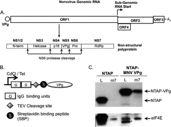
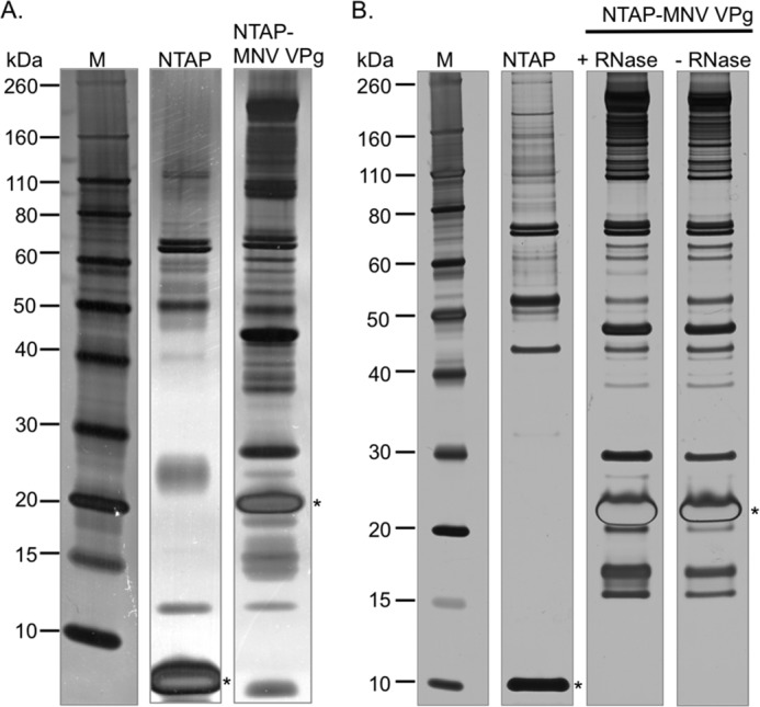
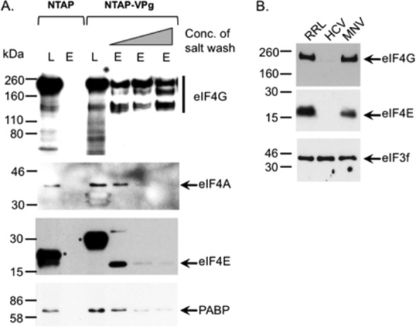
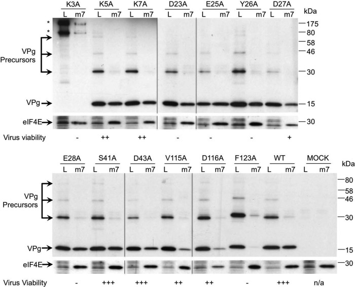
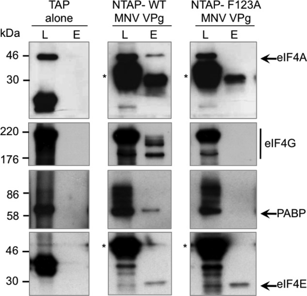
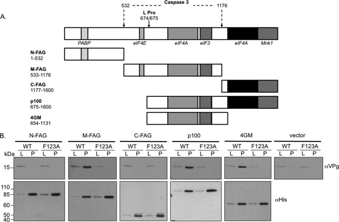
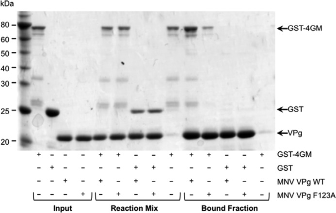
Viruses have evolved a variety of mechanisms to usurp the host cell translation machinery to enable translation of the viral genome in the presence of high levels of cellular mRNAs. Noroviruses, a major cause of gastroenteritis in man, have evolved a mechanism that relies on the interaction of translation initiation factors with the virus-encoded VPg protein covalently linked to the 5' end of the viral RNA. To further characterize this novel mechanism of translation initiation, we have used proteomics to identify the components of the norovirus translation initiation factor complex. This approach revealed that VPg binds directly to the eIF4F complex, with a high affinity interaction occurring between VPg and eIF4G. Mutational analyses indicated that the C-terminal region of VPg is important for the VPg-eIF4G interaction; viruses with mutations that alter or disrupt this interaction are debilitated or non-viable. Our results shed new light on the unusual mechanisms of protein-directed translation initiation.

Keywords: Eukaryotic Translation Initiation Factor 4E (eIF4E); Norovirus; Plus-stranded RNA Virus; Translation; Translation Initiation Factor; VPg; Virus; eIF4G.


Figures

FIGURE 1.
FIGURE 1.
Generation of cell lines expressing TAP-VPg. A, schematic representation of the norovirus genome. The positions of the three major open reading frames (ORF1, -2, and -3) and the murine norovirus-specific open reading frame 4 (ORF4). The position and subgenomic RNA start site and the ORF1 polyprotein cleavage sites are highlighted. The NS1-NS7 nomenclature as proposed by Sosnovtsev (50) has been used to illustrate the identities of the mature cleavage products along with the previously described nomenclature (N-term, helicase, RdRp etc.). B, the domain layout of the TAP-tag is illustrated graphically highlighting the positions of the two protein G IgG binding domains, the tobacco etch virus (TEV) protease cleavage site, and the streptavidin-binding peptide along with the streptavidin binding peptide. C, m7GTP-Sepharose chromatography of cells expressing the NTAP tag or TAP-VPg fusion protein. Lysates from induced cells were incubated with m7GTP-Sepharose to enrich the eIF4F complex. Both lysates (L) and the elutions (m7) from the m7GTP-Sepharose were analyzed by Western blot for eIF4E and the presence of the protein G binding domain present in the TAP tag. An asterisk is used to highlight a TAP-MNV VPg degradation product present in the cell lysate.
FIGURE 2.
FIGURE 2.
Proteomic analysis of the norovirus translation initiation factor complex. Final elutions after the tandem affinity purification procedure are detailed under “Experimental Procedures,” prepared from either cells expressing the NTAP tag alone or the NTAP-VPg fusion protein. Panel A contains the final elutions obtained from the pMEP4-based cadmium chloride inducible expression system, whereas panel B displays the results obtained using the pCDNA4:TO tetracycline inducible system. In the latter case, the purification was performed in the presence and absence of ribonuclease (RNase). An asterisk is used to highlight the position of the bait protein after tobacco etch virus protease digestion and biotin-mediated elution.
FIGURE 3.
FIGURE 3.
VPg interacts with eIF4G. A, cells expressing either the NTAP fusion tag or NTP-MNV VPg were used to perform tandem affinity purification as described under “Experimental Procedures”; however, before biotin elution the samples were washed with buffer containing 125 mm, 500 mm, or 1 m sodium chloride. After biotin elution, samples were concentrated, separated by SDS-PAGE, and then analyzed by Western blotting. Both lysates (L) and the elutions (E) were analyzed for the presence of eIF4G, eIF4A, eIF4A, and PABP. Asterisks are use to highlight the positions of the TAP tag and the TAP-VPg fusion proteins detectable in the lysate due to the presence of protein G binding domains. B, translation initiation complexes were purified from rabbit reticulocyte lysates (RRL) programmed with either the HCV IRES or VPg-linked MNV RNA by RNA-affinity purification as described under “Experimental Procedures.” Samples were subsequently analyzed by Western blot for the presence of various translation initiation factors with a sample of the input rabbit reticulocyte lysates used as a control.
FIGURE 4.
FIGURE 4.
Mutations in the C terminus of VPg affect initiation factor binding and virus viability. BSRT7 cells infected with a fowlpox virus expressing T7 RNA polymerase were transfected with full-length cDNA clones of MNV containing either WT or various VPg mutants. 24 h post transfection cells were lysed, and the resulting lysate (L) was then subjected to m7GTP-Sepharose affinity chromatography. After purification, the proteins associated with m7GTP-Sepharose beads (m7) were separated by SDS-PAGE and analyzed by Western blotting with antisera to MNV VPg and eIF4E. Asterisks highlight the position of high molecular mass VPg-containing precursors formed as a result of incorrect polyprotein processing. Note that all Western blots were performed at the same time, and identical exposures were used to generate the figure shown. The m7GTP-Sepharose data presented is a single representative dataset from at least three independent repeats. The effect of VPg mutations on virus replication was also summarized in this figure. Virus viability is expressed as virus yield 24 h post transfection relative to wild type (+++) as assayed by >5 independent experiments. Typical yields of wild type virus were 1–5 × 10⁴ TCID50 units. −, no virus detected; +, up to 100 TCID50 detected; ++, up to 1000 TCID50; +++, up to WT levels of virus detected, typically >10,000 TCID50 per ml.
FIGURE 5.
FIGURE 5.
The F123A mutation abolishes the VPg-eIF4G interaction. Tandem affinity purification was performed on human 293T cells transiently transfected with plasmids containing the TAP tag alone, TAP-wild type MNV VPg (WT), or TAP-MNV VPg containing the mutation F123A. Samples of the cell lysate (L) or the eluted purified complex (E) were analyzed by Western blot for eIF4A, eIF4G, PABP, and eIF4E. Asterisks are used to highlight the position of the TAP-VPg fusion proteins detected by the binding of the primary antibody to the protein G domains present in the TAP tag.
FIGURE 6.
FIGURE 6.
VPg interacts with the central domain of eIF4G. A, cleavage map of eIF4GI highlighting the positions for translation factors. The arrow and the corresponding numbers illustrate the locations of the individual protease cleavage sites mediated by the cellular proteins caspase 3 or the viral protease Lpro. The expression constructs used in the assay are also illustrated along with the specific amino acids residues encompassed within the construct. B, nickel-affinity purification of His-eIF4G fragments. Cells previously infected with a foxpox virus expressing T7 RNA polymerase were co-transfected with the various eIF4GI expression constructs (or empty vector) and either a wild type murine norovirus cDNA clone (WT) or one containing the F123A mutation in VPg (F123A). Lysates (L) or the purified complex (P) were prepared and used for nickel-affinity purification followed by Western blotting for the presence of wither VPg or the recombinant protein (His).
FIGURE 7.
FIGURE 7.
VPg binds the central domain of eIF4G via a direct protein-protein interaction. The ability of recombinant eIF4G central domain (4GM) to interact with either wild type MNV VPg (WT) or the F123A MNV VPg mutant was examined using a His-tag pulldown assay. Recombinant GST or GST-4GM was mixed with purified His-tagged WT or F123A MNV VPg. and the resulting complex was purified using cobalt affinity chromatography. Samples of the purified proteins (Input), the mixtures of the proteins before purification (Reaction mix), and the final bound fraction were separated by SDS-PAGE and analyzed by Coomassie Blue staining.
FIGURE 8.
FIGURE 8.
Reduced levels of eIF4E and inhibition of the eIF4E-4G interaction are not required for efficient norovirus replication. A, MNV permissive microglial cells BV2 were transiently transfected with either an empty plasmid vector (EMP) or plasmids expressing HA-tagged derivatives of wild type (WT) or a non-phosphorylatable mutant of 4E-BP1 (MUT). Cells were then infected with MNV1 at a multiplicity of infection of 3 TCID50/cell, and viral protein expression as well as virus titer (B) was determined at various times post infection. C, BV2 cells transfected with either non-targeting siRNA or an siRNA directed against eIF4E were infected with MNV1 at a multiplicity of infection of 10 TCID50/cell, and the effect on viral titer examined at various times post infection (D).
FIGURE 9.
FIGURE 9.
eIF4G is required for efficient norovirus translation and replication. HEK 293T cells were transfected with specific eIF4GI siRNAs or a nonspecific (NS) siRNA. The cells were re-transfected with the siRNA at 24 hours post-transfection (h.p.t.) to improve the knockdown efficiency. Thereafter, the cells were transfected with MNV VPg-linked RNA. Western blots were performed on lysates harvested at 11 and 16 hours post transfection of RNA to examine the efficiency of eIF4GI silencing and the translation of NS7 and GAPDH. The numbers below the figures represent the percentage of protein expression relative to the control, which were quantified by densitometry using ImageJ. The yield of virus was determined in RAW 264.7 cells from two independent experiments, expressed as TCID50/ml and compared with NS control. Transfections were performed in triplicate, and the error bar indicates the S.E. Statistical significance was determined by one-way analysis of variance and is represented by the p values. p < 0.001 (***).
FIGURE 10.
FIGURE 10.
The central domain of eIF4G is sufficient for norovirus translation. 293T cells transfected with siRNA directed against eIF4G (4GI) or non-specific siRNA (NS) were co-transfected with plasmids encoding either empty vector (pCDNA) or the minimal VPg binding domain from eIF4G (pCDNA 4GM). A, Western blots showing successful knockdown of 4G and reconstitution of viral protein expression upon co-transfection with FLAG-tagged 4GM. B, the ability of 4GM to reconstitute viral protein expression in eIF4G depleted cells is represented as % reconstitution, relative to a nonspecific siRNA-treated control as described under “Experimental Procedures.” Quantification was performed on a Li-Cor Odyssey imager on data obtained from quadruplicate independent biological samples. Error bars represent S.E. with significance determined by one-way analysis of variance (****, p = <0.0001).

Similar articles

Cited by

References

    1. Reineke L. C., Lloyd R. E. (2011) Animal virus schemes for translation dominance. Curr. Opin. Virol. 1, 363–372 - PMC - PubMed
    1. Firth A. E., Brierley I. (2012) Non-canonical translation in RNA viruses. J. Gen. Virol. 93, 1385–1409 - PMC - PubMed
    1. Gebauer F., Hentze M. W. (2004) Molecular mechanisms of translational control. Nat. Rev. Mol. Cell Biol. 5, 827–835 - PMC - PubMed
    1. Lamphear B. J., Yan R., Yang F., Waters D., Liebig H. D., Klump H., Kuechler E., Skern T., Rhoads R. E. (1993) Mapping the cleavage site in protein synthesis initiation factor eIF-4γ of the 2A proteases from human coxsackievirus and rhinovirus. J. Biol. Chem. 268, 19200–19203 - PubMed
    1. Mader S., Lee H., Pause A., Sonenberg N. (1995) The translation initiation factor eIF-4E binds to a common motif shared by the translation factor eIF-4γ and the translational repressors 4E-binding proteins. Mol. Cell. Biol. 15, 4990–4997 - PMC - PubMed

Publication types