. 2014 Jul 25;289(30):20422-33.
doi: 10.1074/jbc.M114.559054.

Biochemical and spatial coincidence in the provisional Ser/Thr protein kinase interaction network of Mycobacterium tuberculosis

Biochemical and spatial coincidence in the provisional Ser/Thr protein kinase interaction network of Mycobacterium tuberculosis

Christina E Baer et al. J Biol Chem. .

Abstract

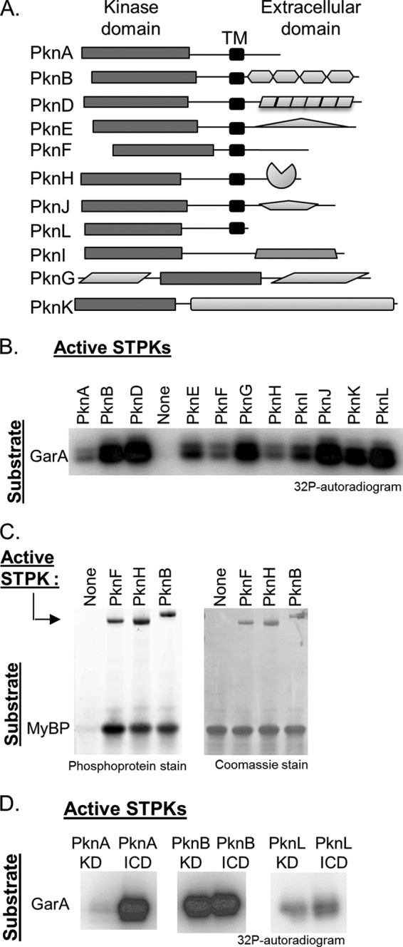
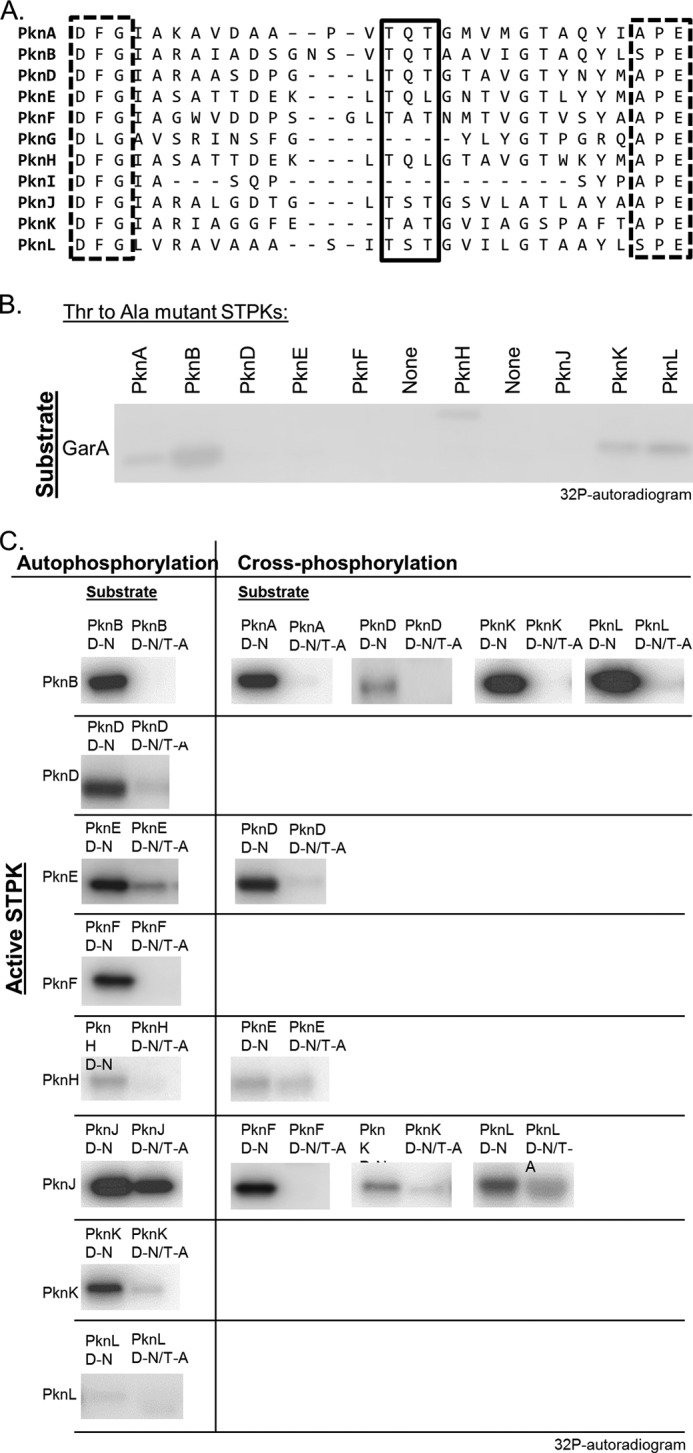
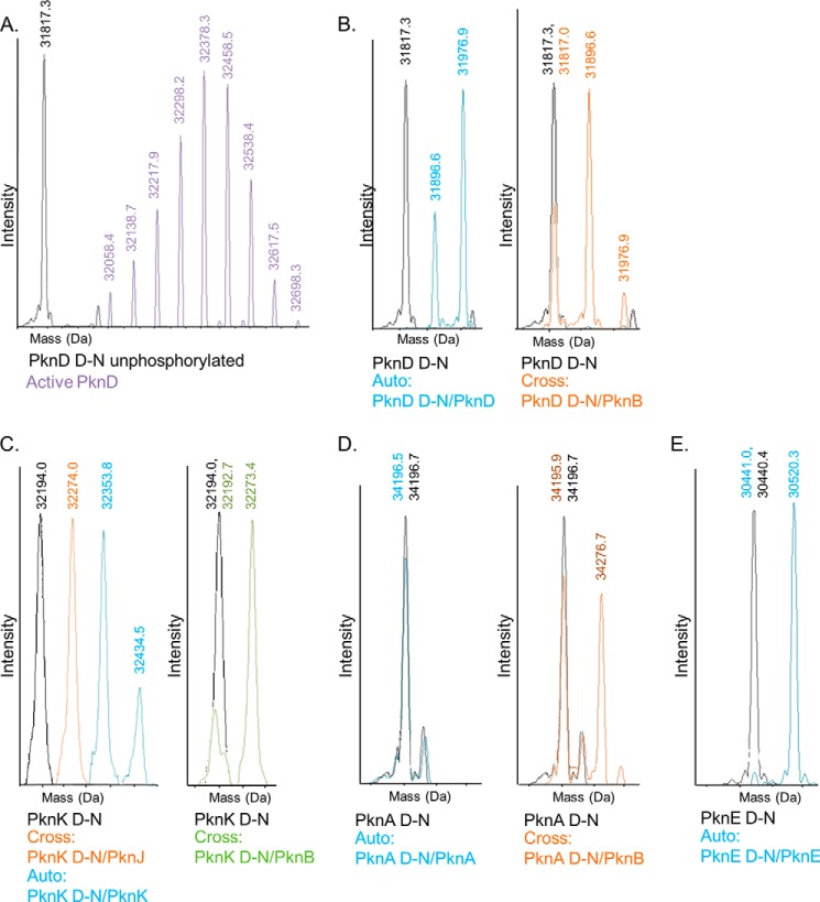
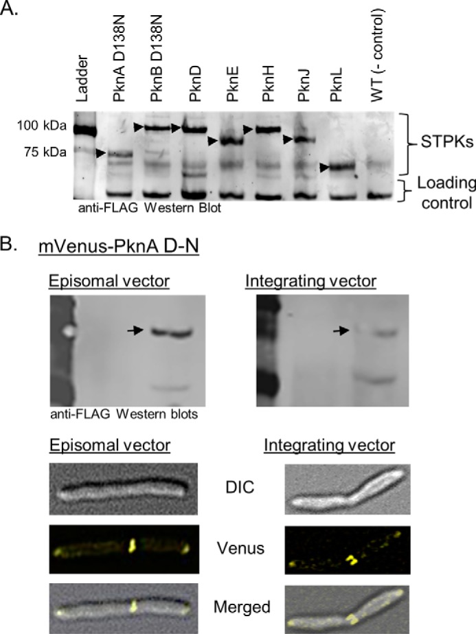
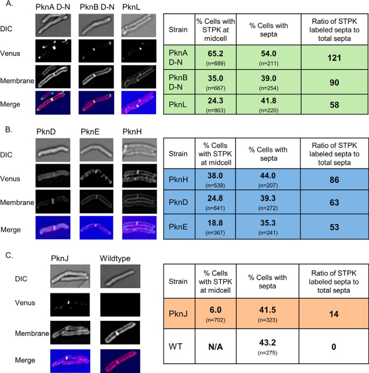
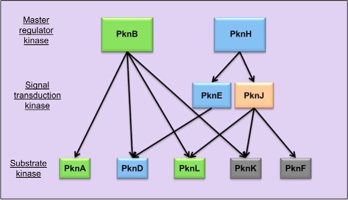
Many Gram-positive bacteria coordinate cellular processes by signaling through Ser/Thr protein kinases (STPKs), but the architecture of these phosphosignaling cascades is unknown. To investigate the network structure of a prokaryotic STPK system, we comprehensively explored the pattern of signal transduction in the Mycobacterium tuberculosis Ser/Thr kinome. Autophosphorylation is the dominant mode of STPK activation, but the 11 M. tuberculosis STPKs also show a specific pattern of efficient cross-phosphorylation in vitro. The biochemical specificity intrinsic to each kinase domain was used to map the provisional signaling network, revealing a three-layer architecture that includes master regulators, signal transducers, and terminal substrates. Fluorescence microscopy revealed that the STPKs are specifically localized in the cell. Master STPKs are concentrated at the same subcellular sites as their substrates, providing additional support for the biochemically defined network. Together, these studies imply a branched functional architecture of the M. tuberculosis Ser/Thr kinome that could enable horizontal signal spreading. This systems-level approach provides a biochemical and spatial framework for understanding Ser/Thr phospho-signaling in M. tuberculosis, which differs fundamentally from previously defined linear histidine kinase cascades.


Figures

FIGURE 1.
FIGURE 1.
11 M. tuberculosis STPKs display trans-phosphorylation activity on multiple substrates. A, domain architecture of the 11 M. tuberculosis STPKs. Soluble, active KDs were obtained for PknA, PknB, PknD, PknE, PknF, PknH, PknJ, PknK, and PknL. PknG and PknI are only active as full-length soluble enzymes. Shapes indicate predicted or experimentally verified folded domains (42). TM, transmembrane. B, autoradiogram showing the reactions of the 11 STPK constructs incubated with the substrate protein GarA and [γ-32P]ATP (top). The STPK constructs phosphorylate GarA. C, PknF and PknH efficiently phosphorylate the model substrate MyBP in comparison with PknB. Phosphoproteins were visualized using Pro-Q Diamond Phosphoprotein Gel Stain. Active STPK autophosphorylation is visible (upper bands) as is trans-phosphorylation on the MyBP substrate; equal protein loading was confirmed by Coomassie stain. D, trans-phosphorylation activity of KD and full-length ICD constructs was compared using GarA as a substrate. The autoradiogram confirms that the PknA ICD is more active than the PknA KD as reported previously (38), but that activity does not differ for the two PknB and PknL constructs.
FIGURE 2.
FIGURE 2.
M. tuberculosis STPKs display specific intermolecular phosphorylation patterns. A, autoradiograms showing the reactions of each active STPK construct (top) with the 11 inactivated substrate kinases (left). Each inactive Asp-to-Asn mutant kinase construct was incubated with each of the 11 M. tuberculosis STPKs in [γ-32P]ATP transfer assays. His-MBP tags on the active kinases and tagless inactive kinases enable separation of the proteins in each reaction by SDS-PAGE. Assays were imaged by autoradiography in parallel. Products on the diagonal reflect efficient autophosphorylation. Off-diagonal bands in each column indicate cross-phosphorylation. B, PknA ICD was tested against the 11 Asp-to-Asn mutant kinases. No additional inter-kinase interactions were observed. C, PknG and PknI KDs were not phosphorylated by any of the other kinases. The full-length PknG and PknI constructs are cross-phosphorylated by multiple kinases, presumably on sites outside of the catalytic domain. D, autoradiographs were quantified in ImageJ and normalized to the autophosphorylation signal observed for each kinase. PknA and PknL do not efficiently autophosphorylate; therefore, these two kinases are not included in this graph. The PknH-PknE interaction was the lowest detected by LC-MS and was used as the lower boundary. Low intensity interactions at or below this cutoff are depicted by gray columns.
FIGURE 3.
FIGURE 3.
M. tuberculosis STPK interactions are substrate-specific. Inter-kinase phosphorylation occurs in a substrate-specific pattern and is not due to differences in enzyme activity. PknB, PknD, and PknH, three STPKs with differing activity, were incubated with the PknB and PknD Asp-to-Asn mutant kinases. Time points taken at 30, 60, and 120 min and 24 h after reaction initiation display the same relative inter-kinase phosphorylation patterns (lower band) when Western-blotted with a phosphothreonine antibody. Active STPK autophosphorylation was similar at each time point (upper bands). Coomassie-stained gels demonstrate equal substrate protein concentrations in all reactions.
FIGURE 4.
FIGURE 4.
Activation loop threonines are the primary sites of intermolecular phosphorylation. A, sequence alignment (47) of the M. tuberculosis STPK activation loop region highlights the conserved Thr residues (boxed). B, mutation of the conserved activation loop threonines in nine of the STPKs markedly reduces or abolishes phospho-transfer activity. Reactions and autoradiography were performed in parallel and under identical conditions to active control kinase in Fig. 1B. C, autoradiograms showing representative autophosphorylation (left) and cross-phosphorylation (right) reactions of substrate (D-N) and activation loop double Thr mutants (D-N/T-A). The activation loop substitutions reduced or abolished phosphorylation.
FIGURE 5.
FIGURE 5.
Inter-STPK phosphorylation is highly site-specific. Mass spectra of STPK constructs following auto- and cross-phosphorylation reactions. The unphosphorylated substrate (D-N) mass distribution is shown in black in each panel. A mass increase of 80 Da is equivalent to one phosphoryl group. The reactions are labeled with the substrate (D-N)/active STPK constructs. A, compared with the unmodified substrate PknD KD, the wild-type, active PknD KD purified from E. coli is a heterogeneous mixture of autophosphorylation states, ranging from 3 to 11 phosphates. B, PknD KD autophosphorylates PknD D-N at one or two sites (blue). PknB cross-phosphorylates PknD D-N at 0–2 sites (orange). C, PknK KD autophosphorylates 2–3 sites (left, blue). The PknJ (left, orange) and PknB (right, green) KDs cross-phosphorylate PknK D-N at one site. D, PknA KD does not autophosphorylate (left), but is cross-phosphorylated on one site by PknB (middle, orange). E, PknE KD autophosphorylates in trans at a single site.
FIGURE 6.
FIGURE 6.
Expression of FLAG-mVenus-STPK reporter fusions in M. smegmatis. A, anti-FLAG Western blot of M. smegmatis lysates containing each indicated full-length STPK fused to FLAG-mVenus expressed in the pDE43-MEK episomal vector. The common band at the bottom of the blot serves as a loading control. Arrows indicate mVenus-kinase fusions. B, Western blots (top) and microscopy (bottom) that compare protein expression levels for the FLAG-Venus-PknA D-N reporter construct in an episomal (left) and integrating vector (right). Western blots were exposed in parallel. Differences in expression level do not change the apparent localization at the poles and septum. DIC, differential interference contrast.
FIGURE 7.
FIGURE 7.
Subcellular localization of M. tuberculosis STPKs in M. smegmatis. Differential interference contrast (DIC), fluorescence (mVenus and FM4-64) microscopy and merged images for the M. smegmatis strains carrying mVenus fusions of the indicated full-length STPKs. Tables present statistical analyses of kinase localization. The STPKs localize in three distinct patterns in vivo. A, PknA, PknB, and PknL localize to the poles and midplane (green). The individual kinases persist at the midplane/septum for varying lengths of time throughout the cell cycle, as represented by the percentage of cells with a fluorescent signal at the septa compared with the cells without visible septa. FM4-64 staining for septa confirms that, with the exception of mVenus-PknA D-N, expression of the STPKs does not alter the cell cycle. B, PknD, PknE, and PknH localize to the peripheral membrane and the midplane (blue). C, PknJ is only found at the midplane of a small percentage of cells (orange).
FIGURE 8.
FIGURE 8.
Provisional M. tuberculosis STPK interaction network. Arrows indicate intermolecular phosphorylation in vitro. STPKs with polar and midplane localization are depicted in green. STPKs with membrane and midplane localization are colored blue. PknJ, localizing only transiently to the septa, is colored orange. PknF and PknK, depicted in gray, were characterized biochemically, but expression of the mVenus fusions in M. smegmatis was not detected. The master STPKs, PknB and PknH, only undergo autophosphorylation and contain folded extracellular domains that receive environmental signals. In contrast, PknA and PknL not only fail to autophosphorylate, they also lack folded extracellular receptor domains. However, PknB efficiently phosphorylates the PknL and PknA KDs, and these three kinases co-localize in vivo. PknH localizes along the cell membrane and also initiates a cascade of phosphorylation of other STPKs, PknE and PknD, that also reside along the cell membrane. PknD contains a folded extracellular domain, but phosphorylation by other kinases suggests that it may be activated by other kinases as well as external signals that trigger autophosphorylation.

References

    1. Wehenkel A., Bellinzoni M., Graña M., Duran R., Villarino A., Fernandez P., Andre-Leroux G., England P., Takiff H., Cerveñansky C., Cole S. T., Alzari P. M. (2008) Mycobacterial Ser/Thr protein kinases and phosphatases: physiological roles and therapeutic potential. Biochim. Biophys. Acta 1784, 193–202 - PubMed
    1. Skerker J. M., Prasol M. S., Perchuk B. S., Biondi E. G., Laub M. T. (2005) Two-component signal transduction pathways regulating growth and cell cycle progression in a bacterium: a system-level analysis. PLoS Biol. 3, e334. - PMC - PubMed
    1. Chang L., Karin M. (2001) Mammalian MAP kinase signalling cascades. Nature 410, 37–40 - PubMed
    1. Cole S. T., Brosch R., Parkhill J., Garnier T., Churcher C., Harris D., Gordon S. V., Eiglmeier K., Gas S., Barry C. E., 3rd, Tekaia F., Badcock K., Basham D., Brown D., Chillingworth T., Connor R., Davies R., Devlin K., Feltwell T., Gentles S., Hamlin N., Holroyd S., Hornsby T., Jagels K., Krogh A., McLean J., Moule S., Murphy L., Oliver K., Osborne J., Quail M. A., Rajandream M. A., Rogers J., Rutter S., Seeger K., Skelton J., Squares R., Squares S., Sulston J. E., Taylor K., Whitehead S., Barrell B. G. (1998) Deciphering the biology of Mycobacterium tuberculosis from the complete genome sequence. Nature 393, 537–544 - PubMed
    1. Fernandez P., Saint-Joanis B., Barilone N., Jackson M., Gicquel B., Cole S. T., Alzari P. M. (2006) The Ser/Thr protein kinase PknB is essential for sustaining mycobacterial growth. J. Bacteriol. 188, 7778–7784 - PMC - PubMed

Publication types

MeSH terms

Substances

LinkOut - more resources