. 2014 Jul;42(12):7762-75.
doi: 10.1093/nar/gku474. Epub 2014 Jun 13.

PARP-2 and PARP-3 are selectively activated by 5' phosphorylated DNA breaks through an allosteric regulatory mechanism shared with PARP-1

Affiliations

PARP-2 and PARP-3 are selectively activated by 5' phosphorylated DNA breaks through an allosteric regulatory mechanism shared with PARP-1

Marie-France Langelier et al. Nucleic Acids Res. 2014 Jul.

Abstract

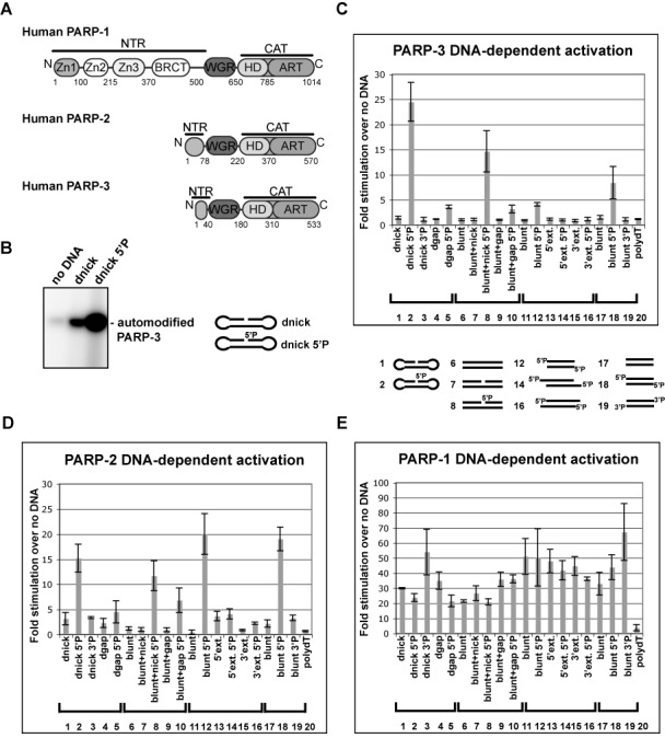
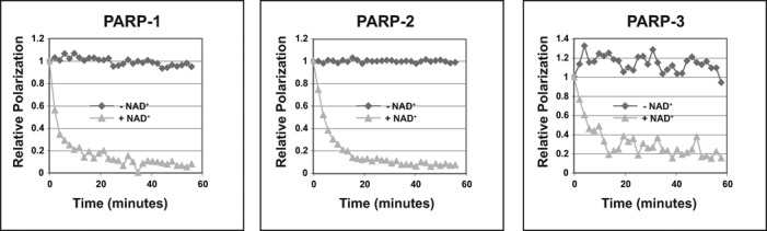
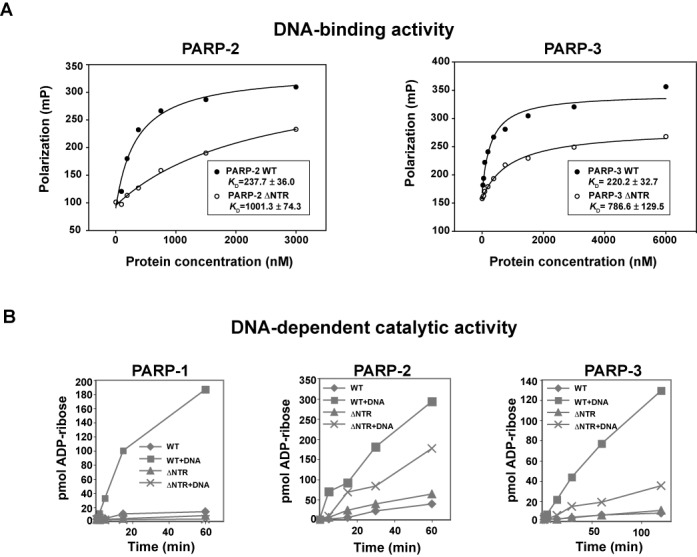
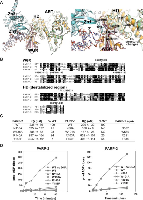
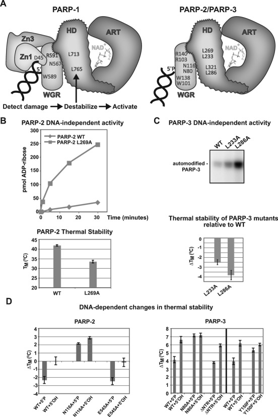
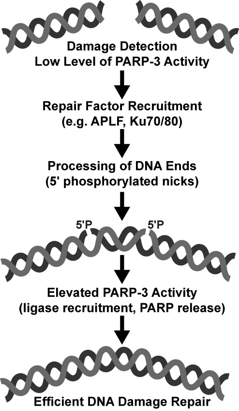
PARP-1, PARP-2 and PARP-3 are DNA-dependent PARPs that localize to DNA damage, synthesize poly(ADP-ribose) (PAR) covalently attached to target proteins including themselves, and thereby recruit repair factors to DNA breaks to increase repair efficiency. PARP-1, PARP-2 and PARP-3 have in common two C-terminal domains-Trp-Gly-Arg (WGR) and catalytic (CAT). In contrast, the N-terminal region (NTR) of PARP-1 is over 500 residues and includes four regulatory domains, whereas PARP-2 and PARP-3 have smaller NTRs (70 and 40 residues, respectively) of unknown structural composition and function. Here, we show that PARP-2 and PARP-3 are preferentially activated by DNA breaks harboring a 5' phosphate (5'P), suggesting selective activation in response to specific DNA repair intermediates, in particular structures that are competent for DNA ligation. In contrast to PARP-1, the NTRs of PARP-2 and PARP-3 are not strictly required for DNA binding or for DNA-dependent activation. Rather, the WGR domain is the central regulatory domain of PARP-2 and PARP-3. Finally, PARP-1, PARP-2 and PARP-3 share an allosteric regulatory mechanism of DNA-dependent catalytic activation through a local destabilization of the CAT. Collectively, our study provides new insights into the specialization of the DNA-dependent PARPs and their specific roles in DNA repair pathways.


Figures

Figure 1.
Figure 1.
PARP-2 and PARP-3 are selectively activated by 5′ phosphorylated DNA breaks. (A) Domain architecture of PARP-1, PARP-2 and PARP-3. The WGR and CAT domains are conserved, while the N-terminal regions (NTRs) are distinct. (B) Radioactive assay showing PARP-3 automodification activity in the absence or presence of DNA. Protein (1.5 μM) and DNA (2.4 μM) were incubated for 1 h in the presence of 25 μM NAD+ (5 μM 32P-NAD+, 20 μM NAD+). dnick and dnick 5′P are dumbbell templates containing a region of 19 base pairs (bp) with a 5′ phosphorylated or non-phosphorylated nick after bp 10 and 4 nucleotide turns at the extremities. (C) Colorimetric assay showing stimulation of PARP-3 DNA-dependent activity by a panel of DNA structures (60 nM protein, 480 nM DNA, 1 h time point). Stimulation is calculated as the ratio of activity measured in the presence versus absence of DNA. The average of three independent experiments is shown with associated standard deviations. Templates 1–5 are dumbbells derived from the dnick template described in (B). The dnick 3′P has a phosphate on the 3′ terminus. The dgap templates have a one-nucleotide gap instead of the nick. Templates 6–10 are 47 bp duplexes (blunt), with either a nick after bp 24 (blunt + nick) or a one-nucleotide gap (blunt + gap). Templates 11–16 are based on a 26-bp palindromes (blunt), with a two-nucleotide 5′ or 3′ extension (5′ext. or 3′ext.). Templates 17–19 are 28 duplexes template with either a 5′ OH (blunt), a 5′P (blunt 5′P) or a 3′P (blunt 3′P). Template 20 is a single-stranded DNA containing 16 dTs. See Supplementary Figure S1 for more details on DNA templates. (D and E) Same as (C) for PARP-2 and PARP-1 with a 15-min time point.
Figure 2.
Figure 2.
PARP-1, PARP-2 and PARP-3 release from DNA upon production of PAR. Fluorescence polarization release assay. Saturating amount of proteins were incubated with a mixture of unlabeled and labeled DNA probe and incubated for 30 min at RT. PARP-1: 200 nM protein, 100 nM DNA (28-bp duplex 5′P, template 18). PARP-2: 2.5 μM protein, 1.25 μM DNA (28-bp duplex 5′P, template 18). PARP-3: 4.0 μM protein and 1 μM DNA (47-bp duplex with 5′P nick, template 8). 1 mM NAD+ was added to start the ADP-ribosylation reaction and fluorescence polarization was measured over a time course. Relative polarization represents the ratio of the polarization measured at time × over the polarization measured at time zero, which was set to one. Representative curves of three replicates are shown.
Figure 3.
Figure 3.
The NTRs of PARP-2 and PARP-3 are not strictly required for DNA binding and activation. (A) Fluorescence polarization DNA binding experiment for PARP-2 WT, PARP-3 WT and their respective NTR deletions using a fluorescently labeled 5′ phosphorylated DNA probe (5 nM). PARP-2 assay was performed using the 28-bp duplex 5′P (template 18). PARP-3 assay was performed using the 47-bp duplex with 5′P nick (template 8). The KD is an average of three independent experiments with associated standard deviation. (B) Colorimetric assay showing the activity of ΔNTR-PARP-1, ΔNTR-PARP-2 and ΔNTR-PARP-3 compared to WT using 60 nM protein and 480 nM DNA. PARP-2 assay was performed using the 28-bp duplex 5′P (template 18). PARP-1 and PARP-3 assays used the dnick 5′P template (template 2). Representative curves of the three replicates are shown.
Figure 4.
Figure 4.
Structure-guided mutagenesis of the PARP-2 and PARP-3 WGR domains. (A) Structural alignment between the NMR structure of the PARP-3 WGR domain (PDB: 2EOC; magenta), a crystal structure of the PARP-3 CAT (HD/ART) (PDB: 3C49; green) and the PARP-1/DNA crystal structure (PDB: 4DQY; Zn1 and Zn3 in teal, WGR-CAT in orange). (B) Sequence alignment of human PARP-1, PARP-2 and PARP-3 WGR domain, and a region of the HD. Residues targeted for mutagenesis are marked with an asterisk. (C) The DNA binding affinity of PARP-2 and PARP-3 WGR mutants was determined by fluorescence polarization. The KD reported represents an average of three independent experiments with associated standard deviation. Examples of binding curves are shown in Supplementary Figure S4. The PARP-2 assay used the fluorescently labeled 28-bp duplex 5′P (template 18, 5 nM). The PARP-3 assay used the fluorescently labeled 47-bp duplex with 5′P nick (template 8, 5 nM). (D) Colorimetric assay showing the activity of PARP-2 and PARP-3 WGR mutants using 60 nM protein, 480 nM DNA (PARP-2: 28-bp duplex 5′P, template 18; PARP-3: dnick 5′P, template 2). Representative curves of the three replicates are shown.
Figure 5.
Figure 5.
PARP-2 and PARP-3 share with PARP-1 a conserved mechanism of activation through HD destabilization. (A) Model for PARP-1, PARP-2 and PARP-3 DNA-dependent allosteric mechanism of activation. Left, PARP-1 regulatory domains (Zn1, Zn3, WGR) collapse onto DNA and form interdomain contacts that create destabilizing changes in the HD of the CAT and lead to ART activation. Right, our results suggest that the same mechanism of activation exists for PARP-2 and PARP-3; however, the WGR is their primary regulatory domain. Mutation of specific residues in the HD hydrophobic core mimic the effect of DNA binding by decreasing thermal stability and increasing catalytic activity. Residues that are involved in critical protein–protein or protein–DNA interactions are indicated (PARP-2 residues listed above PARP-3 residues). (B) Top: DNA-independent automodification activity of PARP-2 L269A mutant compared to PARP-2 WT using the colorimetric assay (60 nM protein, 25 μM NAD+). Bottom: thermal stability of PARP-2 WT and HD mutant L269A determined by DSF (5 μM protein). The TM represents the average of three independent experiments. The error bars represent standard deviations. (C) Top: DNA-independent automodification activity of PARP-3 HD mutants using the radioactive assay with 1.5 μM PARP-3 WT or mutants and 2.5 μM NAD+ (2.0 μM NAD+: 0.5 μM NAD+ 32P). Bottom: relative thermal stability of PARP-3 HD mutants determined by DSF (5 μM protein). The average of three independent experiments are shown with associated standard deviations. (D) Thermal stability of PARP-2 and PARP-3 WT or mutants in the presence of DNA (5 μM protein, 2.5 μM DNA). The ΔTM represents the difference between the average TM obtained in the absence of DNA (three replicates with associated standard deviations) and the average TM obtained in the presence of DNA (three replicates with associated standard deviations) (PARP-2: 28-bp duplex 5′P; PARP-3: 47-bp duplex with 5′P nick).
Figure 6.
Figure 6.
Model for PARP-3 activation during NHEJ. In the proposed model, a low level of PARP-3 activation after damage detection contributes to the recruitment of repair machinery. The DNA ends are processed during repair leading to the final substrate for DNA ligation, a phosphorylated nick (5′P). An elevated level of PARP-3 activation in the presence of the phosphorylated break could then act to recruit the ligase complex for the end-joining reaction, or could release PARP-3 so that DNA ligase can access the break. Based on our biochemical analysis, PARP-2 could function similarly in other repair pathways.

References

    1. D'Amours D., Desnoyers S., D'Silva I., Poirier G.G. Poly(ADP-ribosyl)ation reactions in the regulation of nuclear functions. Biochem. J. 1999;342:249–268. - PMC - PubMed
    1. Ame J.C., Spenlehauer C., de Murcia G. The PARP superfamily. Bioessays. 2004;26:882–893. - PubMed
    1. Hottiger M.O., Hassa P.O., Luscher B., Schuler H., Koch-Nolte F. Toward a unified nomenclature for mammalian ADP-ribosyltransferases. Trends Biochem. Sci. 2010;35:208–219. - PubMed
    1. Kleine H., Poreba E., Lesniewicz K., Hassa P.O., Hottiger M.O., Litchfield D.W., Shilton B.H., Luscher B. Substrate-assisted catalysis by PARP10 limits its activity to mono-ADP-ribosylation. Mol. Cell. 2008;32:57–69. - PubMed
    1. Ame J.C., Rolli V., Schreiber V., Niedergang C., Apiou F., Decker P., Muller S., Hoger T., Menissier-de Murcia J., de Murcia G. PARP-2, A novel mammalian DNA damage-dependent poly(ADP-ribose) polymerase. J. Biol. Chem. 1999;274:17860–17868. - PubMed

Publication types

MeSH terms