. 2014 Aug;80(16):5086-97.
doi: 10.1128/AEM.01296-14. Epub 2014 Jun 13.

Identification of Mn(II)-oxidizing bacteria from a low-pH contaminated former uranium mine

Affiliations

Identification of Mn(II)-oxidizing bacteria from a low-pH contaminated former uranium mine

Denise M Akob et al. Appl Environ Microbiol. 2014 Aug.

Abstract

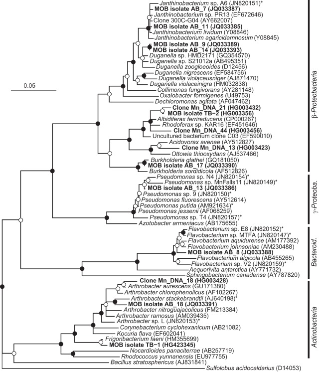
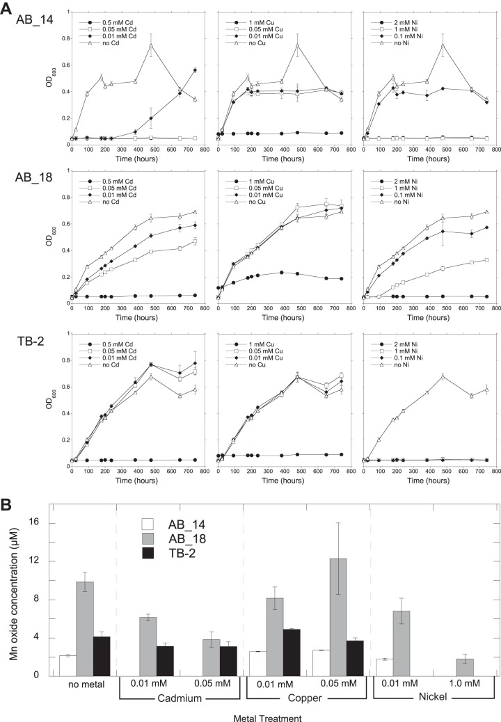
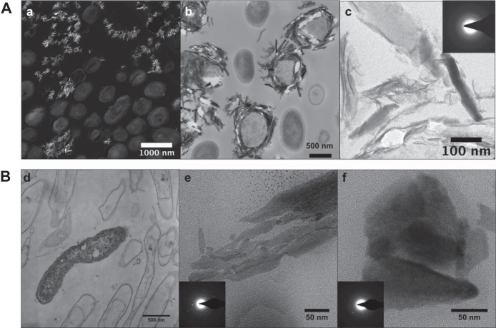
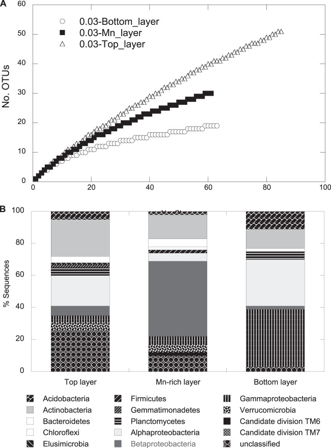
Biological Mn oxidation is responsible for producing highly reactive and abundant Mn oxide phases in the environment that can mitigate metal contamination. However, little is known about Mn oxidation in low-pH environments, where metal contamination often is a problem as the result of mining activities. We isolated two Mn(II)-oxidizing bacteria (MOB) at pH 5.5 (Duganella isolate AB_14 and Albidiferax isolate TB-2) and nine strains at pH 7 from a former uranium mining site. Isolate TB-2 may contribute to Mn oxidation in the acidic Mn-rich subsoil, as a closely related clone represented 16% of the total community. All isolates oxidized Mn over a small pH range, and isolates from low-pH samples only oxidized Mn below pH 6. Two strains with different pH optima differed in their Fe requirements for Mn oxidation, suggesting that Mn oxidation by the strain found at neutral pH was linked to Fe oxidation. Isolates tolerated Ni, Cu, and Cd and produced Mn oxides with similarities to todorokite and birnessite, with the latter being present in subsurface layers where metal enrichment was associated with Mn oxides. This demonstrates that MOB can be involved in the formation of biogenic Mn oxides in both moderately acidic and neutral pH environments.


Figures

FIG 1
FIG 1
Neighbor-joining phylogenetic tree of 16S rRNA gene sequences of Mn(II)-oxidizing bacterial isolates and related clone sequences obtained from Mn-rich soil (indicated by boldface type). Isolates are designated by MOB followed by the strain number; clones are designated by Mn_DNA followed by the clone number. Known MOB are designated with an asterisk. Sulfolobus acidocaldarius was used as the outgroup. Branch points supported by bootstrap resampling (1,000 replicates) are indicated by filled circles (bootstrap values of >90%) and open circles (bootstrap values of >50%). Bar, 0.05 change per nucleotide position. Bacteroid., Bacteroidetes; γ-Proteob., Gammaproteobacteria.
FIG 2
FIG 2
Growth of (A) and Mn oxide production by (B) strains AB_14, AB_18, and TB-2 in the presence of various concentrations of Cd, Cu, and Ni. Growth over time was monitored by OD600 measurements of isolates cultured in liquid medium (AB_14 and TB-2 at pH 5.5, AB_18 at pH 7). Mn oxides were measured using the LBB colorimetric assay after 742 h of growth. All values are means of triplicate cultures minus the concentrations of control cultures (uninoculated media) ± standard deviations. Omitted values indicate no growth under the experimental conditions.
FIG 3
FIG 3
Elemental distributions of strains AB_14 (pH 5.5) (A) and AB_9 (pH 7) (B) grown in BM medium with or without iron, determined using LA-ICP-MS. The graphs show the signal intensities of Mn depending on the position of bacterial biomass on the agar plate (gray bars) and the availability of Fe. The intensities were normalized to an internal standard (carbon).
FIG 4
FIG 4
TEM images of AB_18 and AB_14 cells and Mn oxides. (A) AB_18. (a) STEM high-angle annular dark-field (HAADF) image (the brighter areas indicate higher Z elements); (b) TEM image of Mn oxide minerals surrounding some cells; (c) TEM image of a cluster of minerals and their selected area diffraction patterns, showing some crystallinity (inset). (B) AB_14. TEM images with diffraction patterns (insets) of live and mostly dead bacteria with few obvious minerals around them (d) or aggregates of Mn oxide particles (e and f). Diffraction areas were larger than the images shown.
FIG 5
FIG 5
Rarefaction curves (A) and frequencies of bacterial phylogenetic lineages (B) for the 16S rRNA gene clone libraries obtained for the three subsurface layers, Mn-rich, top, and bottom at the GTF site. A total of 85, 61, and 63 clones were analyzed for the top, Mn-rich, and bottom layers, respectively. Phylogenetic affiliations of OTUs (based on a >97% sequence similarity cutoff) were determined by using mothur against the SILVA database. Frequencies were based on the total number of clones associated with OTUs of sequenced representatives at the phylum level or class level for Proteobacteria.

Similar articles

Cited by

References

    1. Morgan JJ. 2005. Kinetics of reaction between O2 and Mn(II) species in aqueous solutions. Geochim. Cosmochim. Acta 69:35–48. 10.1016/j.gca.2004.06.013 - DOI
    1. Tebo BM, Bargar JR, Clement BG, Dick GJ, Murray KJ, Parker D, Verity R, Webb SM. 2004. Biogenic manganese oxides: properties and mechanisms of formation. Annu. Rev. Earth Planet. Sci. 32:287–328. 10.1146/annurev.earth.32.101802.120213 - DOI
    1. Tebo B, Clement B, Dick G. 2007. Biotransformations of manganese, p 1223–1238 In Hurst CJ. (ed), Manual of environmental microbiology, 3rd ed. ASM Press, Washington, DC
    1. Geszvain K, Butterfield C, Davis R, Madison A, Lee S, Parker D, Soldatova A, Spiro T, Luther G, Tebo B. 2012. The molecular biogeochemistry of manganese(II) oxidation. Biochem. Soc. Trans. 40:1244–1248. 10.1042/BST20120229 - DOI - PubMed
    1. Nealson KH. 2006. The manganese-oxidizing bacteria, p 222–231 In Balows A, Trüper HG, Dworkin M, Harder W, Schleifer K-H. (ed), The prokaryotes. Springer, New York, NY

Publication types

LinkOut - more resources