. 2014 Aug;80(16):5068-77.
doi: 10.1128/AEM.01039-14. Epub 2014 Jun 13.

Epithelial adhesion mediated by pilin SpaC is required for Lactobacillus rhamnosus GG-induced cellular responses

Affiliations

Epithelial adhesion mediated by pilin SpaC is required for Lactobacillus rhamnosus GG-induced cellular responses

Courtney S Ardita et al. Appl Environ Microbiol. 2014 Aug.

Abstract

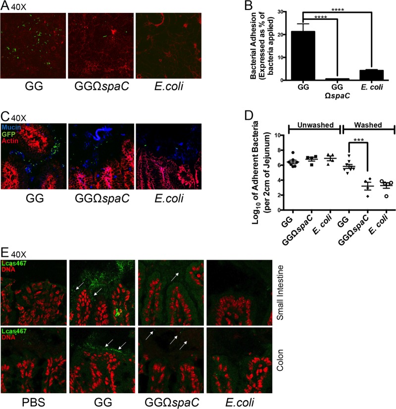
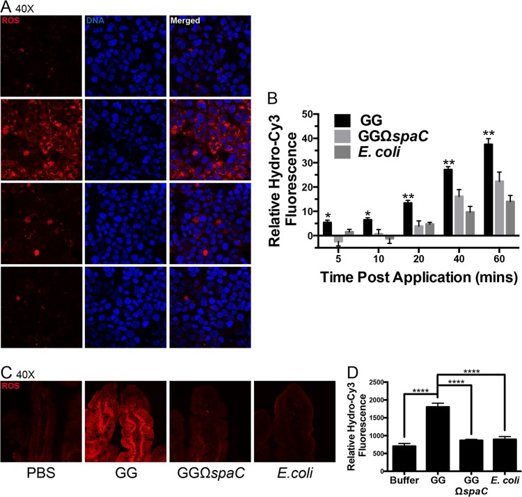
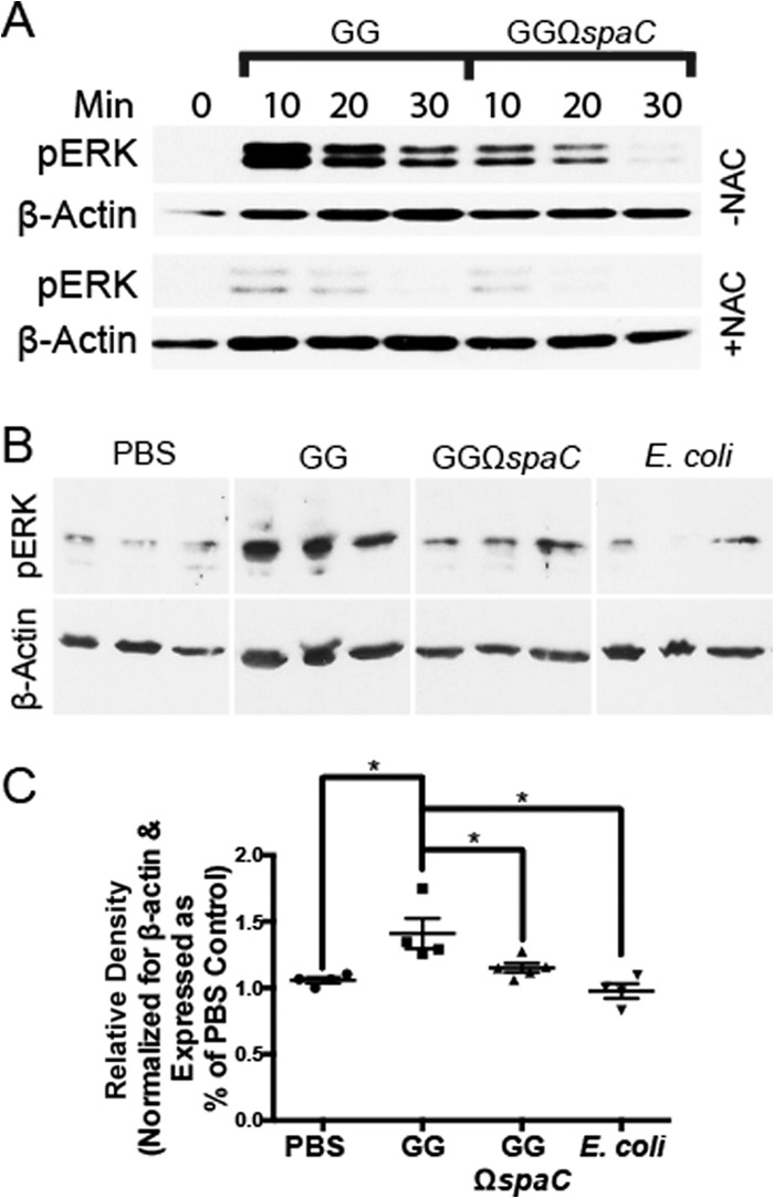
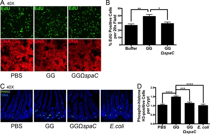
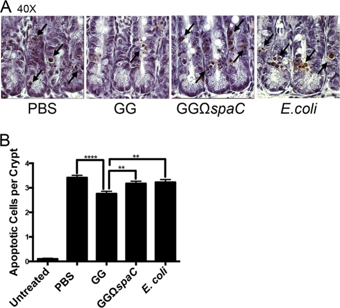
Lactobacillus rhamnosus GG is a widely used probiotic, and the strain's salutary effects on the intestine have been extensively documented. We previously reported that strain GG can modulate inflammatory signaling, as well as epithelial migration and proliferation, by activating NADPH oxidase 1-catalyzed generation of reactive oxygen species (ROS). However, how strain GG induces these responses is unknown. Here, we report that strain GG's probiotic benefits are dependent on the bacterial-epithelial interaction mediated by the SpaC pilin subunit. By comparing strain GG to an isogenic mutant that lacks SpaC (strain GGΩspaC), we establish that SpaC is necessary for strain GG to adhere to gut mucosa, that SpaC contributes to strain GG-induced epithelial generation of ROS, and that SpaC plays a role in strain GG's capacity to stimulate extracellular signal-regulated kinase/mitogen-activated protein kinase (ERK/MAPK) signaling in enterocytes. In addition, we show that SpaC is required for strain GG-mediated stimulation of cell proliferation and protection against radiologically inflicted intestinal injury. The identification of a critical surface protein required for strain GG to mediate its probiotic influence advances our understanding of the molecular basis for the symbiotic relationship between some commensal bacteria of the gut lumen and enterocytes. Further insights into this relationship are critical for the development of novel approaches to treat intestinal diseases.


Figures

FIG 1
FIG 1
The L. rhamnosus GG SpaC pilin protein is required for adherence to cultured intestinal epithelial cells and murine intestinal mucosa. (A) Adhesion of GG, GGΩspaC, and E. coli strains to cultured, confluent Caco-2 intestinal epithelial cells. Bacteria (2 × 10⁷ CFU) were stained with a fluorescent, cell-permeant dye; coincubated with epithelial cells in a chamber slide format for 1 h; gently washed with HBSS; and then prepared for confocal microscopy as described in Materials and Methods. Green, bacteria; red, cellular actin. Representative results are shown. (B) Quantification of cell-adherent bacteria from confocal images averaged from 20 random fields. Experiments were repeated at least 3 times with similar results. ****, P < 0.0001. Error bars show calculated standard errors of the means. (C) Adhesion of GFP-expressing GG, GGΩspaC, and E. coli strains to murine intestinal mucosa. Bacteria (2 × 10¹⁰ CFU) were fed to mice by oral gavage, 1 h after which a 2-cm section of the proximal jejunum was removed, washed, frozen in OCT medium, mounted, and stained for confocal microscopy. Blue, mucus; green, GFP-expressing bacteria; red, cellular actin. (D) Quantification of mucosa-attached bacteria. Two-centimeter sections of proximal jejunum tissue were obtained from mice treated as described for panel C. This tissue was then washed, homogenized, and plated on antibiotic-containing medium. Error bars show calculated standard errors of the means (n ≥ 4). ***, P < 0.001. (E) FISH of murine intestines using a Lactobacillus-specific probe, Lcas467 (white arrows). Samples were taken 24 h after mice were given the last of 3 daily doses of 2 × 10⁹ CFU of bacteria and prepared as described in Materials and Methods.
FIG 2
FIG 2
L. rhamnosus GGΩspaC is compromised for bacterium-induced cellular ROS generation. (A) Cellular ROS generation induced by GG, GGΩspaC, and E. coli strains within Caco-2 intestinal epithelial cells. Cell monolayers were preloaded with hydro-Cy3, washed, and incubated with 1 × 10⁸ CFU bacteria in a chamber slide format for 1 h, after which the slide was mounted and visualized by confocal microscopy. Representative results are shown. Red, ROS; blue, DNA. (B) Quantification of cellular ROS induction in Caco-2 cells by the GG, GGΩspaC, or E. coli strain. Cell monolayers grown in 96-well microplates were preloaded with hydro-Cy3, washed, and incubated with 1 × 10⁸ CFU bacteria. ROS was measured at various time points up to 1 h in a fluorescence microplate reader. Experiments were repeated at least 3 times. Error bars show calculated standard errors of the means. *, P < 0.05; **, P < 0.01. (C) Cellular ROS generation in the murine intestine induced by the GG, GGΩspaC, or E. coli strain. Hydro-Cy3 was administered i.p. 15 min before oral gavage of 2 × 10⁹ CFU bacteria. One hour after gavage, the mice were sacrificed and their proximal jejunums were prepared for whole mount as described in Materials and Methods and examined by confocal microscopy. Representative images at ×40 magnification are shown. Red, ROS. (D) Quantification of ROS in the murine intestine induced by the GG, GGΩspaC, or E. coli strain as shown in panel C. Average ROS fluorescence intensity was measured at 5 random fields within the epithelia and averaged. Five mice were examined per treatment. Error bars show calculated standard errors of the means (n = 5). ****, P < 0.0001.
FIG 3
FIG 3
The SpaC pilin subunit is required for efficient L. rhamnosus GG-dependent ERK phosphorylation. (A) Phosphorylation of cellular ERK within cultured epithelial cells after contact by strain GG or GGΩspaC. Bacteria (1 × 10⁸ CFU) were applied apically to polarized T84 cell monolayers grown on transwell inserts and incubated for various times before immunoblotting for phospho-ERK and β-actin (as described in Materials and Methods). NAC, N-acetyl-cysteine. (B) Phosphorylation of cellular ERK in murine colonic epithelial cells after treatment with the GG, GGΩspaC, or E. coli strain. Colon epithelial cells were harvested from mice 7 min after intrarectal injections with 1 × 10⁶ CFU bacteria. Images were taken from the same blot but rearranged for clarity. (C) Quantification of ERK phosphorylation as shown in panel B. The amount of phosphorylated ERK was normalized for β-actin and compared to a sample treated with PBS. Error bars show calculated standard errors of the means (n ≥ 4). *, P < 0.05.
FIG 4
FIG 4
L. rhamnosus GG-induced cellular proliferation is SpaC dependent. (A) Detection of EdU-positive cells in cultured epithelial cells following contact by either the GG or GGΩspaC strain. Bacteria (1 × 10⁸ CFU) were applied to semiconfluent Caco-2 cell monolayers and assayed for proliferation as described in Materials and Methods. Representative results are shown. Green, EdU; red, DNA. (B) Quantification of cellular proliferation after contact of Caco-2 cell monolayers by strain GG or GGΩspaC as shown in panel A. The average number of EdU-positive cells was quantified as a ratio of the total number of cells counted at 10 random fields. Experiments were repeated at least 3 times. Error bars show calculated standard errors of the means. *, P < 0.05; **, P < 0.01. (C) Visualization of phospho-histone H3 (PHH3)-positive proliferating epithelial cells in the murine proximal small intestine following administration of the GG, GGΩspaC, or E. coli strain. Bacteria (2 × 10⁹ CFU) were given by oral gavage, and cell staining was performed as described in Materials and Methods. Green, PHH3; blue, DNA. (D) Quantification of small intestinal epithelial proliferation as shown in panel C. The average number of PHH3-positive cells is expressed as a ratio of the number of crypts counted in 5 random fields for each mouse. Error bars show calculated standard errors of the means (n ≥ 4). ***, P < 0.001; ****, P < 0.0001.
FIG 5
FIG 5
L. rhamnosus GG-induced in vivo cellular protection is SpaC dependent. (A) Visualization of TUNEL-positive apoptotic cells (black arrows) in murine jejunum following administration of the GG, GGΩspaC, or E. coli strain and irradiation treatment. Mice were orally gavaged with 2 × 10⁹ CFU bacteria once daily for 4 days and then exposed to 12-Gy whole-body irradiation. Cell staining was performed as described in Materials and Methods. (B) Quantification of apoptosis in the jejunum as shown in panel A and expressed as average number of apoptotic cells per jejunal crypt. Error bars show calculated standard errors of the means (50 crypts per mouse, n = 4). **, P < 0.01; ****, P < 0.0001.

References

    1. Hooper LV, Gordon JI. 2001. Commensal host-bacterial relationships in the gut. Science 292:1115–1118. 10.1126/science.1058709 - DOI - PubMed
    1. Neish AS. 2009. Microbes in gastrointestinal health and disease. Gastroenterology 136:65–80. 10.1053/j.gastro.2008.10.080 - DOI - PMC - PubMed
    1. Thomas CM, Versalovic J. 2010. Probiotics-host communication: modulation of signaling pathways in the intestine. Gut Microbes 1:148–163. 10.4161/gmic.1.3.11712 - DOI - PMC - PubMed
    1. Pull SL, Doherty JM, Mills JC, Gordon JI, Stappenbeck TS. 2005. Activated macrophages are an adaptive element of the colonic epithelial progenitor niche necessary for regenerative responses to injury. Proc. Natl. Acad. Sci. U. S. A. 102:99–104. 10.1073/pnas.0405979102 - DOI - PMC - PubMed
    1. Sartor RB. 2008. Microbial influences in inflammatory bowel diseases. Gastroenterology 134:577–594. 10.1053/j.gastro.2007.11.059 - DOI - PubMed

Publication types

MeSH terms

LinkOut - more resources