

Progress in assessing air pollutant risks from in vitro exposures: matching ozone dose and effect in human airway cells
- PMID: 24928893
- PMCID: PMC4833104
- DOI: 10.1093/toxsci/kfu115
Progress in assessing air pollutant risks from in vitro exposures: matching ozone dose and effect in human airway cells
Abstract
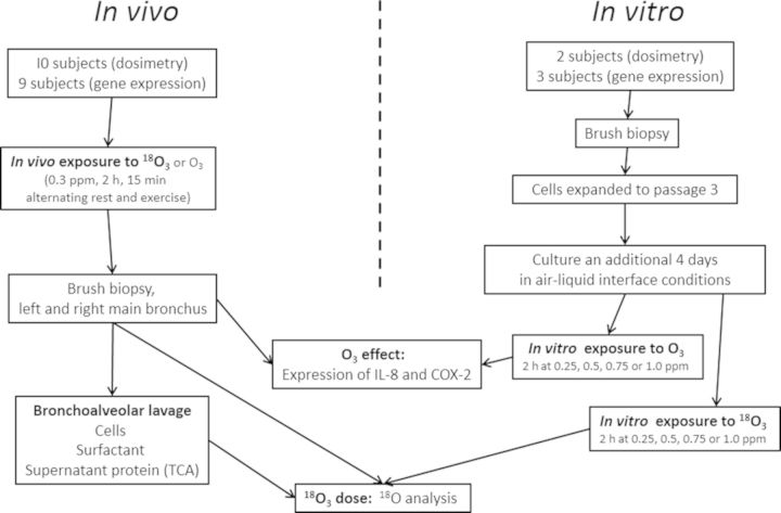
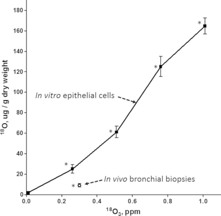
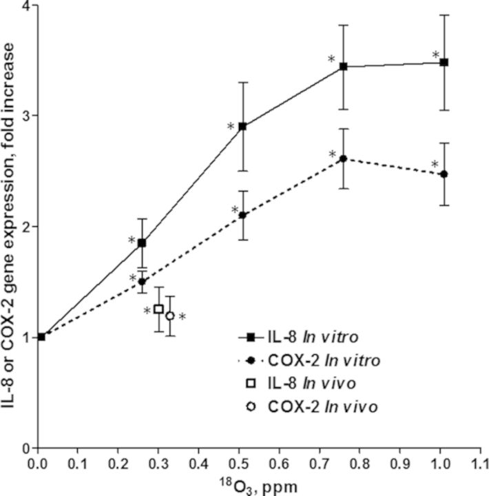
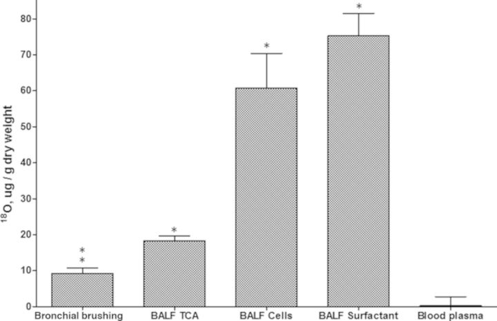
In vitro exposures to air pollutants could, in theory, facilitate a rapid and detailed assessment of molecular mechanisms of toxicity. However, it is difficult to ensure that the dose of a gaseous pollutant to cells in tissue culture is similar to that of the same cells during in vivo exposure of a living person. The goal of the present study was to compare the dose and effect of O3 in airway cells of humans exposed in vivo to that of human cells exposed in vitro. Ten subjects breathed labeled O3 ((18)O3, 0.3 ppm, 2 h) while exercising intermittently. Bronchial brush biopsies and lung lavage fluids were collected 1 h post exposure for in vivo data whereas in vitro data were obtained from primary cultures of human bronchial epithelial cells exposed to 0.25-1.0 ppm (18)O3 for 2 h. The O3 dose to the cells was defined as the level of (18)O incorporation and the O3 effect as the fold increase in expression of inflammatory marker genes (IL-8 and COX-2). Dose and effect in cells removed from in vivo exposed subjects were lower than in cells exposed to the same (18)O3 concentration in vitro suggesting upper airway O3 scrubbing in vivo. Cells collected by lavage as well as previous studies in monkeys show that cells deeper in the lung receive a higher O3 dose than cells in the bronchus. We conclude that the methods used herein show promise for replicating and comparing the in vivo dose and effect of O3 in an in vitro system.
Keywords: bronchoalveolar lavage; epithelial cells; extrapolation; in vivo versus in vitro dose; ozone.
Published by Oxford University Press on behalf of Toxicological Sciences 2014. This work is written by (a) US Government employee(s) and is in the public domain in the US.
Figures
References
-
- Barry B. E., Miller F. J., Crapo J. D. Effects of inhalation of 0.12 and 0.25 ppm ozone on proximal alveolar region of juvenile and adult rats. Lab. Invest. 1985;53:692–704. - PubMed
-
- Devlin R. B., Duncan K. E., Jardim M., Schmitt M. T., Rappold A. G., Diaz-Sanchez D. Controlled exposure of healthy young volunteers to ozone causes cardiovascular effects. Circulation. 2012;126:104–111. - PubMed
-
- Devlin R. B., McDonnell W. F., Becker S., Madden M. C., McGee M. P., Perez R., Hatch G., House D. E., Koren H. S. Time-dependent changes of inflammatory mediators in the lungs of humans exposed to 0.4 ppm ozone for 2 hr: A comparison of mediators found in bronchoalveolar lavage fluid 1 and 18 hr after exposure. Toxicol. Appl. Pharm. 1996;138:176–185. - PubMed
-
- Devlin R. B., McKinnon K. P., Noah T., Becker S., Koren H. S. Ozone-induced release of cytokines and fibronectin by alveolar macrophages and airway epithelial cells. Am. J. Physiol. 1994;266(6 Pt 1):L612–L619. - PubMed
-
- Gerrity T. R., McDonnell W. F., House D. E. The relationship between delivered ozone dose and functional responses in humans. Toxicol. Appl. Pharm. 1994;124:275–283. - PubMed
Publication types
MeSH terms
Substances
LinkOut - more resources
Full Text Sources
Other Literature Sources
Medical
Research Materials
