. 2014 Aug 15;127(Pt 16):3603-13.
doi: 10.1242/jcs.153163. Epub 2014 Jun 13.

A nuclear ubiquitin-proteasome pathway targets the inner nuclear membrane protein Asi2 for degradation

Affiliations

A nuclear ubiquitin-proteasome pathway targets the inner nuclear membrane protein Asi2 for degradation

Mirta Boban et al. J Cell Sci. .

Abstract

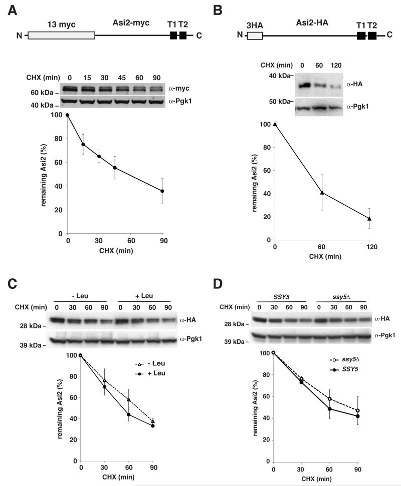
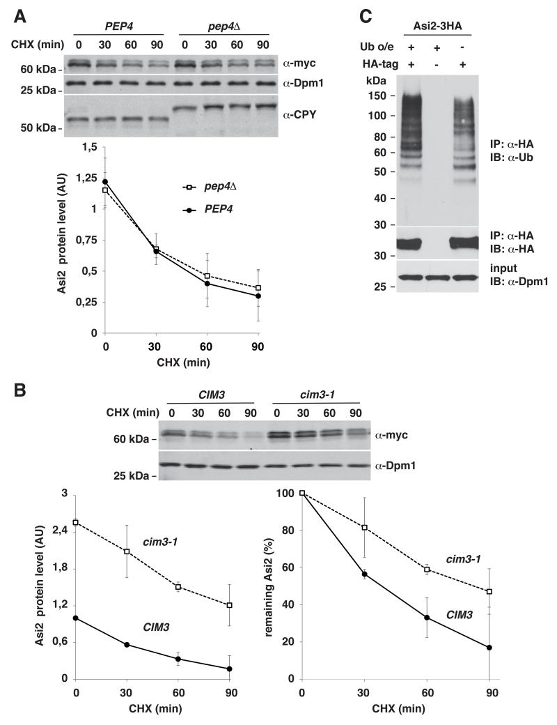
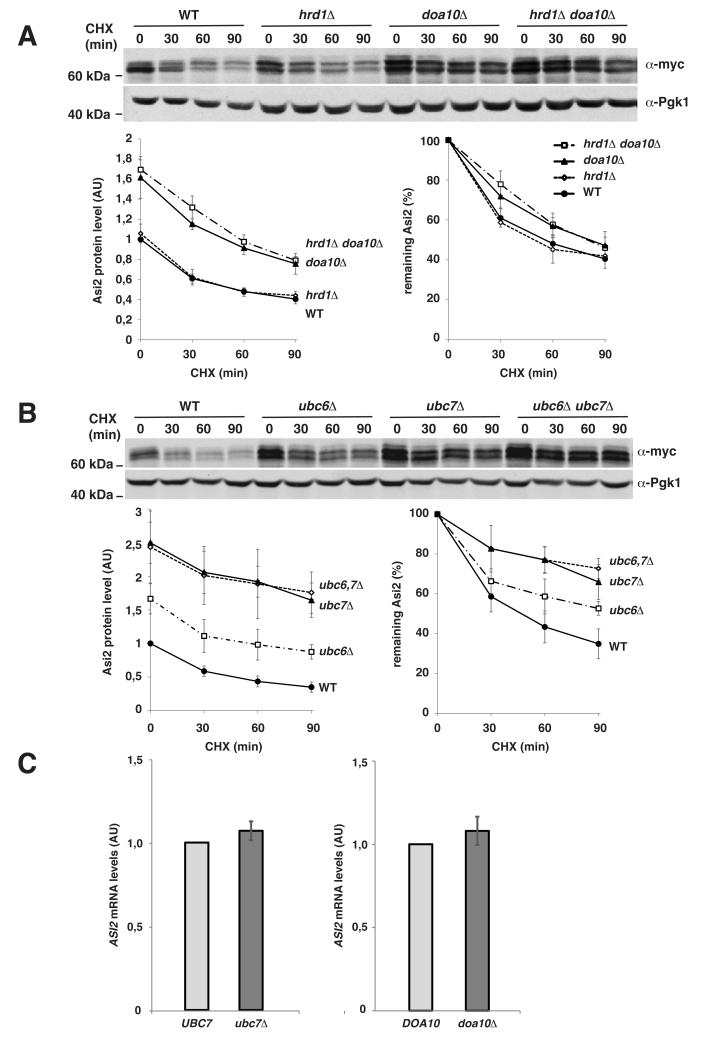
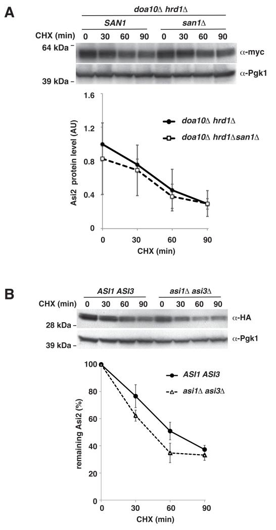
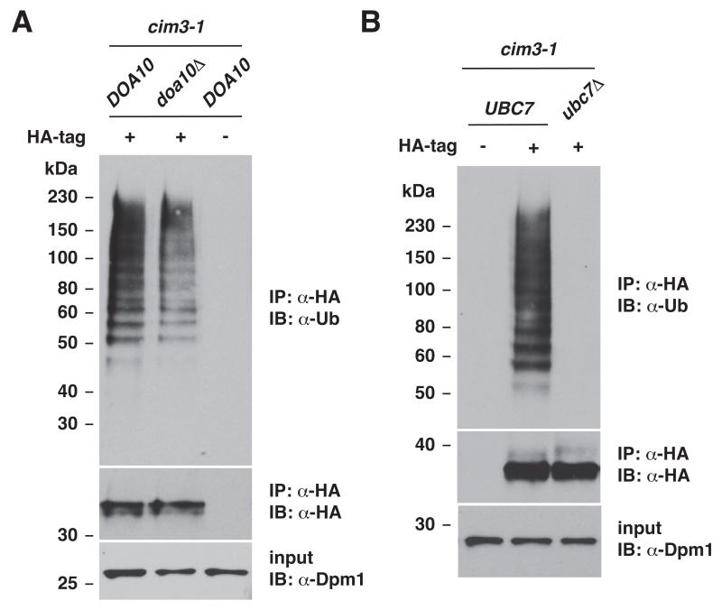
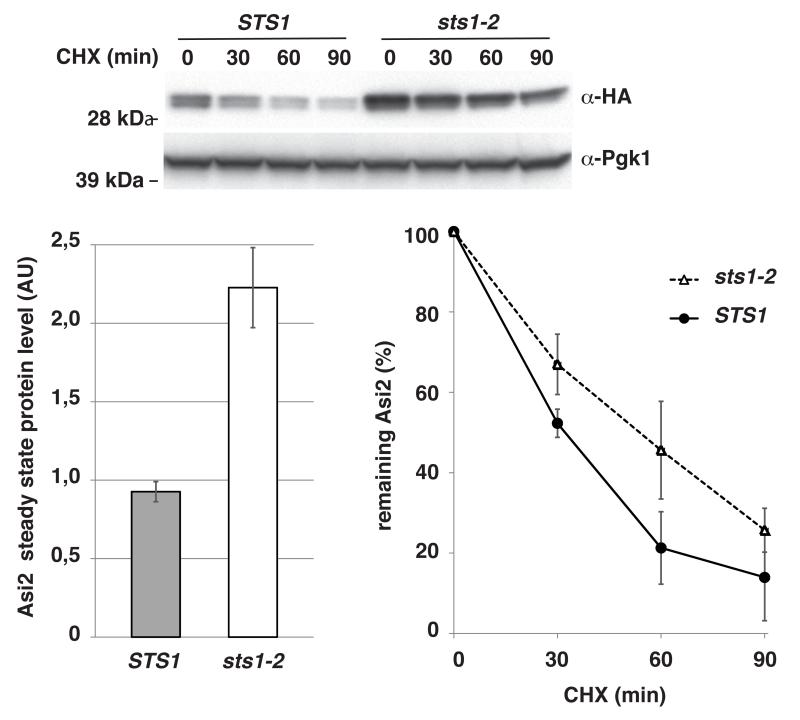
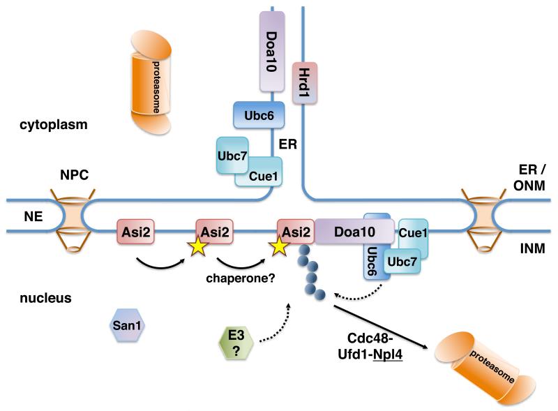
The nuclear envelope consists of inner and outer nuclear membranes. Whereas the outer membrane is an extension of the endoplasmic reticulum, the inner nuclear membrane (INM) represents a unique membranous environment containing specific proteins. The mechanisms of integral INM protein degradation are unknown. Here, we investigated the turnover of Asi2, an integral INM protein in Saccharomyces cerevisiae. We report that Asi2 is degraded by the proteasome independently of the vacuole and that it exhibited a half-life of ∼45 min. Asi2 exhibits enhanced stability in mutants lacking the E2 ubiquitin conjugating enzymes Ubc6 or Ubc7, or the E3 ubiquitin ligase Doa10. Consistent with these data, Asi2 is post-translationally modified by poly-ubiquitylation in a Ubc7- and Doa10-dependent manner. Importantly Asi2 degradation is significantly reduced in a sts1-2 mutant that fails to accumulate proteasomes in the nucleus, indicating that Asi2 is degraded in the nucleus. Our results reveal a molecular pathway that affects the stability of integral proteins of the inner nuclear membrane and indicate that Asi2 is subject to protein quality control in the nucleus.

Keywords: ERAD; Nuclear membrane; Nuclear proteasome; Protein degradation; Ubiquitylation.


Figures

Fig. 1
Fig. 1. Asi2 protein is constitutively turned over
Asi2 protein stability was assessed by cycloheximide (CHX) chase. Cells were harvested at indicated time points after CHX addition and assayed by immunoblotting with anti-myc, anti-HA and anti-Pgk1 antibodies. Pgk1 is a stable protein used as a control. Graphs represent percentage of remaining Asi2 after CHX addition. Average values and standard deviation of indicated number of independent samples are shown. P-values are listed in Table IV, supplementary material. 13myc and 3HA epitope-tagged Asi2 constructs are represented schematically in A and B. Epitope tags and transmembrane segments (T1, T2) are indicated. (A) CHX chase of Asi2-myc (pMB55) in asi2Δ strain (MBY163). Asi2-myc protein half-life: 43 min (n=3). (B) CHX chase of Asi2-HA (pMS1) expressed in asi2Δ strain (MBY163). Asi2-HA protein half-life: 51 min. (n=3). (C) Cells (PLY1340) expressing Asi2-HA (pMS01) were grown in the SD medium and where indicated, 1.3 mM L-leucine was added for 1 hour to induce the SPS sensor. CHX chase was performed. Asi2 protein half-life: 50 min (SD+Leu) and 63 min (SD-Leu), n=3. (D) Asi2-HA (pMS01) stability was examined by CHX chase in SSY5 (PLY1340) and ssy5Δ (PLY1632) strain lacking functional SPS sensor. Asi2 protein half-life: 56 min (SSY5, n=2) and 63 min (ssy5Δ, n=3).
Fig. 2
Fig. 2. Asi2 protein is degraded by the ubiquitin-proteasome system
Cycloheximide (CHX) chase (A, B) was performed as described in Fig. 1. Graphs represent Asi2 protein levels in each strain (arbitrary units, AU) or percentage of remaining Asi2. Average values and standard deviation of three independent samples are shown. P-values are listed in Table IV, supplementary material. (A) Asi2 protein degradation is independent of PEP4. CHX chase of Asi2-myc (pMB55) in a PEP4 (MBY163) and pep4Δ strain (MBY167) was assayed by immunoblotting with anti-myc and anti-Dpm1 antibodies. Cleavage of vacuolar carboxypeptidase Y (CPY), a Pep4 substrate, was examined using anti-CPY antibody. Dpm1 is a stable protein used as a control. (B) Asi2 protein is stabilized in a cim3-1 mutant. CHX chase of Asi2-myc (pMB55) expressed in CIM3 (MBY178) and cim3-1 thermosensitive mutant (MBY179). Growing cultures (27°C) were concentrated to OD600 1.5, incubated for 15 min at 27°C and following a 30 min incubation at 37°C, CHX was added. Immunoblotting was done as in (A). Asi2 protein half-life: 35 min (CIM3) and 81 min (cim3-1). (C) Asi2 protein is poly-ubiquitylated. Immunoprecipitation of HA-tagged Asi2 (pMB3) or untagged Asi2 (pMB128) expressed in cim3-1 strain (PLY1348) carrying ubiquitin-overexpression (Ub o/e) plasmid (myc-Ub/LEU2 2μ) or empty vector (pRS315) was performed using anti-HA antibody (IP: α-HA). Immunoblot (IB) analysis was done using anti-HA, anti-ubiquitin and anti-Dpm1 antibodies.
Fig. 3
Fig. 3. Asi2 is targeted for degradation by Doa10, Ubc6 and Ubc7-dependent ubiquitylation machinery
(A, B) Immunoblot analysis of cycloheximide (CHX) chase was done as described in Fig. 1. Graphs show Asi2 protein levels in each strain (arbitrary units, AU) and percentage of remaining Asi2. Data represent average values (n=3). Standard deviation is indicated. P-values are listed in Table IV, supplementary material. (A) Asi2 stability in doa10Δ and hrd1Δ mutants. Asi2-myc (pMB108) was expressed in WT (MBY163), hrd1Δ (MBY164), doa10Δ (MBY165) and doa10Δ hrd1Δ (MBY166) strains. Asi2 protein half-life: 36 min (WT), 39 min (hrd1Δ), 53 min (doa10Δ) and 67 min (doa10Δ hrd1Δ). (B) Asi2 stability in ubc6Δ and ubc7 Δ mutants. Asi2-myc (pMB55) was expressed in WT (MBY159), ubc6 Δ (MBY160), ubc7Δ (MBY161) and ubc6Δ ubc7Δ (MBY162) strains. Asi2 protein half-life: 36 min (WT), 45 min (ubc6Δ), 86 min (ubc7Δ) and 86 min (ubc6Δ ubc7 Δ). (C) ASI2 mRNA levels were unaffected in ubc7Δ and doa10Δ mutants. Relative ASI2 mRNA levels were determined using RT-PCR and compared in UBC7 (MBY159) versus ubc7Δ (MBY161), and DOA10 (MBY163) versus doa10Δ (MBY165) strains expressing Asi2-HA from pMS01 plasmid. Data represents relative ASI2 mRNA concentration (arbitrary units, AU). Average value and standard deviation of two independent samples is shown.
Fig. 4
Fig. 4. Asi2 degradation is not mediated by San1, Asi1 or Asi3
Cycloheximide (CHX) chase was carried out as described in Fig. 1. Immunoblotting was performed with anti-myc, anti-HA and anti-Pgk1 antibodies. Data represent average values of three independent samples. Standard deviation is indicated. P-values are listed in Table IV, supplementary material. (A) Asi2 protein levels are unaffected in mutant lacking ubiquitin ligase San1. Asi2-myc (pMB108) stability in doa10Δ hrd1Δ (MBY166) and doa10Δ hrd1Δ san1Δ (MPY143) mutants was analyzed. (B) Asi2 protein degradation rate is increased in a mutant lacking Asi1 and Asi3. Asi2-HA (pMS01) stability in ASI1 ASI3 (PLY1340) and asi1Δ asi3Δ (PLY1346) was analyzed. Asi2 protein half-life: 64 min (ASI1 ASI3) and 40 min (asi1Δ asi3Δ).
Fig. 5
Fig. 5. Asi2 ubiquitylation is decreased in mutants lacking Doa10 and Ubc7
Immunoblot analysis of immunoprecipitation experiments. (A) Strains cim3-1 DOA10 (PLY1348) and cim3-1 doa10 Δ (MBY226) carrying ubiquitin-overexpression plasmid (myc-Ub/LEU2 2μ) and either a plasmid expressing epitope-tagged Asi2-HA (pMB3) or a plasmid expressing untagged Asi2 (pMB128) were analyzed. Plasmid pMB128 expressing untagged Asi2 serves as a control for unspecific immunoprecipitation. Immunoprecipitation was performed using anti-HA antibody (IP: α-HA) and immunoblotting (IB) was done using anti-HA or anti-ubiquitin antibodies, as indicated. To verify that a similar amount of protein was used as a starting material for the immunoprecipitation, input samples immunoblotted with anti-Dpm1 antibody are shown. (B) Experiment was done as in (A) using cim3-1 DOA10 (PLY1348) and cim3-1 ubc7Δ (MBY225) strains.
Fig. 6
Fig. 6. Asi2 protein is stabilized in the sts1-2 mutant exhibiting impaired proteasome accumulation in the nucleus
Cycloheximide (CHX) chase of Asi2-HA (pMS01) in a STS1 (NA10) and sts1-2 (NA25) strain was assessed by immunoblotting with anti-HA and anti-Pgk1 antibodies. Cells were grown at 26°C to OD600 0.8, transferred to medium pre-warmed to 37°C (OD600 0.7) and incubated at 37°C for 4 hours before CHX was added. Steady state protein levels at t=0 (p = 0.009, two-tailed T-test, type 3) and the percentage of remaining Asi2 after CHX addition are shown. Data represent average values (n=3). Standard deviation is indicated. P-values of data in time course are listed in Table IV, supplementary material. Asi2 protein half-life: 31 min (STS1) and 45 min (sts1-2).
Fig. 7
Fig. 7. A molecular model for INM protein Asi2 degradation
A putative Asi2 degradation signal (yellow star) may be exposed upon damage or upon changes of its molecular environment at the INM. INM-localized Asi2 is poly-ubiquitylated (blue circles) by the components of ERAD pathway localized in the nucleus (E3 ligase Doa10 and E2 enzymes Ubc6 and Ubc7). The potential involvement of other E3 ligases and chaperones, which may facilitate Asi2 ubiquitylation, is indicated. ER-localized Hrd1 E3 ligase and nuclear San1 are not involved in Asi2 degradation. Poly-ubiquitylated Asi2 is targeted to the nuclear proteasomes, presumably in a manner dependent on the activity of the Cdc48-Ufd1-Npl4 complex (Hitchcock et al., 2003).

References

    1. Ammerer G, Hunter CP, Rothman JH, Saari GC, Valls LA, Stevens TH. PEP4 gene of Saccharomyces cerevisiae encodes proteinase A, a vacuolar enzyme required for processing of vacuolar precursors. Mol. Cell. Bio.J. 1986;6:2490–2499. - PMC - PubMed
    1. Anderson DJ, Hetzer MW. The life cycle of the metazoan nuclear envelope. Curr. Opin. Cell Biol. 2008;20:386–392. - PMC - PubMed
    1. Andréasson C, Heessen S, Ljungdahl PO. Regulation of transcription factor latency by receptor-activated proteolysis. Genes Dev. 2006;20:1563–1568. - PMC - PubMed
    1. Andréasson C, Ljungdahl PO. Receptor-mediated endoproteolytic activation of two transcription factors in yeast. Genes Dev. 2002;16:3158–3172. - PMC - PubMed
    1. Antebi A, Fink GR. The yeast Ca(2+)-ATPase homologue, PMR1, is required for normal Golgi function and localizes in a novel Golgi-like distribution. Mol. Biol. Cell. 1992;3:633–654. - PMC - PubMed

Publication types

MeSH terms

Substances