. 2014 Jul 15;193(2):519-28.
doi: 10.4049/jimmunol.1301668. Epub 2014 Jun 13.

Real-time detection of CTL function reveals distinct patterns of caspase activation mediated by Fas versus granzyme B

Affiliations

Real-time detection of CTL function reveals distinct patterns of caspase activation mediated by Fas versus granzyme B

Jinzhu Li et al. J Immunol. .

Abstract

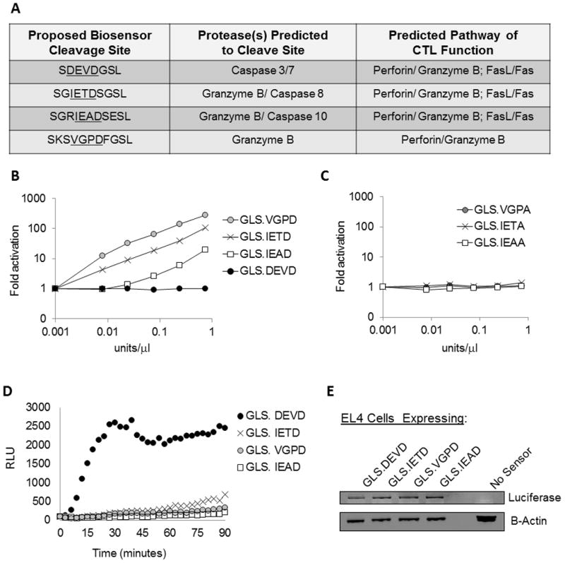
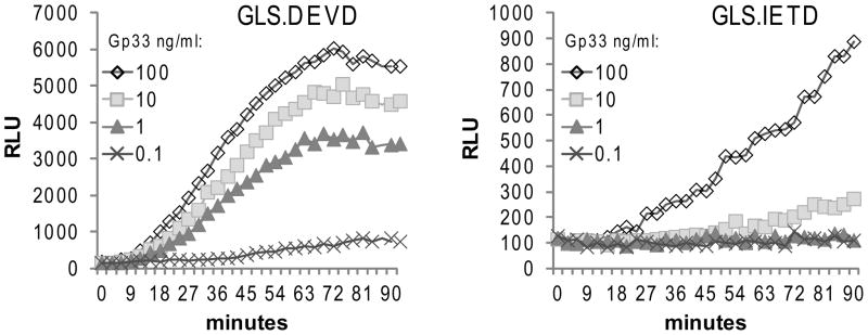
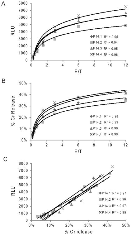
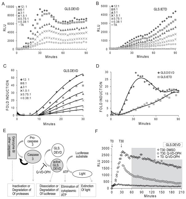
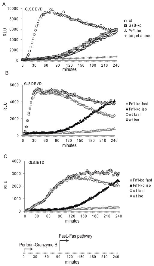
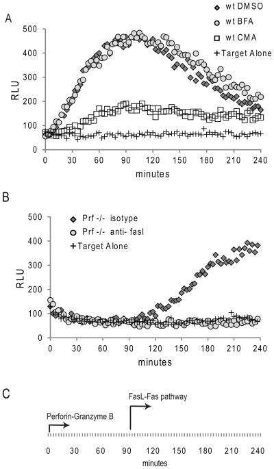
Activation of caspase-mediated apoptosis is reported to be a hallmark of both granzyme B- and Fas-mediated pathways of killing by CTLs; however, the kinetics of caspase activation remain undefined owing to an inability to monitor target cell-specific apoptosis in real time. We have overcome this limitation by developing a novel biosensor assay that detects continuous, protease-specific activity in target cells. Biosensors were engineered from a circularly permuted luciferase, linked internally by either caspase 3/7 or granzyme B/caspase 8 cleavage sites, thus allowing activation upon proteolytic cleavage by the respective proteases. Coincubation of murine CTLs with target cells expressing either type of biosensor led to a robust luminescent signal within minutes of cell contact. The signal was modulated by the strength of TCR signaling, the ratio of CTL/target cells, and the type of biosensor used. Additionally, the luciferase signal at 30 min correlated with target cell death, as measured by a (51)Cr-release assay. The rate of caspase 3/7 biosensor activation was unexpectedly rapid following granzyme B- compared with Fas-mediated signal induction in murine CTLs; the latter appeared gradually after a 90-min delay in perforin- or granzyme B-deficient CTLs. Remarkably, the Fas-dependent, caspase 3/7 biosensor signal induced by perforin-deficient human CTLs was also detectable after a 90-min delay when measured by redirected killing. Thus, we have used a novel, real-time assay to demonstrate the distinct pattern of caspase activation induced by granzyme B versus Fas in human and murine CTLs.


Figures

Figure 1
Figure 1. Design of caspase and granzyme B biosensors to measure CTL function
(A) We developed biosensors which directly measure granzyme B cleavage and/or caspase cleavage upon expression in target cells killed by CTL. The biosensors include a circularly-permuted luciferase containing a protease cleavable site that allows activation of the luciferase only upon cleavage. Shown is the engineered protease cleavage site, the proteases predicted to cleave the site, and the pathways predicted to activate the biosensor following CTL-target cell recognition. (B and C) In order to demonstrate granzyme B dependence upon the newly created biosensors, recombinant protein was generated in vitro and monitored for activation upon the addition of murine granzyme B. Granzyme B activated only the cleavage sites containing the required P1 aspartic acid residue (B); whereas the GLS.DEVD biosensor and control biosensors containing an alanine substitution for the P1 aspartic acid were inactive (B & C). (D) Biosensors were introduced into EL4 target cells by retroviral transduction and cells were sorted for GFP expression. In vitro CTLs were generated by antigen (LCMV gp33–41) stimulation of splenocytes from P14 TCR transgenic mice. CTLs were co-incubated with a biosensor-expressing EL4 target cells at effector/target ratios of 6:1, and luciferase function was measured as the amount of relative light units (RLU) emitted. Data shown is from one cell line, representative of 3 independent experiments. (E) Protein lysates analyzed by western blot using antibodies recognizing luciferase and actin demonstrated equivalent biosensor expression in each cell line.
Figure 2
Figure 2. Strength of antigen recognition affects the induction of biosensors by ex vivo CTL
Ex vivo CTL from C57BL/6 mice infected with LCMV for 7 days were co-incubated with luciferin substrate-loaded EL4 cloned target cells expressing GLS.DEVD or GLS.IETD. EL4 cells were pulsed with gp33 peptide at titrating concentrations and co-incubated with ex vivo CTL at splenocyte/EL4 target ratio of 12:1. Luminescence (RLU) was measured every 3 minutes up to 90 minutes. Data is from one mouse, representative of 8 mice.
Figure 3
Figure 3. Biosensor signal correlates with gold standard 51Cr release assay
In vitro CTLs were generated by antigen stimulation of splenocytes from 4 individual P14 TCR transgenic mice. CTLs were either co-incubated with biosensor expressing, cloned EL4 cells or 51Cr loaded EL4 target cells at effector/target ratios ranging from 12:1 to 0.19:1. 51Cr release was measured after 4 hours. (A) RLU at 30 minutes of the luminescence assay for all 4 mice with titration of E/T ratio; (B) 51Cr release percentage for all 4 mice with titration of E/T ratio; Best fit curves were logarithmic for A and B. (C) Correlation between biosensor assay and 51Cr assay. Data is representative of 3 independent experiments.
Figure 4
Figure 4. Caspase 3/7 activation of biosensor is rapid and transient from CTL induction
CTLs derived in vitro from a representative P14 mouse were co-incubated with substrate-loaded target EL4 expressing GLS.DEVD (A) or GLS.IETD (B) as described in Figure 3. (C and D) Fold induction of GLS.DEVD and GLS.IETD. RLU were graphed as a fold induction (signal generated with CTL/signal generated by target alone) at each time point up to 90 minutes. C) The signal limited to the first 30 minutes of the GLS.DEVD assay is shown in higher detail to demonstrate the dependence of the fold induction on E/T. (D) Comparison of GLS.DEVD and GLS.IETD signals for the first 90 minutes illustrate the distinct kinetics of caspase 3/7 and granzyme B/caspase 8 activation. Data is representative of 3 independent experiments. (E) Model to show the potential mechanisms for acquisition and loss of GLS.DEVD signal. (F) Caspase dependency of GLS.DEVD activity. Using the same assay as described in A and B, CTL were co-incubated with peptide loaded EL4 cells and luminescence monitored from GLS.DEVD out to 4 hours. A pan caspase inhibitor, Q-VD-OPH, was added at the start of the assay, T0, or at 30 minutes, T30. There was complete inhibition of GLS.DEVD when Q-VD-OPH was added at T0. Following addition at T30, there was an immediate dramatic loss of signal to 60 minutes compared to cells exposed to DMSO, illustrating the dependence of the biosensor on caspase signaling from 0–60 minutes. There is a caspase independent loss of activity from 60–240 minutes (delineated by gray box) with nearly identical degradation in the presence or absence of caspase inhibition. (A–D); experiments utilized cloned EL4 cells expressing GLS.IETD or GLS.DEVD. (F) experiments utilized polyclonal, GFP sorted EL4 cells expressing GLS.DEVD.
Figure 5
Figure 5. Caspase 3/7 activation mediated by perforin deficient murine CTL is detectable following late phase Fas-mediated induction
(A) Ex vivo CTL were generated in wild type (WT) C57BL/6, perforin deficient (prf1 ko), and granzyme B deficient knockout (GzB ko) mice infected with LCMV for 7 days. Splenocyte/EL4 target ratio of 12:1 is shown for a representative mouse (4 mice/group tested in 3 independent assays). Evaluation of RLU out to 240 minutes reveals a late activation of the GLS.DEVD (A) and GLS.IETD biosensors (not shown) in mice lacking perforin-mediated granzyme B delivery. (B and C) P14 in vitro CTL from WT and perforin deficient mice were also tested as effectors. Addition of anti-FasL antibody to the reaction prevented the delayed rise in GLS.DEVD (B) and GLS.IETD (C) signal in perforin deficient mice, illustrating the delayed onset of the death receptor pathway (≥ 90 minutes). Target cells were polyclonal EL4, sorted by GFP following retroviral transduction of luciferase biosensors.
Figure 6
Figure 6. Redirected killing: human CTL utilize secretory granule and Fas-mediated pathways to activate Caspase 3/7 in target cells conjugated by anti-CD3
(A) HVS transformed human CTL were co-incubated at 1:1 with GLS.DEVD expressing, cloned P815 cells, pre-incubated with anti-CD3 antibody. P815 cells without added effectors are shown as a negative control. Evaluation of RLU out to 240 minutes reveals early activation of GLS.DEVD in P815 incubated with wild type (wt) CTL contrasting with (B) late activation following co-incubation with perforin deficient CTL (Prf −/−). The early activation in the wt cell line was prevented by the addition of concanamycin A (CMA) but not brefeldin (BFA), demonstrating the dependence upon the secretory granule pathway. Addition of anti-FasL antibody to the PRF −/− CTL prevented the delayed rise in GLS.DEVD (B) confirming the role of the delayed onset death receptor pathway. Shown is a representative figure from 3 independent experiments utilizing these two HVS lines.

References

    1. Jenkins MR, Griffiths GM. The synapse and cytolytic machinery of cytotoxic T cells. Curr Opin Immunol. 2010;22:308–313. - PMC - PubMed
    1. Dustin ML, Long EO. Cytotoxic immunological synapses. Immunol Rev. 2010;235:24–34. - PMC - PubMed
    1. de Saint Basile G, Menasche G, Fischer A. Molecular mechanisms of biogenesis and exocytosis of cytotoxic granules. Nat Rev Immunol. 2010;10:568–579. - PubMed
    1. Praper T, Sonnen AF, Kladnik A, Andrighetti AO, Viero G, Morris KJ, Volpi E, Lunelli L, Dalla Serra M, Froelich CJ, Gilbert RJ, Anderluh G. Perforin activity at membranes leads to invaginations and vesicle formation. Proc Natl Acad Sci U S A. 2011;108:21016–21021. - PMC - PubMed
    1. Thiery J, Keefe D, Boulant S, Boucrot E, Walch M, Martinvalet D, Goping IS, Bleackley RC, Kirchhausen T, Lieberman J. Perforin pores in the endosomal membrane trigger the release of endocytosed granzyme B into the cytosol of target cells. Nat Immunol. 2011;12:770–777. - PMC - PubMed

Publication types

MeSH terms