. 2014 Jul 15;193(2):889-900.
doi: 10.4049/jimmunol.1303389. Epub 2014 Jun 13.

Investigation of soluble and transmembrane CTLA-4 isoforms in serum and microvesicles

Affiliations

Investigation of soluble and transmembrane CTLA-4 isoforms in serum and microvesicles

Laura Esposito et al. J Immunol. .

Abstract

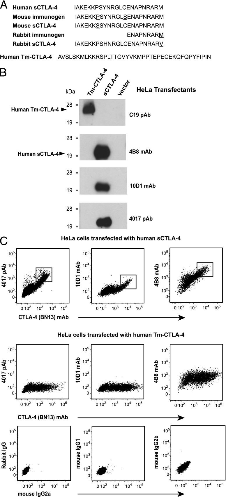
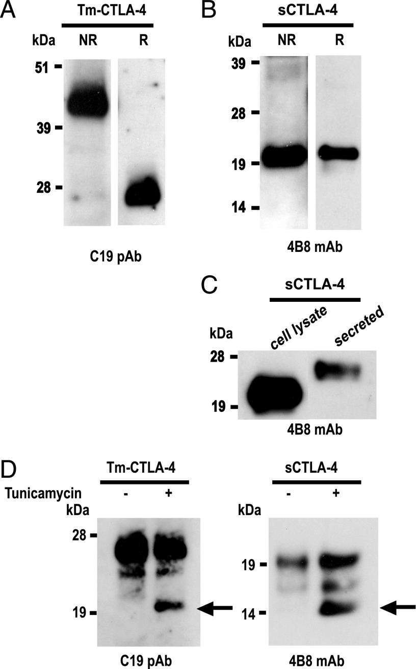
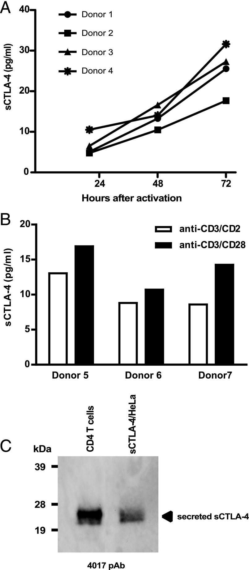
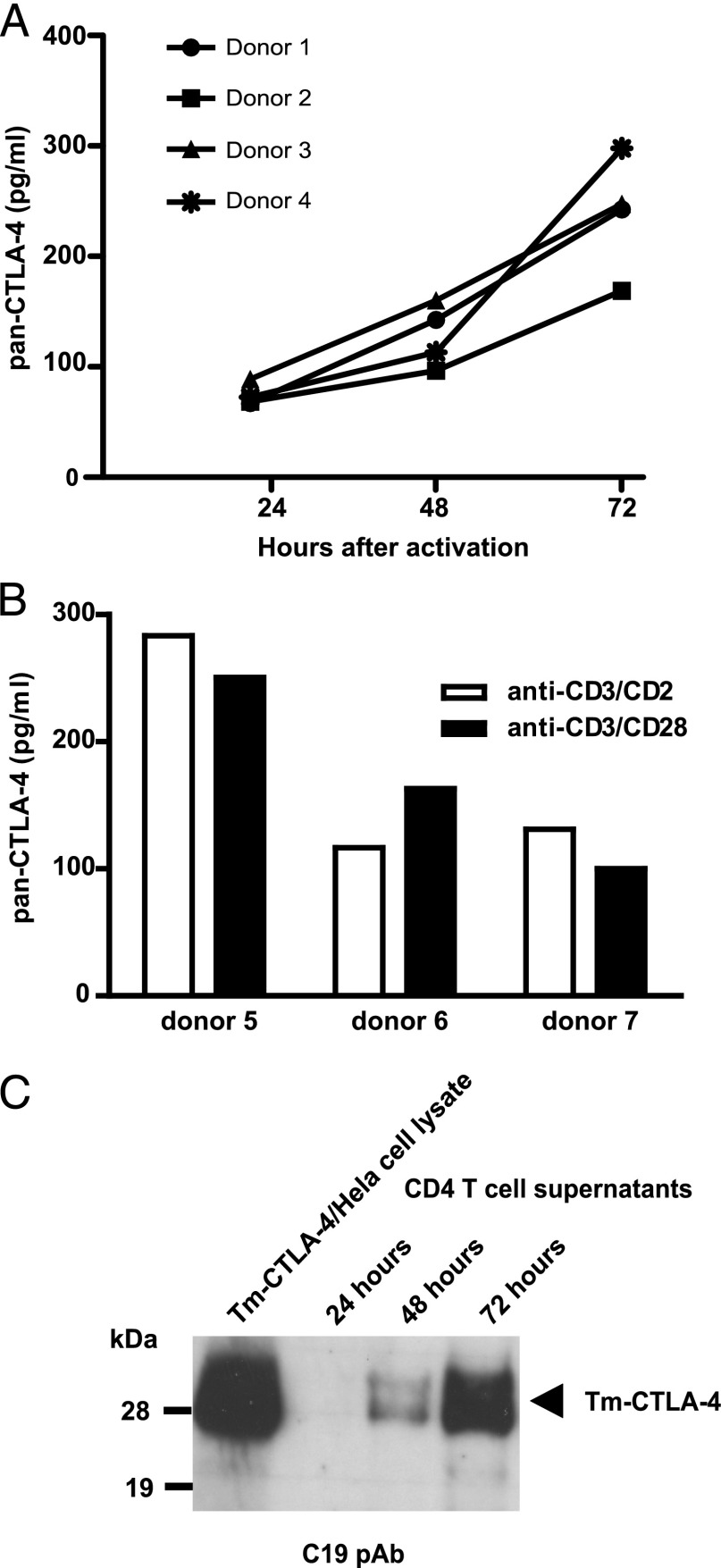
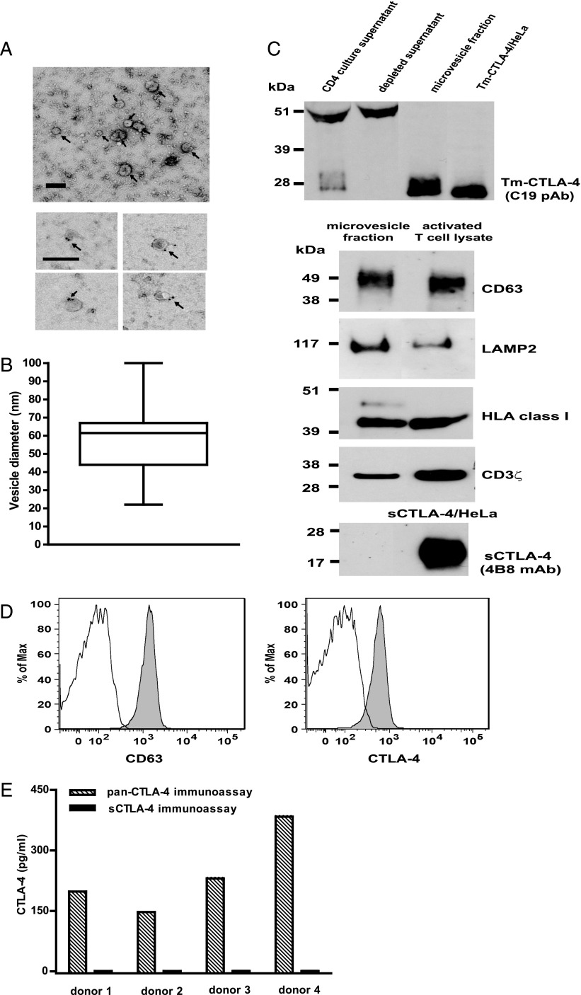
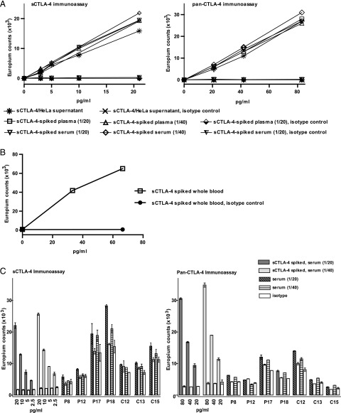
Expression of the CTLA-4 gene is absolutely required for immune homeostasis, but aspects of its molecular nature remain undefined. In particular, the characterization of the soluble CTLA-4 (sCTLA-4) protein isoform generated by an alternatively spliced mRNA of CTLA4 lacking transmembrane-encoding exon 3 has been hindered by the difficulty in distinguishing it from the transmembrane isoform of CTLA-4, Tm-CTLA-4. In the current study, sCTLA-4 has been analyzed using novel mAbs and polyclonal Abs specific for its unique C-terminal amino acid sequence. We demonstrate that the sCTLA-4 protein is secreted at low levels following the activation of primary human CD4(+) T cells and is increased only rarely in the serum of autoimmune patients. Unexpectedly, during our studies aimed to define the kinetics of sCTLA-4 produced by activated human CD4(+) T cells, we discovered that Tm-CTLA-4 is associated with microvesicles produced by the activated cells. The functional roles of sCTLA-4 and microvesicle-associated Tm-CTLA-4 warrant further investigation, especially as they relate to the multiple mechanisms of action described for the more commonly studied cell-associated Tm-CTLA-4.


Figures

FIGURE 1.
FIGURE 1.
Development of Abs specific for the sCTLA-4 isoform. (A) Human (NP_001032720.1), rabbit, and mouse (ENSMUSP00000095327) amino acid sequences at the C-terminal region of sCTLA-4 are aligned with the synthetic peptides used for immunization of rabbits (P1_rabbit) and mice (P2_mouse). The cytoplasmic tail of TM-CTLA-4 is shown for comparison at the bottom of the panel (P16410, UniProt). Mouse and human nucleotide sequences encoding sCTLA-4 have been confirmed in our laboratory by sequencing. Rabbit amino acid sequence was determined from the deposited Tm-CTLA-4 nucleotide sequence (CTLA4_rabbit ENSOCUG00000017274) after alignment with human sCTLA-4 nucleotide sequence (NM_001037631.2). The 9-aa peptide used to immunize rabbits differs at a single residue from the rabbit sequence, a methionine for valine substitution at position 174 of rabbit sCTLA-4. The 23-aa synthetic peptide having the human sequence used to immunize mice differs at position 158 from the endogenous mouse sequence; a proline replaces serine. A serine for cysteine substitution in the 23-aa peptide was made at the residue corresponding to position 165 of the mature sCTLA-4 protein to prevent the formation of disulfide bonds in the peptide preparation and the peptide-KLH conjugate used for immunization. (B) The 4B8 mAb, 10D1 mAb, and 4017 polyclonal Ab validation by Western blot under reducing conditions on detergent lysates of HeLa cells expressing human recombinant Tm-CTLA-4, sCTLA-4, and vector only. Specific detection of Tm-CTLA-4 is performed with CTLA-4 (C19) polyclonal Ab. (C) sCTLA-4 Ab specificity was confirmed by flow cytometry using HeLa cells transfected with sCTLA-4 (top row) or Tm-CTLA-4 (middle row). As a control, HeLa cells transfected with Tm-CTLA-4 were stained with isotype-matched control Abs (bottom row); similar results were obtained using HeLa cells transfected with sCTLA-4 (data not shown). All experiments are representative of at least three independent observations.
FIGURE 2.
FIGURE 2.
sCTLA-4 is secreted and expressed as a glycosylated monomer. (A) Human Tm-CTLA-4 protein is detected as a dimer under nonreducing conditions (NR) and as a monomer under reducing conditions (R) in lysates from HeLa cells transfected with human Tm-CTLA-4 cDNA. (B) Lysates from HeLa cells transfected with human sCTLA-4 cDNA contain sCTLA-4 protein that migrates as a monomer in both nonreducing and reducing conditions. (C) Under reducing conditions, sCTLA-4 protein secreted into the culture medium of HeLa cells transfected with sCTLA-4 cDNA migrates more slowly than sCTLA-4 present in cell lysates. (D) N-glycan addition contributes to the molecular masses of Tm-CTLA-4 and sCTLA-4. Following culture with or without tunicamycin, detergent lysates from HeLa cells transfected with Tm-CTLA-4 or sCTLA were analyzed by Western blot under reducing conditions using C19 polyclonal Ab or 4B8 mAb, respectively. Arrows indicate the protein species lacking N-linked glycans. All experiments are representative of at least three independent observations.
FIGURE 3.
FIGURE 3.
(A) In vitro activation of CD4+ T cells induces secretion of sCTLA-4. CD4+ T cells of four healthy donors were stimulated in vitro with anti-CD3/CD2 mAb and culture supernatants harvested at the indicated time points. CD4+ T cells (3 × 10⁶ cells/ml) from the four donors were cultured on separate days. Isotype controls were negative for all samples (data not shown). (B) Secretion of sCTLA-4 by CD4+ T cells (1 × 10⁶ cells/ml) of three independent donors stimulated for 72 h in vitro with either anti-CD3/CD2 mAb or anti-CD3/CD28 microbeads. (C) Detection of sCTLA-4 following immunoprecipitation and Western blot analysis. sCTLA-4 protein in culture supernatants from activated CD4+ T cells and from HeLa cells transfected with sCTLA-4 was immunoprecipitated using 4B8 mAb, followed by 4017 polyclonal Ab immunoblotting. No sCTLA-4 was immunoprecipitated with the corresponding IgG2b isotype control (data not shown). Western blot gel is representative of more than three independent experiments.
FIGURE 4.
FIGURE 4.
Immunoassay and Western blot analysis of Tm-CTLA-4 present in culture supernatants from activated CD4+ T cells. (A) Culture supernatants from 3 × 10⁶ cells/ml CD4+ T cells activated with anti-CD3/CD2 mAbs in vitro from four donors cultured on different days were harvested at the indicated time points and analyzed using the pan–CTLA-4 immunoassay. (B) Analyses of supernatants of CD4+ T cells (1 × 10⁶ cells/ml) of three donors cultured on different days stimulated in vitro for 72 h with either anti-CD3/CD2 mAbs or anti-CD3/CD28 microbeads. (C) Release of Tm-CTLA-4 in a time-dependent manner in culture supernatants of activated CD4+ T cells was confirmed by Western blot analysis (reducing gel) using Tm-CTLA-4 C terminus C19 polyclonal Ab. Western blot gel is representative of at least four independent experiments.
FIGURE 5.
FIGURE 5.
Characterization of exosome-like vesicles secreted by activated CD4+ T cells. (A) Top panel, Representative electron micrographs of microvesicles, as indicated by arrows, showing vesicles with low electron density as well as characteristic shape and size of exosomes (scale bar, 100 nm). Bottom panel, Anti-CD63 immunogold labeling of CD4+ T cell–derived microvesicles. Arrows indicate immunogold-positive labeling (scale bar, 100 nm). (B) The size distribution of microvesicles is plotted as a box plot; the box spans the 25th and 75th percentile, the horizontal line represents the median diameter, and the whiskers mark the extremes of the distribution (n = 24 microvesicles). (C) Top panel, Western blot analysis (reducing gel) of activated CD4+ T cell supernatant before and after ultracentrifugation, microvesicle fraction isolated from the T cell supernatant, and detergent lysates from Tm-CTLA-4/HeLa cells with anti–Tm-CTLA-4 C19 polyclonal Ab; bottom panels, Western blot analysis of microvesicle fraction isolated from the T cell supernatant with anti-CD63, anti-HLA class I, anti-CD3ζ (nonreducing gels), anti-LAMP2, and anti–sCTLA-4 Abs (reducing gels). CD4+ T cell or sCTLA-4/HeLa detergent lysates were used as positive controls. (D) Microvesicle fraction derived from activated CD4+ T cells was coated onto latex beads, surface stained with anti-CD63 and anti-CTLA-4 (BN13) (filled gray histogram) Abs or the corresponding isotype controls (open histogram), and analyzed by flow cytometry. (E) sCTLA-4 is not expressed in exosomal-like vesicles. The microvesicle fractions were isolated from supernatants of activated CD4+ T cells of four donors after 3 d of stimulation with anti-CD3/CD2 mAb and then tested in the pan–CTLA-4 and sCTLA-4 immunoassays. All experiments are representative of at least three independent observations.
FIGURE 6.
FIGURE 6.
Detection of sCTLA-4 protein in plasma and serum samples. (A) Analysis by sCTLA-4 (left panel) and pan–CTLA-4 (right panel) immunoassays of 2-fold serial dilutions of recombinant sCTLA-4 spiked into 1/20 and 1/40 dilutions of plasma and serum samples. An isotype-matched mouse IgG2a aκ capture Ab is used as a negative control. Serial dilutions of culture supernatant of sCTLA-4 HeLa cell transfectants in standard assay buffer were included in each assay. (B) Detection of recombinant sCTLA-4 spiked into human whole blood. Mouse IgG2a aκ isotype-matched Ab used as capturing reagent served as a negative control. (C) Analysis of sCTLA-4 in serum samples from autoimmune patients and controls using the sCTLA-4 immunoassay (left panel) and the pan–CTLA-4 immunoassay (right panel). Serial dilutions of recombinant sCTLA-4 were spiked into serum diluted 1:20 (dark gray columns) and 1:40 (light gray columns), and representative examples from a total of 61 donors are shown (see Supplemental Fig. 3 for results from all donors), as follows: 4 GD patients and 3 age- and sex-matched, healthy volunteers, sera diluted 1/20 (dark shaded columns) and 1/40 (light shaded columns). For all samples, including the spiked serum samples, the mouse IgG2aκ isotype control is shown (open columns). Columns, mean of duplicate measurements; bars, ±SD.

References

    1. Tivol E. A., Borriello F., Schweitzer A. N., Lynch W. P., Bluestone J. A., Sharpe A. H. 1995. Loss of CTLA-4 leads to massive lymphoproliferation and fatal multiorgan tissue destruction, revealing a critical negative regulatory role of CTLA-4. Immunity 3: 541–547. - PubMed
    1. Walunas T. L., Lenschow D. J., Bakker C. Y., Linsley P. S., Freeman G. J., Green J. M., Thompson C. B., Bluestone J. A. 1994. CTLA-4 can function as a negative regulator of T cell activation. Immunity 1: 405–413. - PubMed
    1. Waterhouse P., Penninger J. M., Timms E., Wakeham A., Shahinian A., Lee K. P., Thompson C. B., Griesser H., Mak T. W. 1995. Lymphoproliferative disorders with early lethality in mice deficient in Ctla-4. Science 270: 985–988. - PubMed
    1. Read S., Greenwald R., Izcue A., Robinson N., Mandelbrot D., Francisco L., Sharpe A. H., Powrie F. 2006. Blockade of CTLA-4 on CD4+CD25+ regulatory T cells abrogates their function in vivo. J. Immunol. 177: 4376–4383. - PMC - PubMed
    1. Read S., Malmström V., Powrie F. 2000. Cytotoxic T lymphocyte-associated antigen 4 plays an essential role in the function of CD25(+)CD4(+) regulatory cells that control intestinal inflammation. J. Exp. Med. 192: 295–302. - PMC - PubMed

Publication types

MeSH terms