. 2014 Aug 15;307(4):H493-503.
doi: 10.1152/ajpheart.00091.2014.

Diversity of mitochondria-dependent dilator mechanisms in vascular smooth muscle of cerebral arteries from normal and insulin-resistant rats

Diversity of mitochondria-dependent dilator mechanisms in vascular smooth muscle of cerebral arteries from normal and insulin-resistant rats

Prasad V G Katakam et al. Am J Physiol Heart Circ Physiol. .

Abstract

Mitochondrial depolarization following ATP-sensitive potassium (mitoKATP) channel activation has been shown to induce cerebral vasodilation by generation of mitochondrial reactive oxygen species (ROS), which sequentially promotes frequency of calcium sparks and activation of large conductance calcium-activated potassium channels (BKCa) in vascular smooth muscle (VSM). We previously demonstrated that cerebrovascular insulin resistance accompanies aging and obesity. It is unclear whether mitochondrial depolarization without the ROS generation enhances calcium sparks and vasodilation in phenotypically normal [Sprague Dawley (SD); Zucker lean (ZL)] and insulin-resistant [Zucker obese (ZO)] rats. We compared the mechanisms underlying the vasodilation to ROS-dependent (diazoxide) and ROS-independent [BMS-191095 (BMS)] mitoKATP channel activators in normal and ZO rats. Arterial diameter studies from SD, ZL, and ZO rats showed that BMS as well as diazoxide induced vasodilation in endothelium-denuded cerebral arteries. In normal rats, BMS-induced vasodilation was mediated by mitochondrial depolarization and calcium sparks generation in VSM and was reduced by inhibition of BKCa channels. However, unlike diazoxide-induced vasodilation, scavenging of ROS had no effect on BMS-induced vasodilation. Electron spin resonance spectroscopy confirmed that diazoxide but not BMS promoted vascular ROS generation. BMS- as well as diazoxide-induced vasodilation, mitochondrial depolarization, and calcium spark generation were diminished in cerebral arteries from ZO rats. Thus pharmacological depolarization of VSM mitochondria by BMS promotes ROS-independent vasodilation via generation of calcium sparks and activation of BKCa channels. Diminished generation of calcium sparks and reduced vasodilation in ZO arteries in response to BMS and diazoxide provide new insights into mechanisms of cerebrovascular dysfunction in insulin resistance.


Figures

Fig. 1.
Fig. 1.
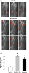
BMS-191095 (BMS) induced mitochondrial-depolarization and vasodilation. Mitochondrial depolarization and vasodilation in response to BMS in endothelium-denuded cerebral arteries from SD rats are shown. A: typical images of tetramethylrhodamine ethyl ester (TMRE) fluorescence in the presence of vehicle (DMSO; n = 7 arteries) and BMS (50 μmol/l; n = 8 arteries) showing decreased TMRE fluorescence in response to BMS vs. vehicle indicating mitochondrial depolarization. B: cumulative data of percent change from TMRE fluorescence at baseline before the application of BMS/vehicle are represented in the bar graph. C: vascular responses to BMS (10, 50, and 100 μmol/l) showing dose-dependent increase in vasodilation to BMS (n = 6–14 arteries). *Significant difference in vasodilation to BMS response (P < 0.05) compared with vehicle treatment, which had no effect arterial diameter. D: vascular responses to BMS (50 μmol/l) in the presence of iberiotoxin (n = 6 arteries) and MnTBAP (n = 5 arteries). Data are presented as means ± SE. *Significant difference in corresponding baseline BMS response (P < 0.05).
Fig. 2.
Fig. 2.
Mitochondrial depolarization with and without reactive oxygen species (ROS) generation. A: representative electron spin resonance (ESR) spectra of SD rat aortas incubated for 60 min at 37°C with 200 μmol/l 1-hydroxy-3-methoxycarbonyl-2,2,5,5-tetramethyl-pyrrolidine (CMH), a spin trap for superoxide radical. The vertical axis represents signal intensity in arbitrary units, and the horizontal axis represents the magnetic field (G). A characteristic CMH signal with 3 peaks was detected in rat aortas incubated with CMH and various drugs. Representative ESR spectra of the basal (vehicle treated, n = 7), 50 μmol/l BMS (n = 7), and 100 μmol/l diazoxide (n = 9) are shown in A. B: cumulative data normalized to the dry weight of aortic segments and expressed as arbitrary units as a bar graph are shown. Cotreatment with ROS scavenger SOD reduced the ESR signal amplitude in response to vehicle and diazoxide. Mitochondrial superoxide generation following treatment with the combination of antimycin A and rotenone (inhibitors of respiratory chain complexes) was used as positive control to confirm the superoxide generation. An n represents the number of experiments (2 aortic segments each) that include 2 experiments per treatment per each rat. C: fluorescence images of endothelium-denuded cerebral arteries loaded with MitoSOX, a fluoroprobe for mitochondrial superoxide, are shown. Typical spindle-shaped MitoSOX fluorescence with a central hollow space is seen in the smooth muscle cell around the fluorescence free nucleus. D: bar graph of cumulative MitoSOX fluorescence data. Diazoxide (100 μmol/l) treatment enhanced MitoSOX fluorescence in the vascular smooth muscle (VSM) cells of endothelium-denuded cerebral arteries vs. vehicle (DMSO) treatment indicating increased generation of mitochondrial superoxide. However, treatment with BMS-19195 did not increase MitoSOX fluorescence in VSM cells, indicating that mitochondrial depolarization by BMS-191095 was not accompanied by generation of superoxide from mitochondria. The n value represents average fluorescence intensity of all the cells from n number of arteries that were treated with each treatment. *Significant difference in corresponding response following vehicle treatment (P < 0.05).
Fig. 3.
Fig. 3.
Calcium sparks generation in response to BMS-191095 and diazoxide. A: selected fluorescence images of VSM cells loaded with 5 μmol/l Fluo-4AM from time series image stacks are shown. Region of interest sites of calcium sparks are shown (arrow heads) from vehicle (DMSO) and BMS (50 μmol/l)-treated endothelium-denuded cerebral arteries from SD rats. B: bar graph showing the cumulative data of calcium sparks frequency in response to vehicle, diazoxide, and BMS. *Significant difference in response vs. vehicle (P < 0.05).
Fig. 4.
Fig. 4.
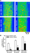
Mitochondrial depolarization in Zucker rat arteries. A: representative images of VSM cells loaded with TMRE are shown from BMS (50 μmol/l) and diazoxide (100 μmol/l) treated cerebral arteries of Zuker lean (ZL) and Zucker obese (ZO) rats. The color range from red to yellow indicates the range of TMRE fluorescence intensity from fully polarized (red) to depolarized (yellow) mitochondria. BMS and diazoxide elicited robust mitochondrial depolarization in ZL arteries indicated by greater yellow relative to red. In contrast, BMS and diazoxide elicited relatively diminished mitochondrial depolarization in ZO arteries compared with ZL arteries, indicated by less yellow relative to red suggestive of impaired mitochondrial depolarization. B: bar graph showing cumulative data of percent decrease in TMRE fluorescence in response to BMS (25 and 50 μmol/l) and diazoxide (100 μmol/l) from baseline before the application of drugs. Data are means ± SE of 6–14 experiments. *Significant difference in response to corresponding treatment in ZL arteries (P < 0.05). An n represents the number of arterial segments and single arterial segment per experiment from each rat was used.
Fig. 5.
Fig. 5.
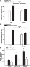
Mitochondria-mediated vasodilation in Zucker rats. A: diameter measurements in response to BMS (50 μmol/l) in endothelium-denuded cerebral arteries of ZL and ZO rats are shown. B: diameter measurements in response to diazoxide (100 μmol/l) in endothelium-denuded cerebral arteries of ZO and ZL are shown. C: bar graph showing cumulative data of calcium sparks frequency in response to vehicle, BMS, and diazoxide in ZL and ZO arteries. *Significant difference in response vs. corresponding response in ZL arteries (P < 0.05). An n represents the number of arterial segments and single arterial segment per experiment from each rat was used.
Fig. 6.
Fig. 6.
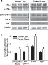
Increased levels of endoplasmic reticulum (ER) stress markers in cerebral arteries from Zucker rats. A: representative images of immunoblots determining the total and phosphorylated binding protein/78 kDa glucose regulated protein (BiP/GRP78), C/EBP-homologous protein that inhibits C/EBP (CHOP), and X box binding protein 1 (XBP1) in the cellular lysates from the cerebral arteries of ZL and ZO rats are shown. B: bar graphs showing cumulative data of immunoband intensity normalized to β-actin in arbitrary units are shown. *Significant differences in immunoband intensity of given protein compared with the immunoband intensity the same protein in ZL arteries (P < 0.05; n = 6 animals per group). IF2α, initiation factor 2α.
Fig. 7.
Fig. 7.
Mechanisms of mitochondrial-depolarization-induced vasodilation. A schematic of the proposed mechanisms underlying the vasodilation induced by the mitochondrial depolarization in the VSM is shown. BMS and diazoxide cause mitochondrial depolarization by activating mitochondrial KATP-sensitive (mitoKATP) channels. Depolarization of mitochondria leads to generation of calcium sparks by activation of ryanodine-sensitive calcium channels (RyR) in the sarcoplasmic reticulum (SR) in microdomains adjacent to mitochondria. Calcium sparks generation in response to diazoxide is ROS dependent. However, an unknown ROS-independent mechanism mediates calcium spark generation in response to BMS-191095. Generation of calcium sparks results in localized elevation of calcium as calcium transients lasting for a few milliseconds leading to activation of adjacent large-conductance calcium-activated potassium channels (BKCa). Potassium efflux through BKCa channels hyperpolarizes the VSM membrane resulting in inactivation of voltage-gated calcium channels leading to decreased global intracellular calcium ([Ca2+]i) and vasodilation. The likely sites affected by the insulin resistance in cerebral arteries of ZO rats are indicated by the numbers 1–3. 1, Previous findings from our studies demonstrated impaired activation of mitoKATP channels in ZO arteries resulting in impaired vasodilation to diazoxide (34). 2, Our current Western blot analysis revealed that ZO arteries display ER stress that may likely contribute to impaired calcium spark generation from intracellular calcium stores. 3, Our previous studies also demonstrated impaired activation of BKCa channels and plasma membrane ATP-sensitive potassium channels in cerebral arteries of ZO rats (16, 17).

Similar articles

Cited by

References

    1. Adebiyi A, McNally EM, Jaggar JH. Sulfonylurea receptor-dependent and -independent pathways mediate vasodilation induced by ATP-sensitive K+ channel openers. Mol Pharmacol 74: 736–743, 2008 - PMC - PubMed
    1. Ahmad N, Wang Y, Haider KH, Wang B, Pasha Z, Uzun O, Ashraf M. Cardiac protection by mitoKATP channels is dependent on Akt translocation from cytosol to mitochondria during late preconditioning. Am J Physiol Heart Circ Physiol 290: H2402–H2408, 2006 - PubMed
    1. Alder NN, Shen Y, Brodsky JL, Hendershot LM, Johnson AE. The molecular mechanisms underlying BiP-mediated gating of the Sec61 translocon of the endoplasmic reticulum. J Cell Biol 168: 389–399, 2005 - PMC - PubMed
    1. Bodalia A, Li H, Jackson MF. Loss of endoplasmic reticulum Ca2+ homeostasis: contribution to neuronal cell death during cerebral ischemia. Acta Pharmacol Sin 34: 49–59, 2013 - PMC - PubMed
    1. Bray GA. The Zucker-fatty rat: a review. Fed Proc 36: 148–153, 1977 - PubMed

Publication types

MeSH terms

LinkOut - more resources