

doi: 10.1007/s00401-014-1307-x.
Epub 2014 Jun 15.
Subventricular spread of diffuse intrinsic pontine glioma
Affiliations
- PMID: 24929912
- PMCID: PMC4161623
- DOI: 10.1007/s00401-014-1307-x
Item in Clipboard
Subventricular spread of diffuse intrinsic pontine glioma
Acta Neuropathol.
2014 Oct.
No abstract available
Conflict of interest statement
Figures
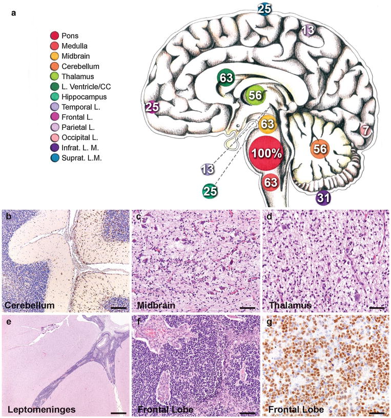
Extent of spread in DIPG. a Neuroanatomical sites and frequency of tumor invasion. Numbers indicate the percentage of cases that exhibit tumor invasion at the indicated anatomical location. The size of the circles marking each anatomical site (color key to the left) illustrates the frequency. CC corpus callosum, infrat. L.M. infratentorial leptomeninges, Suprat. L.M. supratentorial leptomeninges. photomicrographs illustrating b Olig2 + tumor cells infiltrating the cerebellum, c tumor infiltrating the substantia nigra in the midbrain (H&E), d tumor infiltrating the thalamus (H&E), e leptomeningeal spread affecting the temporal lobe (H&E), f tumor in the frontal cortex (H&E) and g Olig2 + tumor cells in the frontal lobe. Immunostains DAB with hematoxylin counterstain. Scale bars 100 μm (b–d, f), 1 mm (e) and 50 μm (g)
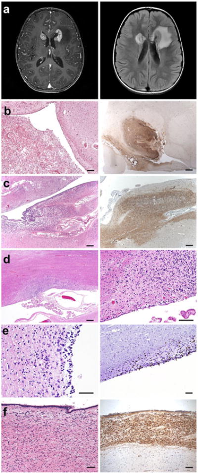
Invasion of the subventricular zone in DIPG (a). MRI images illustrating enhancing lesions (T1 post gadolinium, left image) at the frontal horns of the lateral ventricles with associated edema (FLAIR, right image) in case SU-DIPG-XIII. b H&E (left) and nestin immunostaining (right) of DIPG tumor at the frontal horn of the lateral ventricle in case SU-DIPG-III. c Similar to b in SU-DIPG-V. d H&E demonstrating DIPG tumor at the frontal horn of the lateral ventricle in SU-DIPG-XIII. e H&E stain (left) and Ki67 immunostaining (right image) illustrating tumor infiltration in the lateral ventricle SVZ of SU-DIPG-VI. f H&E (left) and nestin immunostaining (right) of DIPG tumor in the third ventricle of SU-DIPG-IV. Immunostains DAB with hematoxylin counterstain. Scale bars 1.25 mm (b–c, d, left panel), 250 μm (d, right panel; e, f)
References
-
- Donaldson SS, et al. Advances toward an understanding of brainstem gliomas. J Clin Oncol. 2006;24(8):1266–1272. - PubMed
-
- Schwartzentruber J, et al. Driver mutations in histone H3.3 and chromatin remodelling genes in paediatric glioblastoma. Nature. 2012;482(7384):226–231. - PubMed
-
- Mantravadi RV, et al. Brain stem gliomas: an autopsy study of 25 cases. Cancer. 1982;49(6):1294–1296. - PubMed
-
- Gururangan S, et al. Incidence and patterns of neuraxis metastases in children with diffuse pontine glioma. J Neurooncol. 2006;77(2):207–212. - PubMed
Publication types
MeSH terms
Grants and funding
LinkOut - more resources
Full Text Sources
Other Literature Sources
