

The laryngeal motor cortex: its organization and connectivity
- PMID: 24929930
- PMCID: PMC4177508
- DOI: 10.1016/j.conb.2014.05.006
The laryngeal motor cortex: its organization and connectivity
Abstract
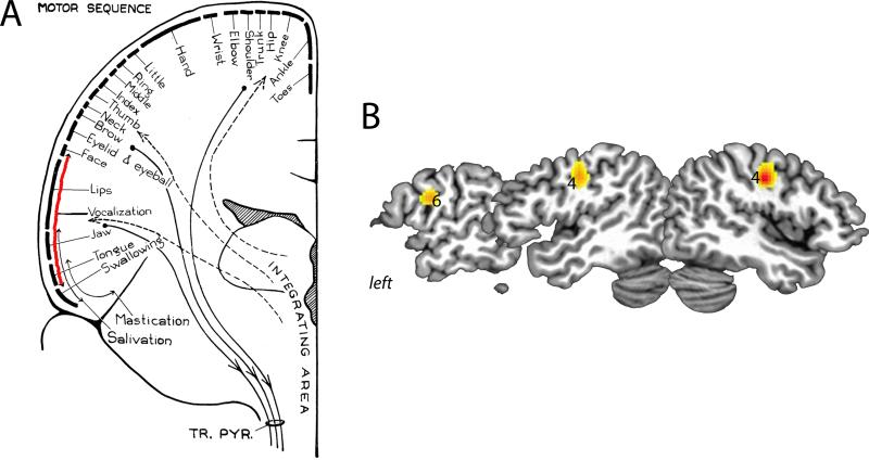
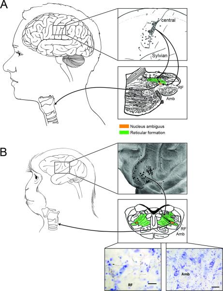
Our ability to learn and control the motor aspects of complex laryngeal behaviors, such as speech and song, is modulated by the laryngeal motor cortex (LMC), which is situated in the area 4 of the primary motor cortex and establishes both direct and indirect connections with laryngeal motoneurons. In contrast, the LMC in monkeys is located in the area 6 of the premotor cortex, projects only indirectly to laryngeal motoneurons and its destruction has essentially no effect on production of species-specific calls. These differences in cytoarchitectonic location and connectivity may be a result of hominid evolution that led to the LMC shift from the phylogenetically 'old' to 'new' motor cortex in order to fulfill its paramount function, that is, voluntary motor control of human speech and song production.
Copyright © 2014 Elsevier Ltd. All rights reserved.
Figures
References
-
- Jürgens U. Neural pathways underlying vocal control. Neurosci Biobehav Rev. 2002;26:235–258. [A compehensive review of literature and hypotheses on the central and peripheral organization of voice control in humans and non-human primates.] - PubMed
-
- Penfield W, Boldrey E. Somatic motor and sensory representation in the cerebral cortex of man as studied by electrical stimulation. Brain. 1937;60:389–443. [Seminal work on the mapping of the cerebral cortex in human patients, which revealed a somatotopic organization of the motor cortex.]
-
- Hast MH, Fischer JM, Wetzel AB, Thompson VE. Cortical Motor Representation of Laryngeal Muscles in Macaca-Mulatta. Federation Proceedings. 1974;33:399–399. [A direct electrical stimulation study of the brain in the rhesus monkey, which described the localization of individual laryngeal muscles in this species.] - PubMed
-
- Woolsey CN, Settlage PH, Meyer DR, Sencer W, Hamuy TP, Travis AM. Patterns of Localization in Precentral and Supplementary Motor Areas and Their Relation to the Concept of a Premotor Area. Research Publications- Association for Research in Nervous and Mental Disease. 1950;30:238–264. - PubMed
Publication types
MeSH terms
Grants and funding
LinkOut - more resources
Full Text Sources
Other Literature Sources
