. 2014 Aug 7;55(3):422-435.
doi: 10.1016/j.molcel.2014.05.012. Epub 2014 Jun 12.

The calcineurin signaling network evolves via conserved kinase-phosphatase modules that transcend substrate identity

Affiliations

The calcineurin signaling network evolves via conserved kinase-phosphatase modules that transcend substrate identity

Aaron Goldman et al. Mol Cell. .

Abstract

To define a functional network for calcineurin, the conserved Ca(2+)/calmodulin-regulated phosphatase, we systematically identified its substrates in S. cerevisiae using phosphoproteomics and bioinformatics, followed by copurification and dephosphorylation assays. This study establishes new calcineurin functions and reveals mechanisms that shape calcineurin network evolution. Analyses of closely related yeasts show that many proteins were recently recruited to the network by acquiring a calcineurin-recognition motif. Calcineurin substrates in yeast and mammals are distinct due to network rewiring but, surprisingly, are phosphorylated by similar kinases. We postulate that corecognition of conserved substrate features, including phosphorylation and docking motifs, preserves calcineurin-kinase opposition during evolution. One example we document is a composite docking site that confers substrate recognition by both calcineurin and MAPK. We propose that conserved kinase-phosphatase pairs define the architecture of signaling networks and allow other connections between kinases and phosphatases to develop that establish common regulatory motifs in signaling networks.


Figures

Figure 1
Figure 1. Characterization of the CN-dependent phosphoproteome
(A) Strategy used to identify CN substrates. (B) Overlap between the 699 phosphorylated peptides enriched in CN-deficient samples identified in distinct screens. Phosphorylated peptides from verified CN substrates are labeled. See also Figure S1A,C and Table S1. (C) Logo of 18 verified PxIxIT motifs used to generate the consensus motif: P[^PG][IVLF][^PG][IVLF][TSHEDQNKR]. See also Figure S1B and Table S2. (D) Overlap between 363 proteins with a predicted accessible PxIxIT motif and 387 proteins with one or more calcineurin-regulated phosphorylated peptide. See also Table S3.
Figure 2
Figure 2. Interaction of candidates with CN
(A) High- or (B) low-expressing GST-candidate fusions were purified from strain JRY11 to assay co-purification with Cna2-STEV-ZZ (αS) and Cna1-GFP (αGFP). Candidates classified as interactors (+), noninteractors (-), or ambiguous (*). See also Table S3.
Figure 3
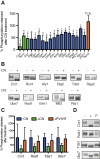
Figure 3. Identification of new CN substrates
(A) In vitro dephosphorylation of GST candidate fusions by CN. “% Phosphorylation retained after CN treatment”: Normalized 32P content of each CN-treated sample was compared to its paired mock-treated sample, which was set to 100%; for each protein, experimental replicates were averaged. Established (green) and new (blue) substrates and non-substrates (red) are shown (n = 2 for Aly1, Rcn1, and Atg13, n ≥ 3 for all others). Error bars represent 1 SD. Data were analyzed by Student's T-test. * p < 0.05, ** p < 0.01, *** p < 0.001, and **** p < 0.0001. See also Table S3. (B) Substrates that showed electrophoretic mobility changes in CN-treated (+) vs. mock-treated (-) samples. (C) Dephosphorylation of Crz1, Rod1, Tda1 and Ubx7 is inhibited by high-affinity PVIVIT peptide. “% Phosphorylation retained after treatment”: Normalized 32P contents of mock-treated, CN-treated, and CN + PVIVIT-treated samples were determined for each experimental replicate and averaged, with the average of mock-treated set to 100%. Error bars represent 1 SD (n = 6 for Crz1, Rod1 and Ubx7, n = 4 for Tda1). (D) Electrophoretic mobility changes in mock- treated (-), CN-treated (+) or CN + PVIVIT treated (P).
Figure 4
Figure 4. Characterization of the CN signaling network
(A) Gene products in CN signaling network grouped by associated cellular processes (see Table S3 and Supplemental Experimental Procedures). CN substrates (rectangles) and interacting proteins (octagons) are shown. Orange lines depict interaction with CN; line width indicates the amount of evidence supporting the interaction (see Table S3). Physical and genetic interactions between network members shown with dotted blue lines. Node size is proportional to connection number. Symbols with thick outlines designate gene products whose interaction with CN is newly identified in this study. (B) GO terms significantly enriched in the network relative to the yeast proteome. P-values of enrichment from hypergeometric distribution are shown. See also Figure S2 and Table S3.
Figure 5
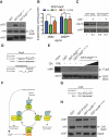
Figure 5. CN negatively regulates Elm1 activity
(A) Immunoblot analysis of extracts from CN-proficient (mock-treated, -FK) or CN-deficient (FK506-treated, +FK) yeast overexpressing GST-Elm1 or GST-Elm1ΔPSIHID. (B) Analysis of GST-Elm1 purified from CN-deficient yeast extracts and treated with buffer (NT), CN, or λ phosphatase (λ) in vitro. (C) Schematic of phosphorylation sites identified in Elm1 (see Table S1). CN-dependent dephosphorylation sites in the C-terminal domain (CTD) are shown (red) as well as the PxIxIT motif (PSIHID) (D) Immunoblot analysis of GST-Elm1 or GST-Elm1-4D (S516D, S519D, S562D, S581D) purified from WT or BY4741::cnb1Δ (Δ) yeast. (E) Kinase activity of GST-Elm1 or GST-Elm1-4D purified from WT or BY4741::cnb1Δ yeast and subsequently treated with (+CN) or without CN (-CN). 32P incorporation into Snf1 substrate shown in arbitrary units (AU). Error bars are 1 SD from triplicate experiments; data analyzed by two-way ANOVA with Bonferroni post-tests. ** p < 0.01, *** p < 0.001, and **** p < 0.0001. (F) β-galactosidase activity of extracts from sak1Δ tos3Δ crz1Δ cells expressing HXT2-lacZ at indicated times following transfer to YPD, pH 8.0. Error bars are 1 SD from triplicate samples; data analyzed by paired, two-way ANOVA. **** p < 0.0001. See also Figure S3A. (G) Growth rate of elm1-as cln1Δ cln2Δ treated with 1-NM-PP1 and/or FK506. Error bars are 1 SD from triplicate samples; data analyzed by one-way ANOVA. ** p < 0.05. See also Figure S3B. (H) Model for dual regulation of genes by CN through Elm1 and Crz1 during alkaline stress. See also Table S4. (I) Model for regulation of Elm1 by Ca2+/CN signaling during G1.
Figure 6
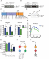
Figure 6. CN inhibits pheromone signaling by dephosphorylating Dig2 and competes with MAPK for substrate binding via overlapping docking sites
(A) Co-purification of GST or GST-Dig295-116 (WT or Mut, see D) with recombinant His-tagged CN (bait). (B) β-galactosidase activity of extracts from pheromone-treated dig1Δ cells containing FUS1-lacZ and DIG2 (JRY16) or DIG2Mut (JRY17) and expressing vector (pRS316) or constitutively active Cna1 (CN*, pCNA1trunc-316, encoding a stop codon at Ser417 to remove the calmodulin binding and autoinhibitory domains of Cna1). Cells were treated with FK506 where indicated. Error bars are 1 SD in triplicate experiments. Data analyzed by Student's T-test. * p < 0.01 and *** p < 0.001. See also Figure S4. (C) Immunoblot analysis of GST-Dig2 or GST-Dig2Mut purified from extracts of pheromone-treated JRY11 cells expressing either pRS314-CNA1trunc (+CN*, constitutively active Cna1) or pRS314-CNA1 (-CN*), with α-phospho-Ser/Thr (αSpTp) and αGST antisera. Relative phosphorylation was quantified by comparing αSpTp to αGST signal and normalized to lane 1. (D) Overlapping PxIxIT/D-site in Dig2. Consensus residues in PxIxIT (*) and D-site (underlined) are shown. Key: Φ, hydrophobic residue; ζ, hydrophilic residue; Ψ, basic residue. (E) Co-purification of recombinant MBP-Fus3 with GST-Dig295-116 (WT or Mut, bait). CN was included in increasing amounts where indicated. (F) Model of CN-dependent regulation of Ste12-activated transcription during mating via Dig2; Ca2+ activates CN, which dephosphorylates Dig2 to inhibit Ste12 activity. Fus3 and CN bind competitively to Dig2 via overlapping PxIxIT/D-site (green box). (G) Overlapping PxIxIT/D-site in human JunB. (H) Co-purification of GST-tagged PVIVIT, Jun B31-48, or Jun B31-48 Mut with His-tagged human CN (bait). See also Table S2.
Figure 7
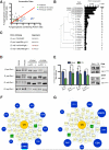
Figure 7. A Conserved set of kinase families act antagonistically to CN in yeast and mammals
(A) Conservation of PxIxIT sites in yeast CN network proteins and sequences that match the scrambled PxIxIT consensus is compared; all sites are predicted to occur in accessible protein domains. Each point represents a single protein. For proteins that contain multiple sites, the average result from all sites is plotted. Proteins with verified PxIxIT sites are labeled. Red line represents equivalent PxIxIT vs. scrambled conservation. Analysis of Rcn1 non-canonical PxIxIT site is described in supplemental experimental procedures. See also Figure S5B. (B) Tally and percentages of confirmed and putative PxIxIT sites from S. cerevisiae that are conserved in other fungal species. Unweighted phylogenetic tree for fungal species is shown to the left. See also Figure S5A. (C) Alignment of Elm1 PxIxIT site with corresponding regions of the homologous proteins from sensu stricto species. Positions that diverge from the PxIxIT consensus sequence are highlighted. See also Figure S5C. (D) Immunoblot analyses with αGST antisera of GST-tagged sensu stricto Elm1 proteins (Elm1-pEGH, pMMC-6, pMMC-7, pJR172), expressed in Ca2+-treated BY4741 with or without FK506 treatment. Each protein was purified, then incubated in vitro with λ phosphatase either in the presence (λ+PPI) or absence (λ) of phosphatase inhibitors. See also Figure S5D-E. (E) In vitro dephosphorylation of GST-Elm1 orthologs purified from BY4741::cnb1Δ and processed as in Figure 3A. S. cer Elm1 and Elm1ΔPSIHID are S. cerevisiae proteins containing or lacking the CN docking site. S. mik Elm1 and Elm1PSIHID are S. mikatae proteins containing either residues VSSHTD or PSIHID at positions 468-473. “% Phosphorylation retained after treatment”: Normalized 32P contents of CN-treated samples were compared to their paired mock-treated samples and averaged, with the average of mock-treated set to 100%. Error bars represent 1 SD (n =3). Data were analyzed by Student's T-test. * p < 0.01 and **** p < 0.0001. Electrophoretic mobility changes of representative samples are depicted on the right. (F-G) Conserved kinases, grouped by family, that were significantly associated both with (F) yeast CN signaling network and (G) mammalian CN substrates. Kinase node size is proportional to its connectivity with CN substrates and interacting proteins. Nodes: CN phosphatase (orange), kinases (blue), conserved substrates (green), CN substrates and interacting proteins (rectangles). Edges: dephosphorylation (orange), phosphorylation (blue), CN targets (dot), kinase targets (arrow). See also Tables S5 and S6 for description of kinase family members and listing of specific interactions.

References

    1. Aramburu J, Garcia-Cozar F, Raghavan A, Okamura H, Rao A, Hogan PG. Selective inhibition of NFAT activation by a peptide spanning the calcineurin targeting site of NFAT. Mol Cell. 1998;1:627–637. - PubMed
    1. Aramburu J, Yaffe MB, Lopez-Rodriguez C, Cantley LC, Hogan PG, Rao A. Affinity-driven peptide selection of an NFAT inhibitor more selective than cyclosporin A. Science. 1999;285:2129–2133. - PubMed
    1. Asano S, Park JE, Yu LR, Zhou M, Sakchaisri K, Park CJ, Kang YH, Thorner J, Veenstra TD, Lee KS. Direct phosphorylation and activation of a Nim1-related kinase Gin4 by Elm1 in budding yeast. J Biol Chem. 2006;281:27090–27098. - PubMed
    1. Bardwell L. A walk-through of the yeast mating pheromone response pathway. Peptides. 2005;26:339–350. - PMC - PubMed
    1. Bhaduri S, Pryciak PM. Cyclin-specific docking motifs promote phosphorylation of yeast signaling proteins by G1/S Cdk complexes. Curr Biol. 2011;21:1615–1623. - PMC - PubMed

Publication types

MeSH terms

Substances

LinkOut - more resources