. 2014 Sep;35(27):7786-99.
doi: 10.1016/j.biomaterials.2014.05.018. Epub 2014 Jun 13.

Inhibition of apoptosis in human induced pluripotent stem cells during expansion in a defined culture using angiopoietin-1 derived peptide QHREDGS

Affiliations

Inhibition of apoptosis in human induced pluripotent stem cells during expansion in a defined culture using angiopoietin-1 derived peptide QHREDGS

Lan T H Dang et al. Biomaterials. 2014 Sep.

Abstract

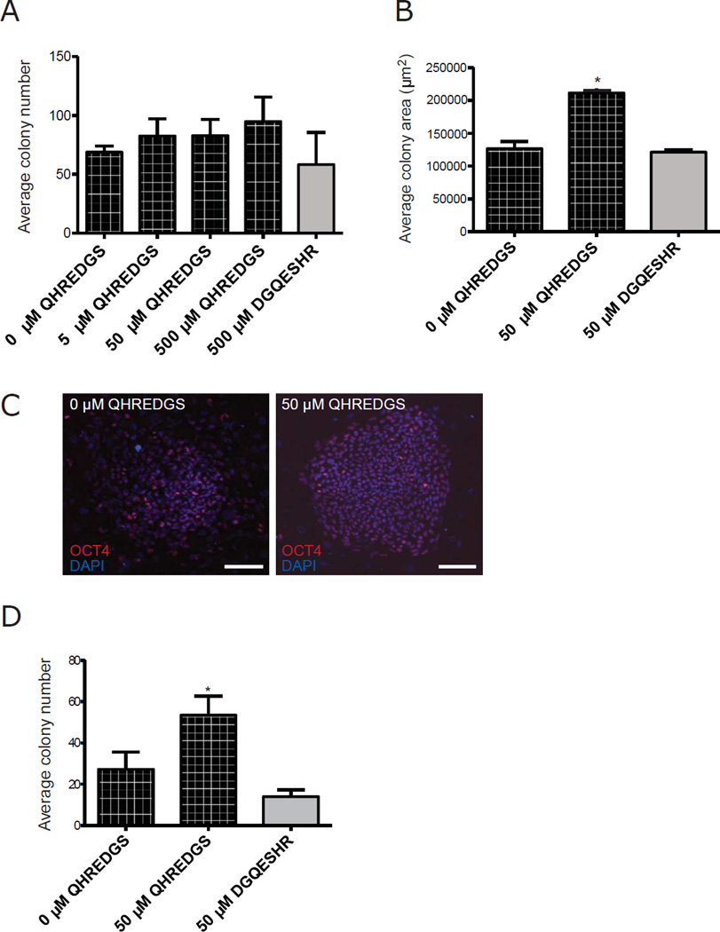
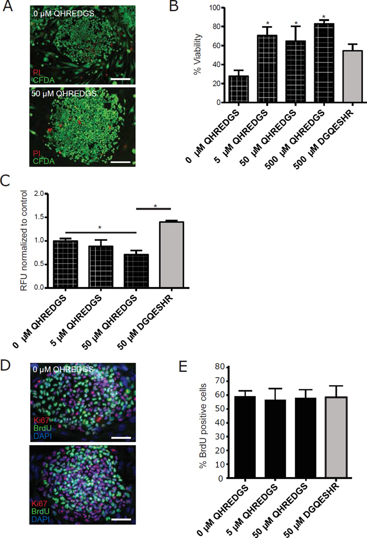
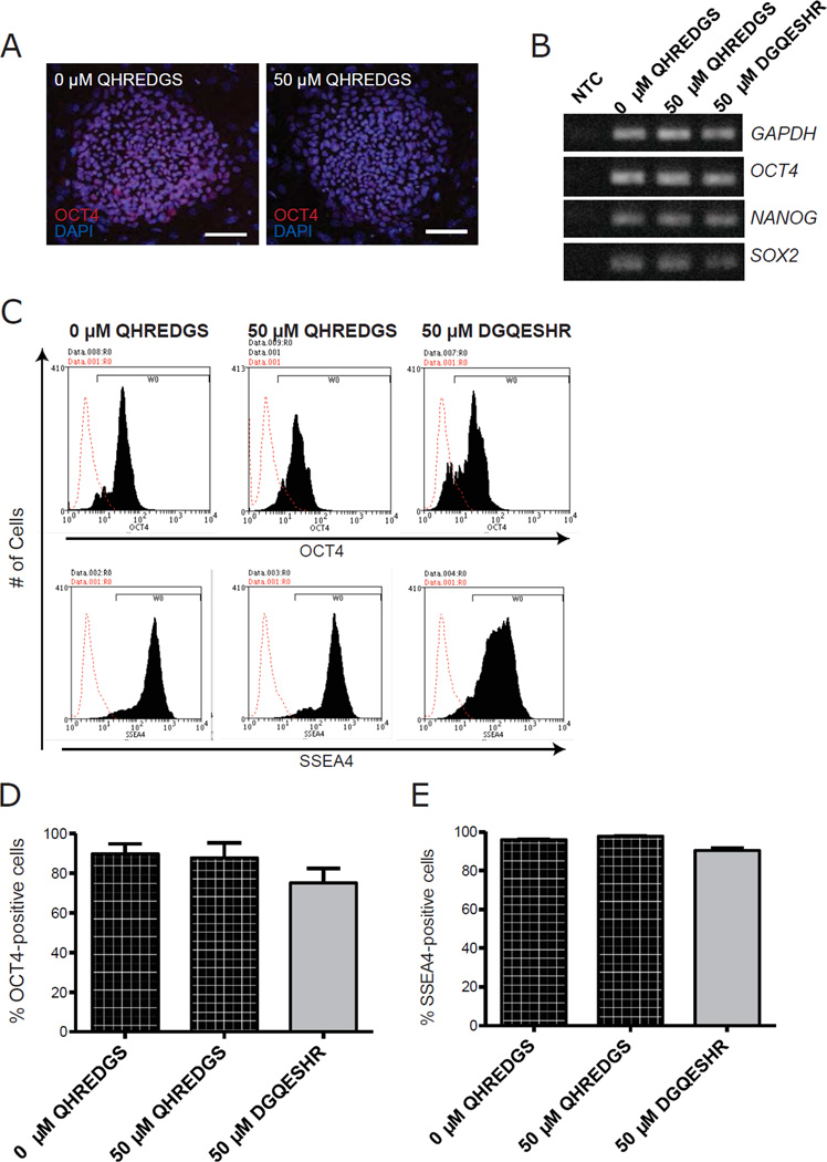
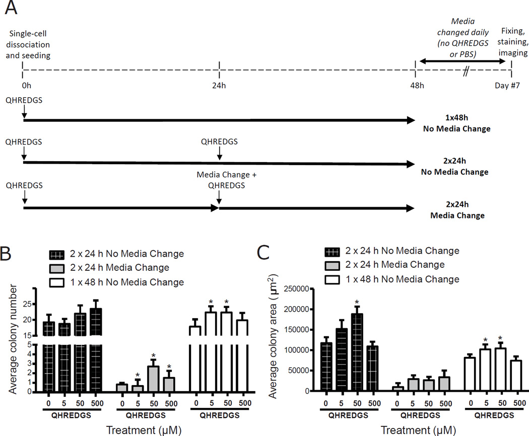
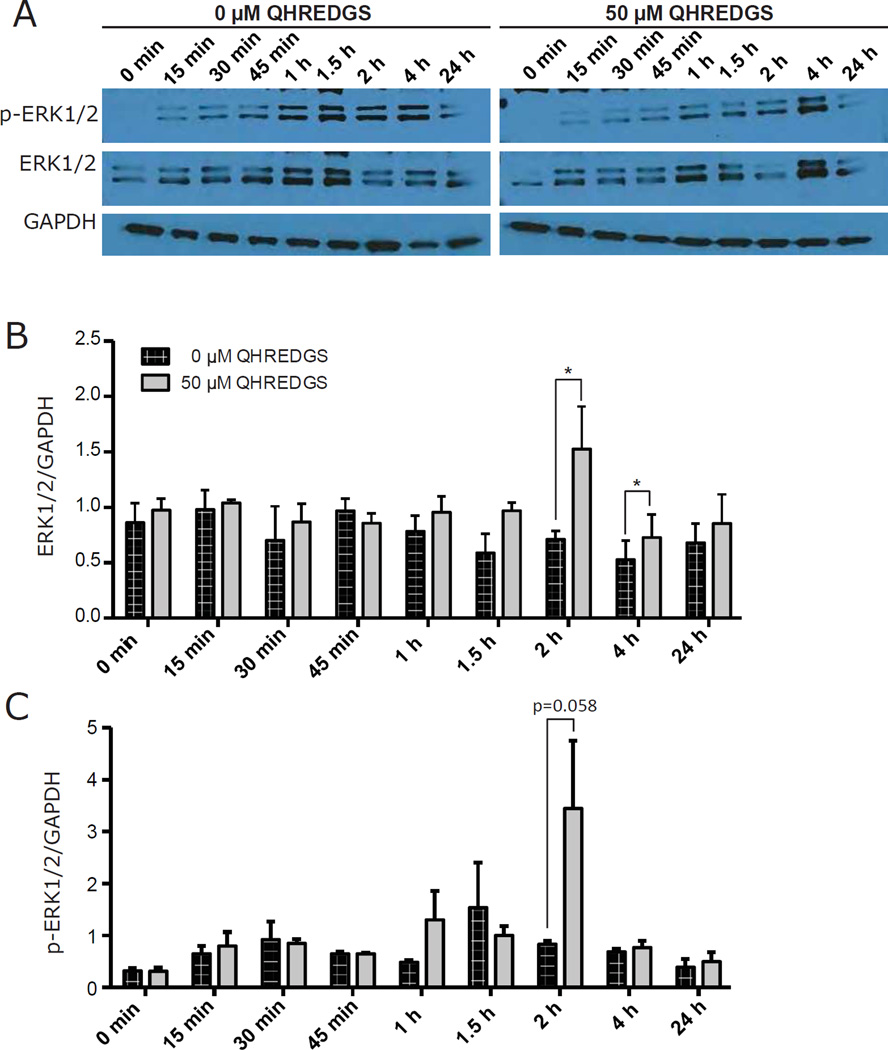
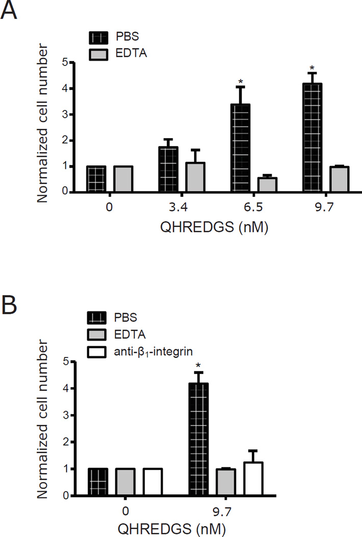
Adhesion molecule signaling is critical to human pluripotent stem cell (hPSC) survival, self-renewal, and differentiation. Thus, hPSCs are grown as clumps of cells on feeder cell layers or poorly defined extracellular matrices such as Matrigel. We sought to define a small molecule that would initiate adhesion-based signaling to serve as a basis for a defined substrate for hPSC culture. Soluble angiopoeitin-1 (Ang-1)-derived peptide QHREDGS added to defined serum-free media increased hPSC colony cell number and size during long- and short-term culture when grown on feeder cell layers or Matrigel, i.e. on standard substrates, without affecting hPSC morphology, growth rate or the ability to differentiate into multiple lineages both in vitro and in vivo. Importantly, QHREDGS treatment decreased hPSC apoptosis during routine passaging and single-cell dissociation. Mechanistically, the interaction of QHREDGS with β1-integrins increased expression of integrin-linked kinase (ILK), increased expression and activation of extracellular signal-regulated kinases 1/2 (ERK1/2), and decreased caspase-3/7 activity. QHREDGS immobilization to polyethylene glycol hydrogels significantly increased cell adhesion in a dose-dependent manner. We propose QHREDGS as a small molecule inhibitor of hPSC apoptosis and the basis of an affordable defined substrate for hPSC maintenance.

Keywords: Adhesion; Angiopoietin-1; Apoptosis; Human pluripotent stem cells; Integrins; QHREDGS.


Figures

Figure 1
Figure 1. QHREDGS increases hiPSC colony number and size during clump and single-cell passaging in serum-free, feeder layer culture conditions
[A–B] BJ1Ds hiPSCs pre-treated for 5 passages with PBS alone (0µM QHREDGS), an increasing concentration of QHREDGS peptide or the scrambled peptide DGQESHR (grey bar) were passaged in clumps in the presence of the treatments for 7 days, then colony number [A] and colony size [B] were determined. [C–D] Human iPSCs were pre-treated for 5 passages with PBS alone (0µM QHREDGS), 50µM QHREDGS or 50µM DGQESHR, dissociated to single-cell and plated at a low density, then cultured for 7 days in the presence of the treatments. [C] Representative Oct4- and DAPI staining images of BJ1D hiPSC colonies pre-treated with PBS (0µM QHREDGS) or 50µM QHREDGS, 7 days after single-cell dissociation (scale bar = 100µm). [D] Colony number determined for BJ1D hiPSCs 7 days after single-cell dissociation. Data presented are the mean ± SEM. P values are derived from Student’s t-test, P < 0.05 considered significant (n=3).
Figure 2
Figure 2. QHREDGS promotes hiPSC survival by inhibiting caspase-dependent apoptosis but does not affect proliferation
BJ1D hiPSCs were pre-treated for 5 passages with PBS alone (0µM QHREDGS), an increasing concentration of QHREDGS peptide or the scrambled peptide DGQESHR, dissociated to single cells and plated at a low density, then cultured for 7 days in the presence of the treatments. [A–B] The cells were stained with Propidium Iodide (PI, dead cells) and Carboxyfluorescein Diacetate Succinimidyl Ester (CFDA, live cells) (scale bar = 150µm). [A] Representative PI/CFPA staining images of BJ1D hiPSC colonies pre-treated with PBS (0µM QHREDGS) or 50µM QHREDGS, 7 days after single-cell dissociation. [B] Quantification of percent viability (live/dead cells). Grey bar denotes scrambled DGQESHR peptide treatment. [C] The cells were assayed for caspase-3/7 in BJ1D hiPSCs. The relative fluorescence units (RFU) per cell is indicative of caspase-3/7 activity. [D–E] The cells were assayed for BrdU incorporation, a measure of proliferation. [D] Representative BrdU, Ki67 and DAPI staining images of BJ1D hiPSC colonies pre-treated with PBS (0µM QHREDGS) or 50µM QHREDGS, 7 days after single-cell dissociation. [E] Quantification of percent BrdU-positive cells. Data presented are the mean ± SEM. P values are derived from Student’s t-test, P < 0.05 considered significant (n=3).
Figure 3
Figure 3. QHREDGS treatment does not affect the pluripotency of hiPSCs in vitro
BJ1D hiPSCs pretreated for 5 passages with PBS alone (0µM QHREDGS), 50µM QHREDGS or 50µM DGQESHR were assessed for their ability to express pluripotency markers. [A] Representative Oct4 and DAPI staining images of BJ1D hiPSC colonies pre-treated with PBS (0µM QHREDGS) or 50 µM QHREDGS, 7 days after single-cell dissociation. [B] Agarose gels showing RT-PCR products generated by primers for the pluripotency markers OCT4, NANOG and SOX2 and the housekeeping gene, GAPDH. No Template Control is denoted by NTC. [C–E] Flow cytometric analysis of PBS (0µM QHREDGS), 50µM QHREDGS and 50µM DGQESHR treated BJ1D hiPSCs. [C] Representative Oct4 (top panels) and SSEA4 histograms (bottom panels). Red denotes secondary antibody only control and black denotes cell population positive for the designated antibody. [D–E] Quantification of Oct4+ [D] and SSEA4+ [E] cells. Data presented are the mean ± SEM. P values are derived from Student’s t-test, P < 0.05 considered significant (n=3).
Figure 4
Figure 4. Long-term QHREDGS treatment does not affect the ability of hiPSCs to differentiate into cells of all three germ layers in vitro and in vivo
[A–B] BJ1D hiPSCs pre-treated for 5 passages with PBS (0µM QHREDGS), 50µM QHREDGS or 50µM DGQESHR were subjected to an embryoid body-based differentiation protocol. [A] Representative images of differentiated cells pre-treated with PBS alone (0µM QHREDGS; top row) or 50µM QHREDGS (bottom row) and stained for SMA (mesoderm), GATA 6 (endoderm) or βIII-tubulin (ectoderm) (n=3). [B] Agarose gels showing RT-PCR products generated by primers for the germ layer markers PAX6 (ectoderm), AFP (endoderm), BRACHYURY (mesoderm), and CDX2 (mesoderm) (n=3). [C–D] BJ1D hiPSCs treated with PBS alone (0µM QHREDGS), 50µM QHREDGS or 50µM DGQESHR for 10 passages were subjected to a teratoma formation assay (n=3). [C] Representative images of teratomas formed from cells pre-treated with PBS alone (0µM QHREDGS) or 50µM QHREDGS. [D] Representative images of teratomas formed from cells pre-treated with PBS alone (0µM QHREDGS), 50µM QHREDGS or 50µM DGQESHR sectioned and stained with H&E. Arrows, ectodermal; asterisks, mesodermal; and arrowheads, endodermal structures.
Figure 5
Figure 5. Single dose, low concentration QHREDGS treatment improves colony number and size of single-cell dissociated hiPSCs in serum-free, feeder-free culture conditions
[A] Short-term QHREDGS treatment protocol. At 0h, BJ1D hiPSCs were single-cell dissociated and seeded onto Matrigel in the presence of PBS alone (0µM QHREDGS) or QHREDGS. Three different treatment schemes were investigated: (1) cells remained undisturbed for 48h at 37°C (1×48h No Media Change); (2) at 24h, an additional dose of PBS or QHREDGS was added to the media and the cells returned to the incubator (2×24h No Media Change); or (3) at 24h, the media was aspirated and replaced, and an additional dose of PBS or QHREDGS was added to the media and the cells returned to the incubator (2×24h Media Change). At 48h, the media was replaced with PBS/QHREDGS-free medium for all groups. The cells were fed daily with QHREDGS-free medium for the following 5 days. On day 7, the cells were fixed and stained for Oct4 and counterstained with DAPI. [B–C] BJ1D hiPSCs were single-cell dissociated and seeded onto Matrigel in the presence of PBS alone (0µM QHREDGS) or QHREDGS (5, 50 or 500µM), as described in [A]. After 7 days, the cells were fixed and stained for Oct4 and the number of Oct4-positive colonies in each well was counted [B] and the area of the colonies was measured [C]. Data presented are the average ± SEM. P values are derived from Student’s t-test, P < 0.05 considered significant (n=3–7).
Figure 6
Figure 6. QHREDGS increases ILK expression
[A–B] BJ1D hiPSCs were single-cell dissociated and seeded onto Matrigel in the presence of PBS (0µM QHREDGS) or 50µM QHREDGS. At various time points, cells were scraped and lysed. Cell lysates were analyzed by Western blot analysis using the denoted antibodies. The resultant bands were quantified by densitometry. [A] Representative images of Western blot membranes probed with denoted antibodies. [B] Quantification of Western blot analysis. Data presented are the average normalized ILK concentration determined from the ILK:GAPDH ratio ± SEM. P values are derived from Student’s t-test, P < 0.05 considered significant (n=3–5).
Figure 7
Figure 7. QHREDGS increases activation of ERK1/2
[A–C] BJ1D hiPSCs were single-cell dissociated and seeded onto Matrigel in the presence of PBS (0µM QHREDGS) or 50µM soluble QHREDGS. At various time points, cells were scraped and lysed. Cell lysates were analyzed by Western blot analysis using the denoted antibodies and the resultant bands were quantified by densitometry. [A] Representative images of Western blot membranes probed with denoted antibodies. [B–C] Quantification of Western blot analysis. Data presented are the average normalized ERK1/2 concentration determined from the ERK1/2:GAPDH ratio ± SEM [B] and the average normalized phospho-ERK1/2 (p-ERK1/2) concentration determined from the p-ERK1/2:GAPDH ratio ± SEM [C]. P values are derived from Student’s t-test, P < 0.05 considered significant (n=3).
Figure 8
Figure 8. QHREDGS binding to β1-integrin receptors is sufficient to promote adhesion of single-cell dissociated hiPSCs to non-fouling PEG hydrogels
[A] BJ1D hiPSCs were single-cell dissociated and incubated with PBS or 10mM EDTA prior to seeding onto PEG hydrogels conjugated with different amounts of QHREDGS peptide. After 6 hours, cells were fixed, stained with DAPI, imaged and the cells adherent to the hydrogel were counted. Data presented are the average normalized values ± SEM (n = 3–5); values were normalized by dividing by the number of cells that adhered to the PEG hydrogel in a given experiment. P values were determined from paired Student’s t-tests, P < 0.05 considered significant. [B] BJ1D hiPSCs were single-cell dissociated and incubated with PBS, 10mM EDTA or 50µg/mL anti-β1-integrin antibody prior to seeding onto PEG hydrogels with or without QHREDGS conjugated. Data presented are the average normalized values ± SEM; values were normalized by dividing by the number of cells that adhered to the PEG hydrogel in a given experiment. P values were determined from paired Student’s t-tests, P < 0.05 considered significant (n = 3–5).
Figure 9
Figure 9. QHREDGS-dependent pro-survival mechanism in hPSCs
Model depicting the postulated mechanism for increased QHREDGS-dependent hPSC survival, wherein the binding of QHREDGS to a β1-integrin receptor on the hPSC surface increases ILK, stimulating the upregulation of the pro-survival ERK1/2 signaling pathway and the eventual downregulation of caspase-dependent apoptotic pathways.

Similar articles

Cited by

References

    1. Watanabe K, Ueno M, Kamiya D, Nishiyama A, Matsumura M, Wataya T, et al. A ROCK inhibitor permits survival of dissociated human embryonic stem cells. Nat Biotechnol. 2007;25:681–686. - PubMed
    1. Ohgushi M, Matsumura M, Eiraku M, Murakami K, Aramaki T, Nishiyama A, et al. Molecular pathway and cell state responsible for dissociation-induced apoptosis in human pluripotent stem cells. Cell Stem Cell. 2010;7:225–239. - PubMed
    1. Chen G, Hou Z, Gulbranson DR, Thomson JA. Actin-myosin contractility is responsible for the reduced viability of dissociated human embryonic stem cells. Cell Stem Cell. 2010;7:240–248. - PMC - PubMed
    1. Xu Y, Zhu X, Hahm HS, Wei W, Hao E, Hayek A, et al. Revealing a core signaling regulatory mechanism for pluripotent stem cell survival and self-renewal by small molecules. Proc Natl Acad Sci U S A. 2010;107:8129–8134. - PMC - PubMed
    1. Li L, Bennett SA, Wang L. Role of E-cadherin and other cell adhesion molecules in survival and differentiation of human pluripotent stem cells. Cell Adh Migr. 2012;6:59–70. - PMC - PubMed

Publication types

MeSH terms