. 2014 Jul 1;20(1):61-72.
doi: 10.1016/j.cmet.2014.05.004. Epub 2014 Jun 12.

The glucose transporter Glut1 is selectively essential for CD4 T cell activation and effector function

Affiliations

The glucose transporter Glut1 is selectively essential for CD4 T cell activation and effector function

Andrew N Macintyre et al. Cell Metab. .

Abstract

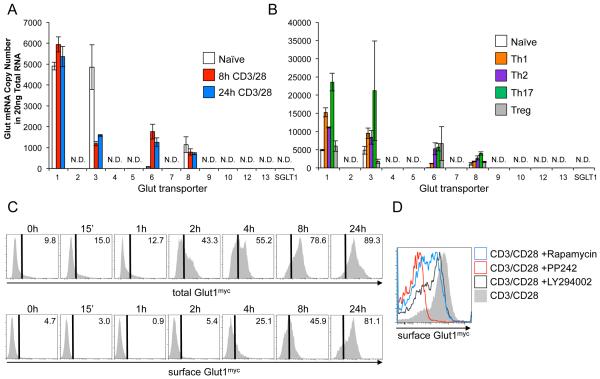
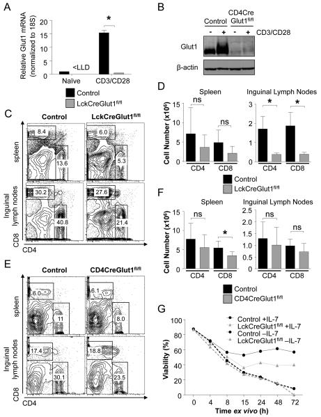
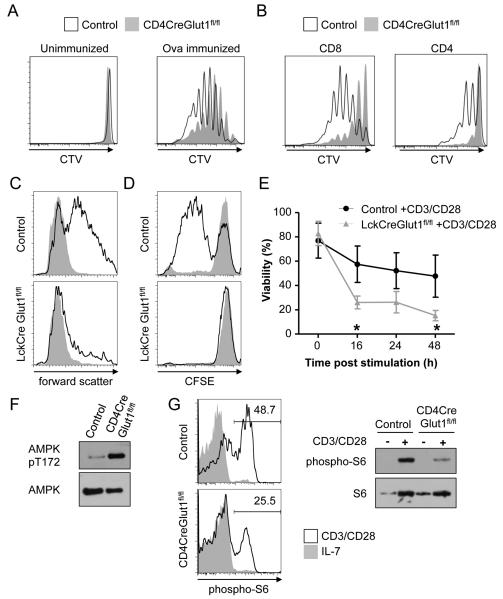
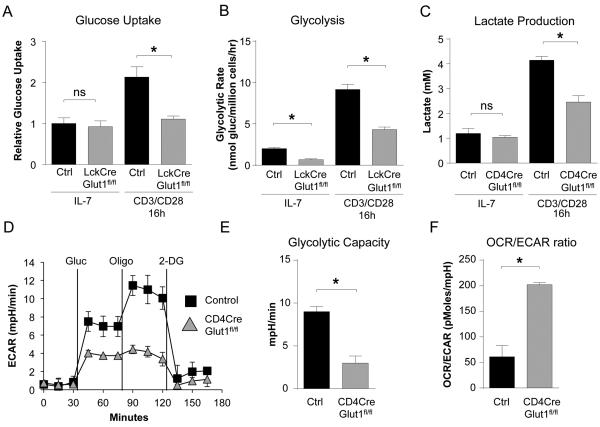
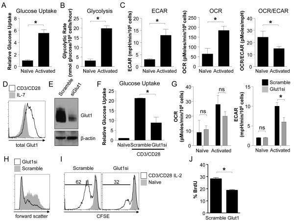
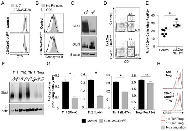
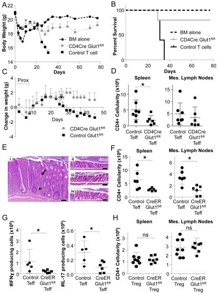
CD4 T cell activation leads to proliferation and differentiation into effector (Teff) or regulatory (Treg) cells that mediate or control immunity. While each subset prefers distinct glycolytic or oxidative metabolic programs in vitro, requirements and mechanisms that control T cell glucose uptake and metabolism in vivo are uncertain. Despite expression of multiple glucose transporters, Glut1 deficiency selectively impaired metabolism and function of thymocytes and Teff. Resting T cells were normal until activated, when Glut1 deficiency prevented increased glucose uptake and glycolysis, growth, proliferation, and decreased Teff survival and differentiation. Importantly, Glut1 deficiency decreased Teff expansion and the ability to induce inflammatory disease in vivo. Treg cells, in contrast, were enriched in vivo and appeared functionally unaffected and able to suppress Teff, irrespective of Glut1 expression. These data show a selective in vivo requirement for Glut1 in metabolic reprogramming of CD4 T cell activation and Teff expansion and survival.


Figures

Figure 1
Figure 1. Glut1 is selectively and rapidly increased in activated murine T cell activation
(A, B) Glut family mRNA copy number in (A) naïve and CD3/CD28-stimulated CD4 murine T cells, and (B) in vitro polarized CD4 T cell subsets. N.D.: not detected. (C, D) Glut1myc expression in CD3/CD28 stimulated CD4 Glut1myc T cells (C) over time and (D) with inhibitors or vehicle control. Mean ± SD from 3 or more independent experiments are shown.
Figure 2
Figure 2. Glut1 is not required for survival of quiescent peripheral T cells
(A, B) Glut1fl/fl and LckCreGlut1fl/fl or CD4CreGlut1fl/fl T cells were rested in IL-7 (naive) or CD3/CD28-stimulated for 16h and Glut1 levels measured by (A) qrtPCR or (B) immunoblot. (C-F) Flow cytometry (C, E) and cell numbers (D, F) are shown for CD4 and CD8 cells from spleen and inguinal lymph nodes of control and (C and D) LckCreGlut1fl/fl or (E and F) CD4CreGlut1fl/fl mice. (G) Viability of isolated control (Glut1fl/+) and LckCreGlut1fl/fl T cells over time in culture ± IL-7. Mean cell count ± SD or representative data from (C, D) 7 and (E, F) 5 animals and (A, B, G) 2 or more independent experiments are shown.
Figure 3
Figure 3. Glut1 is necessary to support activation-induced growth, proliferation and survival
(A, B) Proliferation of CellTrace Violet (CTV) labeled control (Glut1fl/fl) and CD4CreGlut1fl/fl (A) OT-II transgenic T cells on day 3 after adoptive transfer into intact recipients ± immunization with Ovalbumin or (B) T cells 6 days after adoptive transfer into irradiated recipients for homeostatic proliferation. (C) Control (Glut1fl/fl) and LckCreGlut1fl/fl T cells were rested in IL-7 or CD3/CD28-stimulated and cell size (forward scatter) of viable cells was determined by flow cytometry after 24h. (D, E) Control (Glut1fl/+) and LckCreGlut1fl/fl T cells were CFSE-labeled and either rested in IL-7 or CD3/CD28-stimulated and examined by flow cytometry for (D) proliferation at 72 hours or (E) viability over time. (F) Control (Glut1fl/fl) and CD4CreGlut1fl/fl T cells were CD3/CD28-stimulated 16h and analyzed by immunoblot. (G) Control (Glut1fl/fl) and CD4CreGlut1fl/fl T cells were rested in IL-7 or CD3/CD28-stimulated for 10h and analyzed by intracellular flow cytometry and immunoblot. Data are representative of n=3 mice/group (A, B) a minimum of (F, G) 2 or (C-E) 3 experiments. (E) Shows mean ± SD of 3 independent experiments.
Figure 4
Figure 4. Glut1 is required for activation-induced metabolic reprogramming
(A, B) Control (Glut1fl/fl) and LckCreGlut1fl/fl T cells were rested in IL-7 or CD3/CD28-stimulated and (A) glucose uptake or (B) glycolytic rate was measured after 16h. (C-F) Control (Glut1fl/fl) and CD4CreGlut1fl/fl T cells were rested in IL-7 or CD3/CD28-stimulated for 16h. (C) Total lactate produced was measured. (D) Extracellular acidification rate (ECAR) was assessed after the addition of glucose (gluc), oligomycin (oligo), and 2-deoxyglucose (2-DG) at indicated times and (E) glycolytic capacity and (F) Oxygen Consumption Rate (OCR)/ECAR ratio determined. Mean ± SD (n=4) are shown from a minimum of 2 or more independent experiments.
Figure 5
Figure 5. Activation of human T cells triggers Glut1-dependent glycolytic reprogramming
(A-D) Isolated human T cells were rested in IL-7 (naïve) or CD3/CD28-stimulated for 48 hours to measure (A) glucose uptake, (B) glycolytic flux, (C) ECAR and OCR, and (D) Glut1 expression by flow cytometry. (E-G) T cells transfected with scrambled or Glut1-targeted siRNA pools were cultured in IL-7 (naïve) or CD3/CD28-stimulated 48h to measure (E) Glut1 by immunoblot, (F) glucose uptake, (G) and OCR and ECAR. (H-I) T cells were transfected as above, CFSE-labeled and rested in IL-7 (naïve) or CD3/CD28-stimulated for 72h in the presence of IL-2. Flow cytometry measured (H) cell size by forward scatter and (I) proliferation by CFSE dilution. (J) T cells were transfected and stimulated and Bromodeoxyuridine (BrdU) was added for the final 8h of culture. BrdU incorporation was assessed by flow cytometry. Data show mean ± SD and are representative of (A-J) 3-5 or (J) two independent experiments.
Figure 6
Figure 6. Glut1 is required for Teff, but not Treg or CTL, generation and function
(A) CD8 T cells from control (Glut1fl/+) and CD4CreGlut1fl/fl mice were CellTrace Violet (CTV) labeled and rested in IL-7 or CD3/CD28-stimulated + IL-2 for flow cytometric analysis after 48 hours. (B, C) Control (Glut1fl/+) and CD4CreGlut1fl/fl cytotoxic CD8 T lymphocytes (CTL) were analyzed (B) after 3h restimulation by flow cytometry and (C) immunoblot. (D, E) Flow cytometry of control (Glut1fl/fl) and LckCreGlut1fl/fl spleen for FoxP3+ CD4 T cells. (D) Representative plot and (E) cumulative data of fraction of CD4 T cells expressing FoxP3. (F-H) Th1, Th2, Th17, and Treg were induced using control (Glut1fl/+, Glut1fl/fl) and CD4CreGlut1fl/fl CD4 T cells and analyzed by (F) immunoblot, (G) flow cytometry to determine the number of live skewed cells expressing indicated subset markers. (H) Treg function was tested in an in vitro suppression assay. Data are representative or show mean cell count ± SD from (D, E) 5, (F-H) 3, or (A-C) 2 independent experiments.
Figure 7
Figure 7. Glut1 is selectively required for Teff, but not Treg, expansion and function in inflammatory disease
(A, B) GvHD was induced with transplant of T cell-depleted bone marrow (BM) or BM + control (Glut1fl/fl or Glut1fl/+) or CD4CreGlut1fl/fl T cells into allogeneic hosts and (A) body weight and (B) survival were measured over time. (C-D) Naïve control (Glut1fl/fl) or CD4CreGlut1fl/fl T cells were transferred into Rag1−/− hosts and colitis was triggered 2 weeks later via piroxicam (Pirox; day 0 on start of Pirox) exposure and (C) animal weights measured over time, or (D) mice were sacrificed at day 30 and the number of CD4 T cells present in the spleen and mesenteric (mes) lymph nodes determined by flow cytometry. (E-H) Rag1−/− mice were injected with control (CreERGlut1+/+, Glut1fl/fl) or CreERGlut1fl/fl naïve T cells. Colitis was triggered by piroxicam exposure 2 weeks after T cell transfer. Animals were then treated with tamoxifen to activate Cre. (E) H&E histology of proximal colon from mice that received (i) naïve control T cells, (ii) naïve CreERGlut1fl/fl T cells, (iii) naïve control T cells plus control Treg, or (iv) naïve control T cells plus CreERGlut1fl/fl Treg. Bar indicates 100μm; arrow indicates cryptic abscess and arrowhead indicates a granuloma. (F) The number of CD4 or (G) IFNγ or IL-17 producing T cells in the mesenteric (mes) lymph nodes was determined after 4 weeks by flow cytometry. (H) Rag1−/− mice were co-injected with wild type naïve T cells and either control (CreERGlut1+/+, Glut1fl/fl) or CreERGlut1fl/fl nTreg. Mice were treated after 2 weeks with piroxicam and tamoxifen to trigger IBD and activate Cre and CD4 T cells were determined after 4 weeks. Data are representative of 2 (A-B) or 3 (C-H) independent experiments and show mean clinical score ± SEM (A, C) or mean ± SD (D, F-H).

References

    1. Bian L, Josefsson E, Jonsson IM, Verdrengh M, Ohlsson C, Bokarewa M, Tarkowski A, Magnusson M. Dichloroacetate alleviates development of collagen II-induced arthritis in female DBA/1 mice. Arthritis research & therapy. 2009;11:R132. - PMC - PubMed
    1. Brand S. Crohn’s disease: Th1, Th17 or both? The change of a paradigm: new immunological and genetic insights implicate Th17 cells in the pathogenesis of Crohn’s disease. Gut. 2009;58:1152–1167. - PubMed
    1. Byersdorfer CA, Tkachev V, Opipari AW, Goodell S, Swanson J, Sandquist S, Glick GD, Ferrara JL. Effector T cells require fatty acid metabolism during murine graft-versus-host disease. Blood. 2013;122:3230–3237. - PMC - PubMed
    1. Carrette F, Surh CD. IL-7 signaling and CD127 receptor regulation in the control of T cell homeostasis. Semin Immunol. 2012;24:209–217. - PMC - PubMed
    1. Cham CM, Gajewski TF. Glucose availability regulates IFN-gamma production and p70S6 kinase activation in CD8+ effector T cells. J Immunol. 2005;174:4670–4677. - PubMed

Publication types

MeSH terms

LinkOut - more resources